I now have comprehensive material. Let me compile the full detailed response:
WHO 2021 Classification of Lung Tumors: Morphology, IHC, and Molecular Genetics
(Based on Robbins & Kumar Basic Pathology, Fishman's Pulmonary Diseases and Disorders, Sabiston Textbook of Surgery, and Harrison's Principles of Internal Medicine)
Overview and Historical Context
The WHO has classified lung tumors through four major editions (1999, 2004, 2015, 2021). The 2021 WHO classification represents the most significant update, driven by three forces:
- Widespread adoption of targeted therapies requiring precise molecular subtyping
- Recognition that most patients present with advanced disease and small biopsy/cytology specimens
- Improved immunohistochemical profiling and genomic characterization
The 2021 edition specifically expands commentary on small diagnostic samples, modernizes IHC-based subtype classification, and broadens the emphasis on genetic testing. - Sabiston Textbook of Surgery
Approximately 95% of primary lung tumors are carcinomas; the remaining 5% include carcinoids, mesenchymal malignancies, lymphomas, and rare benign lesions such as pulmonary hamartoma. - Robbins & Kumar Basic Pathology
WHO 2021 Major Categories of Malignant Lung Tumors
| Category | Key Subtypes |
|---|
| Adenocarcinoma | AIS, MIA, Invasive (lepidic, acinar, papillary, micropapillary, solid, invasive mucinous) |
| Squamous Cell Carcinoma | Keratinizing, Non-keratinizing, Basaloid |
| Large Cell Carcinoma | Undifferentiated (diagnosis of exclusion) |
| Adenosquamous Carcinoma | Mixed SCC + adenocarcinoma (>10% each) |
| Sarcomatoid Carcinoma | Pleomorphic, Spindle cell, Giant cell, Carcinosarcoma, Pulmonary blastoma |
| Neuroendocrine Tumors | Typical carcinoid, Atypical carcinoid, LCNEC, Small cell carcinoma |
| Salivary Gland-type Tumors | Mucoepidermoid, Adenoid cystic |
1. Adenocarcinoma
Epidemiology and Background
Adenocarcinoma is now the most common histologic subtype of lung carcinoma worldwide, accounting for approximately 50% of all lung cancers. It is the predominant subtype in:
- Women
- Never-smokers
- Patients under 45 years old
- People of Asian descent
It tends to metastasize earlier than squamous cell carcinoma, with a predilection for CNS involvement. Most tumors (75%) are peripherally located. - Sabiston Textbook of Surgery
Precursor Lesions
The 2021 WHO classification recognizes a clear stepwise progression:
- Atypical Adenomatous Hyperplasia (AAH): Cuboidal epithelium with mild nuclear atypia and mild interstitial fibrosis. Lesion <5 mm. The earliest recognized precursor.
- Adenocarcinoma in Situ (AIS): ≤3 cm solitary lesion with pure lepidic growth (tumor cells growing along alveolar septa without invasion, stromal, vascular, or pleural involvement). Replaced the old term "bronchioloalveolar carcinoma" (BAC). Typically nonmucinous. 100% disease-free survival after complete resection.
- Minimally Invasive Adenocarcinoma (MIA): ≤3 cm solitary lesion with predominantly lepidic growth and invasion ≤0.5 cm. Near 100% disease-free survival after resection.
The critical historic revision: the 2015 WHO classification abolished the term BAC entirely, replacing it with AIS and MIA. This was validated by studies showing that even small foci of invasion >5 mm altered prognosis. - Fishman's Pulmonary Diseases and Disorders
Morphology - Invasive Adenocarcinoma
The 2021 WHO classification defines five major growth patterns for invasive non-mucinous adenocarcinoma, classified by predominant pattern:
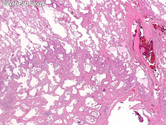
- Lepidic predominant: Tumor cells proliferate along preexisting alveolar walls without destruction. Cuboidal to columnar cells resembling type II pneumocytes. Best prognosis among invasive subtypes.
- Acinar predominant: Gland-forming, round to oval spaces lined by cuboidal to columnar cells. Intermediate prognosis.
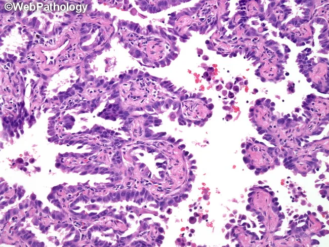
- Papillary predominant: Tumor cells lining true fibrovascular cores.
- Micropapillary predominant: Small papillary tufts lacking fibrovascular cores, floating in alveolar spaces. Associated with worst prognosis among adenocarcinoma subtypes and high rate of lymphovascular invasion.
- Solid predominant: Sheets of polygonal cells with abundant cytoplasm, without acinar, papillary, or lepidic formation, but with intracytoplasmic mucin on special stains.
Special subtypes include:
- Invasive Mucinous Adenocarcinoma (formerly mucinous BAC): Tall columnar goblet cells with basal nuclei and abundant apical mucin, growing in lepidic or acinar pattern. Often multifocal or lobar pneumonic pattern. Associated with KRAS mutations.
- Colloid adenocarcinoma, Fetal adenocarcinoma, Enteric-type adenocarcinoma: Rarer subtypes.
IHC Profile
| Marker | Status |
|---|
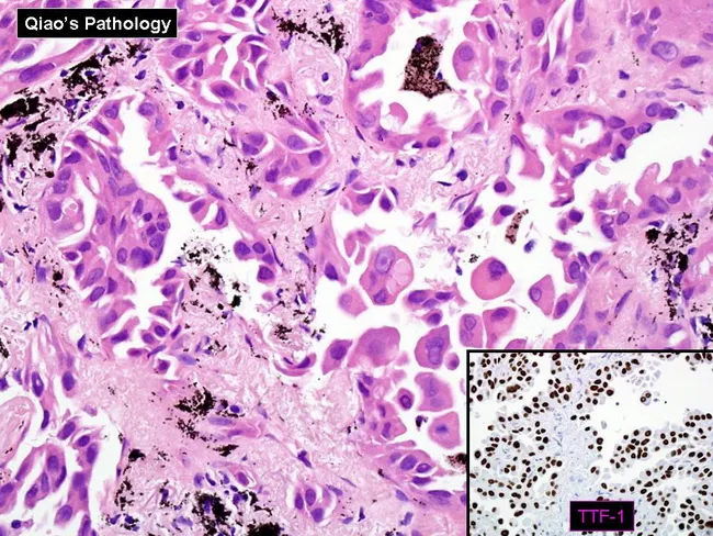
| TTF-1 (Thyroid Transcription Factor-1) | Positive (~75-85% of adenocarcinomas) |
| Napsin A | Positive (highly specific for lung adenocarcinoma) |
| CK7 | Positive |
| CK20 | Negative |
| CDX2 | Negative (positive in enteric and mucinous variants) |
| CEA | Positive |
| p40, p63 | Negative |
| Synaptophysin, Chromogranin | Negative |
TTF-1 and Napsin A together are the most useful panel to confirm lung adenocarcinoma origin. Mucinous adenocarcinomas may be TTF-1 negative but CK7 positive.
Molecular Genetics
Adenocarcinoma has the richest landscape of targetable driver mutations:
| Mutation/Alteration | Frequency | Notes |
|---|
| KRAS | ~30% (mostly smokers) | Most common in Western populations; oncogenic RAS signaling; mucinous subtype |
| EGFR | ~20% overall; up to 50% in Asian/never-smoker women | Exon 19 deletions, Exon 21 L858R point mutation; most common in nonsmokers/women/Asians |
| ALK rearrangement | 4-6% | Fusion with EML4 or other partners; often signet ring morphology; exclusive with EGFR/KRAS |
| ROS1 rearrangement | 1-2% | Shares features with ALK+ tumors; young nonsmokers |
| BRAF V600E | ~2% | Small subset amenable to targeted therapy |
| MET exon 14 skipping | ~3% | Associated with older patients |
| RET rearrangement | ~1-2% | Targetable with RET inhibitors |
| NTRK fusion | Rare | Pan-tumor target |
| STK11 (LKB1) | ~15-30% | Co-occurs with KRAS; associated with immunotherapy resistance |
| TP53 | ~50% | Loss of function |
| 3p deletions | ~80% | Includes VHL, FHIT loci |
- Robbins & Kumar Basic Pathology, Table 11.6
EGFR and ALK mutations are mutually exclusive. EGFR mutations respond remarkably to tyrosine kinase inhibitors (gefitinib, erlotinib, osimertinib), but resistance typically develops within months to a year, often through secondary EGFR mutations (T790M) or bypass signaling. - Robbins
Lung adenocarcinoma, lepidic pattern: neoplastic columnar cells proliferating along preexisting alveolar walls without stromal invasion
2. Squamous Cell Carcinoma (SCC)
Epidemiology
SCC accounts for approximately 30% of all lung cancers. Over 90% occur in cigarette smokers. Two-thirds are centrally located (arising from main/lobar/segmental bronchi). SCC tends to metastasize later than adenocarcinoma and may be more detectable on sputum cytology. - Sabiston Textbook of Surgery
Precursor Lesions
The stepwise progression is best documented in SCC:
- Squamous metaplasia → squamous dysplasia (mild, moderate, severe) → squamous carcinoma in situ → invasive SCC
- An in-situ component in a resected specimen helps confirm primary pulmonary origin vs. metastasis from head and neck. - Fishman's Pulmonary Diseases
Morphology
- Gross: Central endobronchial mass, often with post-obstructive collapse/pneumonitis distal to tumor. Prone to central necrosis and cavitation.
- Microscopic hallmarks: keratinization (keratin pearls), intercellular bridges (desmosomes between cells), stratification.
- Well-differentiated SCC: abundant keratin pearls, easily visible intercellular bridges.
- Moderately differentiated: less keratinization.
- Poorly differentiated: sheets of cells without clear squamous features - requires IHC.
- Basaloid subtype: nests of small cells with peripheral palisading and minimal keratinization; must be distinguished from LCNEC.
- Desmoplastic stroma with nests of infiltrating squamous cell carcinoma is characteristic.
IHC Profile
| Marker | Status |
|---|
| p40 (best) / p63 | Strongly positive (most specific) |
| CK5/6 | Positive |
| SOX2 | Positive |
| Desmoglein-3 | Positive |
| TTF-1 | Negative |
| Napsin A | Negative |
| CK7 | Negative or focal |
p40 is the single most specific marker, preferred over p63 (which can be expressed in small numbers of adenocarcinomas).
Molecular Genetics
SCC has a distinct genomic profile from adenocarcinoma:
| Alteration | Frequency |
|---|
| TP53 | ~80% |
| CDKN2A (p16) deletion/mutation | ~50% |
| SOX2 amplification | ~30% |
| FGFR1 amplification | ~20% |
| PIK3CA | ~15% |
| DDR2 mutation | ~10% |
| PTEN loss | ~10-15% |
| 3p deletions | ~80% |
| RB mutations | ~20% |
| KRAS | Rare |
| EGFR | Rare (in Western patients) |
| ALK | Absent |
SCC is not typically driven by the actionable kinase mutations that define adenocarcinoma therapeutics. It does express parathyroid hormone-related peptide (PTHrP), responsible for hypercalcemia as a paraneoplastic syndrome. - Robbins Table 11.6
3. Small Cell Lung Carcinoma (SCLC)
Epidemiology
SCLC accounts for approximately 15-20% of all lung cancers. It is overwhelmingly associated with heavy smoking (95%). About 80% are centrally located. SCLC is virtually always metastatic at presentation, making surgical resection rarely curative. - Sabiston Textbook of Surgery
Morphology
SCLC is defined by high-grade neuroendocrine morphology:
- Gross: Soft, white-gray central mass, often with extensive mediastinal/hilar lymph node involvement. Necrosis prominent.
- Microscopic:
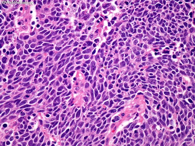
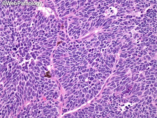
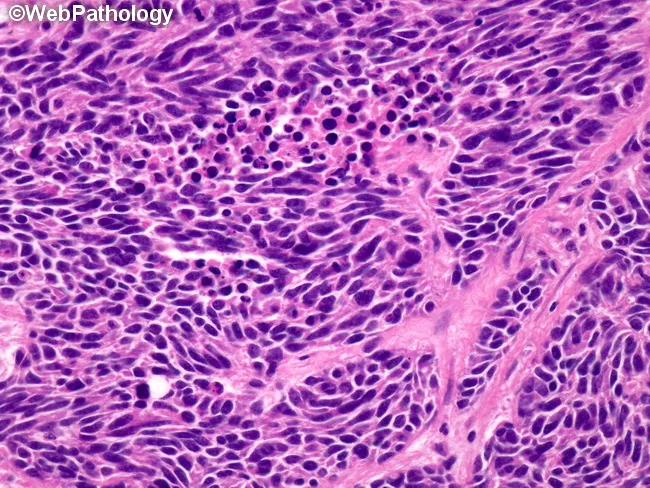
- Small round to oval cells, scant cytoplasm, ill-defined borders
- High nuclear-to-cytoplasmic ratio
- Finely dispersed granular ("salt-and-pepper") chromatin
- Absent or inconspicuous nucleoli
- Nuclear molding (adjacent nuclei deform each other) - characteristic
- Diffuse sheets or clusters; organoid nesting pattern absent (unlike carcinoid)
- Brisk mitotic activity (>10/2 mm², typically very high)
- Extensive necrosis common
- Crush artifact is frequent in small biopsies
- Dense-core neurosecretory granules on electron microscopy
Small cell lung carcinoma: dense sheets of small cells with scant cytoplasm, salt-and-pepper chromatin, nuclear molding, and brisk mitoses
IHC Profile
| Marker | Status |
|---|
| Synaptophysin | Positive |
| Chromogranin A | Positive (can be focal/weak) |
| CD56 (NCAM) | Strongly positive |
| TTF-1 | Positive (~80-90%) |
| CK (low molecular weight, dot-like/paranuclear pattern) | Positive |
| p40, p63 | Negative |
| Ki-67 | >80% (often >90%) |
| PD-L1 | Variable |
Epithelial markers (EMA, CEA, cytokeratin) are present in both SCLC and NSCLC. The key discriminator is neuroendocrine marker expression AND TTF-1 with p40 negativity. - Robbins Table 11.6
Molecular Genetics
SCLC has a unique, highly altered genomic profile:
| Alteration | Frequency |
|---|
| TP53 mutation | ~90% |
| RB1 mutation/deletion | ~90% (near universal) |
| 3p deletions | ~90% |
| MYCL1 amplification | ~30% |
| MYCN amplification | ~5-10% |
| MYC amplification | ~5-10% |
| CREBBP/EP300 | ~15% |
| NOTCH family | ~25% |
| SOX2 amplification | Present |
| KRAS | Rare |
| EGFR | Absent |
| ALK | Absent |
The near-universal RB1 + TP53 loss is the defining molecular signature of SCLC - equivalent to complete inactivation of the two founding tumor suppressor pathways. SCLC has one of the highest mutation burdens of any cancer (due to tobacco carcinogens) yet is paradoxically unresponsive to immune checkpoint inhibitors in most cases. - Robbins Table 11.6
SCLC is associated with production of peptide hormones as paraneoplastic phenomena: ACTH (ectopic Cushing syndrome), ADH (SIADH), gastrin-releasing peptide (GRP), and calcitonin.
4. Large Cell Neuroendocrine Carcinoma (LCNEC)
LCNEC represents a high-grade neuroendocrine carcinoma of the lung with "large cell" (NSCLC-like) morphology:
Morphology:
- Organoid, trabecular, rosette-like, or palisading architecture
- Large cells with moderate-to-abundant cytoplasm (contrast with SCLC)
- Prominent nucleoli
- High mitotic rate (>10/2 mm², typically much higher)
- Widespread necrosis
- Neuroendocrine differentiation confirmed by IHC
IHC: Synaptophysin, chromogranin, CD56 positive. TTF-1 variably positive. p40 negative. Ki-67 >50%.
Molecular genetics: LCNEC can be divided into "SCLC-like" (TP53+RB1) and "NSCLC-like" (TP53+STK11 or KEAP1) subtypes, with therapeutic implications. - Fishman's Pulmonary Diseases
5. Typical and Atypical Carcinoid
Overview: Carcinoid tumors account for <2% of all lung cancers. The 2021 WHO recognizes two subtypes based on histologic grade:
| Feature | Typical Carcinoid (TC) | Atypical Carcinoid (AC) |
|---|
| Mitoses | <2/2 mm² | 2-10/2 mm² |
| Necrosis | Absent | Present (punctate/focal) |
| 5-yr survival | ~90% | ~70% |
| LN metastases | ~5-10% | ~30% |
Morphology:
- Organoid, trabecular, rosette, palisading, or spindle cell patterns
- Uniform cells with moderate granular eosinophilic cytoplasm
- Round to oval nuclei with finely stippled ("salt-and-pepper") chromatin
- Absent or inconspicuous nucleoli
- Vascular stroma
IHC: Synaptophysin, chromogranin A, CD56 strongly positive. TTF-1 variable. Ki-67: TC < 5%, AC 5-20%.
Molecular genetics: Carcinoids are genetically distinct from high-grade neuroendocrine carcinomas. They have low mutation burden, with recurrent mutations in MEN1, PSIP1, ARID1A, and chromatin remodeling genes. EGFR, KRAS, and TP53 mutations are uncommon. - Fishman's Pulmonary Diseases
6. Large Cell Carcinoma
With widespread use of IHC and molecular profiling, large cell carcinoma (LCC) has been dramatically reclassified, dropping from ~10% to only ~1.5% of all lung cancers. The 2021 WHO defines LCC as a strict diagnosis of exclusion:
"An undifferentiated non-small-cell carcinoma that lacks the cytologic, architectural, and immunohistochemical features of SCLC, adenocarcinoma, or squamous cell carcinoma, AND lacks giant cell, spindle cell, or pleomorphic features."
Morphology:
- Large sheets of round to polygonal cells
- Prominent nucleoli
- Abundant pale-staining cytoplasm
- No glandular, squamous, or neuroendocrine differentiation
- No mucin production
- Usually large and peripherally located
IHC: All lineage markers (TTF-1, Napsin A, p40, p63, synaptophysin, chromogranin) are negative by definition.
Molecular genetics: No specific genomic driver; heterogeneous. Some cases harbor KRAS or TP53 mutations.
7. Sarcomatoid Carcinoma
A group of poorly differentiated NSCLCs with sarcoma-like features. Five subtypes recognized by WHO:
- Pleomorphic carcinoma: Contains >10% spindle and/or giant cells, or is exclusively composed of them, WITH an adenocarcinoma/SCC component.
- Spindle cell carcinoma: Purely spindle cells
- Giant cell carcinoma: Purely giant cells with inflammatory infiltrate (hallmark: neutrophilic emperipolesis)
- Carcinosarcoma: True biphasic tumor with malignant epithelial and heterologous sarcomatous components (rhabdomyosarcoma, chondrosarcoma, osteosarcoma)
- Pulmonary blastoma: Resembles embryonic lung; biphasic fetal adenocarcinoma + sarcomatous stroma
IHC: AE1/AE3, EMA positive in epithelial components. Vimentin positive in spindle elements.
Molecular genetics: MET exon 14 skipping mutations occur in ~20-30% of pleomorphic carcinomas - significantly higher than in adenocarcinoma, making this a targetable alteration in this rare subtype. - Fishman's Pulmonary Diseases
8. Adenosquamous Carcinoma
Defined as a carcinoma containing both SCC and adenocarcinoma components, each comprising >10% of the tumor. Incidence ~2-3%.
- More common in smokers but occurs in nonsmokers
- More common peripherally
- Molecular data show both components share similar driver mutations, confirming a clonally identical tumor (not a "collision tumor")
- Prognosis is worse than other NSCLC subtypes
- Targeted therapy selection follows the guidelines for adenocarcinoma genetics
Comparison Table: Key Molecular Features (Robbins Table 11.6)
| Feature | SCLC | Adenocarcinoma | SCC |
|---|
| 3p deletions | ~90% | ~80% | ~80% |
| RB mutations | ~90% | ~20% | ~20% |
| p16/CDKN2A mutations | ~10% | ~30% | ~50% |
| TP53 mutations | ~90% | ~50% | ~80% |
| KRAS mutations | Rare | ~30% | Rare |
| EGFR mutations | Absent | ~20% (nonsmokers, women) | Rare |
| ALK fusions | Absent | 4-6% | Absent |
| NE markers | Present | Absent | Absent |
| Response to chemo/RT | High (but invariably recurs) | Incomplete | Incomplete |
| Checkpoint inhibitor response | Unresponsive | Responsive | Responsive |
| Paraneoplastic hormones | ACTH, ADH, GRP, calcitonin | -- | PTHrP (hypercalcemia) |
General Molecular Targets in Lung Carcinoma (Murray & Nadel's / Robbins)
The clinically relevant molecular targets as of the 2021 WHO era:
| Alteration | Tumor Type | Drug Class |
|---|
| EGFR exon 19 del / L858R | Adenocarcinoma | TKI (erlotinib, gefitinib, osimertinib) |
| EGFR T790M (resistance) | Adenocarcinoma | 3rd gen TKI (osimertinib) |
| ALK fusion | Adenocarcinoma | ALK inhibitor (crizotinib, alectinib, lorlatinib) |
| ROS1 fusion | Adenocarcinoma | ROS1 inhibitor (crizotinib, entrectinib) |
| BRAF V600E | Adenocarcinoma | BRAF+MEK inhibitor (dabrafenib+trametinib) |
| MET exon 14 skipping | Adeno/Pleomorphic carcinoma | MET inhibitor (capmatinib, tepotinib) |
| RET fusion | Adenocarcinoma | RET inhibitor (selpercatinib, pralsetinib) |
| NTRK fusion | Any | TRK inhibitor (larotrectinib, entrectinib) |
| KRAS G12C | Adenocarcinoma | KRAS inhibitor (sotorasib, adagrasib) |
| PD-L1 high expression | NSCLC (all) | Checkpoint inhibitor (pembrolizumab) |
| High TMB | NSCLC | Checkpoint inhibitor |
Paraneoplastic Syndromes Summary
| Syndrome | Tumor Type | Mediator |
|---|
| SIADH | SCLC | ADH |
| Ectopic Cushing's | SCLC | ACTH |
| Eaton-Lambert myasthenic syndrome | SCLC | Anti-VGCC antibodies |
| Hypercalcemia | SCC | PTHrP |
| Hypertrophic pulmonary osteoarthropathy | Adenocarcinoma | Unclear |
| Hypercoagulability (Trousseau) | Adenocarcinoma/mucin-secreting | Mucin activating clotting |
| Peripheral neuropathy, encephalopathy | SCLC | Anti-Hu (ANNA-1) antibodies |
Key Clinical Points
- Pancoast tumor: Apical tumor (any histology) invading brachial/cervical sympathetic plexus → Pancoast syndrome (Horner + C8/T1 pain + rib/vertebral destruction)
- Superior vena cava syndrome: Tumor compression/infiltration of SVC
- Virchow's node (left supraclavicular): Characteristic lymphatic metastasis
- All lung cancer subtypes can metastasize to adrenal glands, liver, brain, and bone (osteolytic); adenocarcinoma has highest predilection for CNS metastases
- Staging: TNM system universally applied; SCLC also uses limited vs. extensive stage clinical staging
Sources:
- Robbins & Kumar Basic Pathology (10th ed.) - Chapter 11, Table 11.5 and 11.6
- Fishman's Pulmonary Diseases and Disorders (6th ed.) - Chapter 111 (Histologic Classification of Lung Tumors)
- Sabiston Textbook of Surgery (21st ed.) - Chapter 110, Pathology section
- Harrison's Principles of Internal Medicine (22nd ed., 2025)
- Murray & Nadel's Textbook of Respiratory Medicine - Clinically relevant molecular targets in lung carcinoma