
Tell me about clavicle fracture and their non surgical management
Reading File
Now let me search the medical library for clavicle fractures in parallel:
Finding Sources
Searching PubMed
"clavicle fracture" AND "conservative treatment"
Good results. Now let me read all three textbook sections in parallel:
Reading File
Reading File
Reading File
Let me get more detailed content from Pye's Surgical Handicraft and search for adult clavicle fracture management:
Reading File
Finding Sources
Finding Sources
Finding Sources
Excellent findings. Now let me read the key sections from Rockwood & Green (the most authoritative fracture reference) and Campbell's:
Reading File
Reading File
Reading File
Reading File
Finding Sources
Finding Sources
Reading File
Reading File
Reading File
Reading File
I now have comprehensive information from multiple authoritative sources. Let me compile the full answer.
Loading Image
I now have all the information needed for a comprehensive answer. Here is the complete response:
Clavicle Fractures and Non-Surgical Management
Anatomy and Why the Clavicle Fractures
The clavicle is the only bony connection between the thorax and the upper limb. It is S-shaped in the coronal plane (medial end convex anteriorly, lateral end concave anteriorly). It is the first bone to ossify (5-6 weeks gestation) but the last to complete ossification (~27 years of age), which is why physeal injuries rather than true SC dislocations are common in young adults.
It is the most commonly fractured bone in children, accounting for ~90% of obstetric fractures. In adults, it represents about 4% of all fractures, making it one of the most frequent injuries seen in emergency departments.
Mechanism of Injury
- Direct blow to the clavicle (most common in adults - fall onto the shoulder or direct impact)
- Indirect force transmitted via a fall on an outstretched hand (FOOSH)
- Birth trauma - direct pressure from the symphysis pubis during delivery
Classification
Allman Classification (most widely used)
| Group | Location | Frequency |
|---|---|---|
| Group I | Middle third (midshaft) | ~80% |
| Group II | Distal (lateral) third | ~15% |
| Group III | Medial (proximal) third | ~5% |
Neer Classification (for lateral/distal fractures)
- Type I - Lateral to the coracoclavicular (CC) ligaments; ligaments intact - usually stable
- Type II - Medial to or through the CC ligaments; displaced - high nonunion risk
- Type III - Intra-articular (AC joint) extension
Robinson Classification (Edinburgh)
An expanded scheme based on displacement and comminution, particularly useful for midshaft fractures, adding subtypes for displacement and comminution that better predict outcomes than Allman alone.
OTA/AO Classification
- 15.1 - Medial end
- 15.2 - Diaphyseal (15.2A simple, 15.2B wedge, 15.2C multifragmentary/comminuted)
- 15.3 - Distal (lateral) end (15.3A extra-articular, 15.3B partial articular, 15.3C complete articular)
Clinical Presentation
- Pain at the clavicle and shoulder, worsening with neck and arm movement
- Visible or palpable deformity, crepitus, edema over the clavicle
- "Drooped" or medially displaced shoulder
- A fractured clavicle in a neonate may only be detected at day 10 when callus becomes visible, or from parental history of crying when the baby is lifted
Neurovascular exam is mandatory given proximity to the subclavian vessels and brachial plexus, particularly in displaced fractures.
Imaging
- Standard AP radiograph of the clavicle - sufficient for most fractures
- Cephalic tilt view (35-40 degrees) - reduces overlap with thoracic structures
- Apical oblique view (ipsilateral rotation 45° + 20° cephalic tilt) - better visualization
- Zanca view (15° cephalic tilt centered on AC joint) - for lateral fractures
- CT scan - for medial fractures involving the SC joint, physeal separations, and polytrauma patients
- Stress views (weight suspended from wrist) - assess CC ligament integrity in distal fractures
- Ultrasound - for obstetric/neonatal fractures
Polytrauma note: Initial supine AP chest radiographs can underestimate displacement by up to 89% compared to upright views. Always repeat proper upright films once the patient's condition allows.
Non-Surgical (Conservative) Management
Indications for Nonoperative Treatment
The majority of clavicle fractures are managed nonoperatively. This is the standard of care for:
- Undisplaced or minimally displaced midshaft fractures
- Most distal third fractures (especially in middle-aged and elderly patients, including Neer Type I)
- All clavicle fractures in newborns (no treatment often needed)
- Clavicle fractures in children and adolescents (sling and swath 4-6 weeks)
- Medial third fractures (unless there is posterior displacement threatening mediastinal structures)
Sling vs. Figure-of-Eight Bandage
This is one of the most well-studied questions in orthopaedics:
The figure-of-eight bandage is no longer recommended as a primary treatment.
"Despite its early popularity, this device never proved to be superior to a simple sling (in terms of cosmesis, functional outcome, or pain)." - Roberts and Hedges' Clinical Procedures in Emergency Medicine
"A figure-of-eight sling is not recommended." - Campbell's Operative Orthopaedics 15th Ed (2026)
Key comparative evidence:
- Andersen et al. found the sling caused less discomfort and fewer complications than figure-of-eight
- A randomized trial showed significantly more pain in the figure-of-eight group on day 1 (VAS 6.8 vs. 5.6, p = .034)
- The figure-of-eight carries a risk of lower trunk brachial plexus palsy if overtightened
- Ersen et al. confirmed higher pain scores and found no difference in clavicular shortening between the two methods
Current recommendation: A simple sling with a padded neckpiece, without any attempt at fracture reduction.
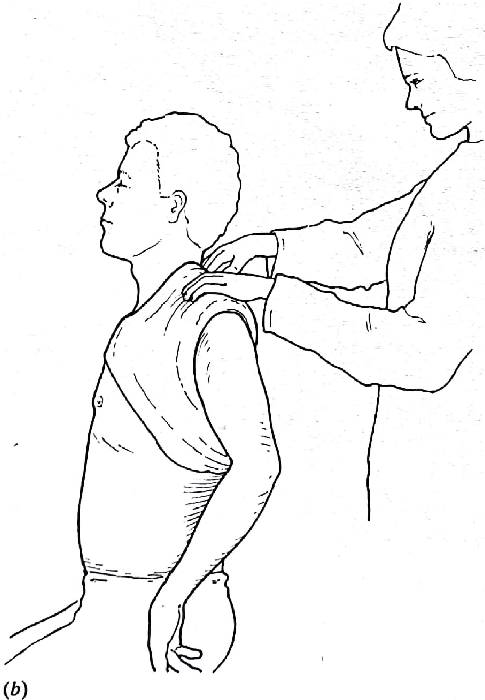
Figure: Application of the figure-of-eight clavicle bandage - now considered historical. (Pye's Surgical Handicraft, 22nd Ed)
Nonoperative Protocol (Current Standard)
Campbell's Operative Orthopaedics / Rockwood and Green protocol:
- Week 0-2: Simple sling for comfort; no attempt at reduction
- ~Week 2: Begin pendulum exercises, followed by progressive active non-weight-bearing shoulder motion up to the horizontal plane
- Week 6: Repeat radiograph - bony union is often only partly visible (callus formation at best); begin strengthening exercises as pain allows, under physiotherapy supervision if appropriate
- If no callus visible at 6 weeks and fracture site is mobile - avoid aggressive activities; consider earlier conversion to operative treatment
- ~1 year: Functional outcome is unlikely to change significantly beyond this point (Schemitsch et al.)
For children/neonates:
- Newborns: no treatment required
- Infants/children: sling and swath for 4-6 weeks
- Note: figure-of-eight splinting is not recommended in children due to risk of brachial plexus palsy
Indications to Consider Operative Treatment (Absolute vs. Relative)
Absolute indications for surgery (nonoperative NOT appropriate):
- Open fracture
- Neurovascular compromise
- Associated floating shoulder (ipsilateral scapular fracture)
- Skin tenting threatening imminent perforation
Relative indications (discuss with patient; early conversion to operative may be considered):
- Displacement >2.0 cm / significant shortening
- Comminuted fracture pattern (especially midshaft)
- Protracted scapula with shoulder girdle weakness
- Neer type II distal fractures (high nonunion rate nonoperatively)
- High-level athlete seeking faster return to activity
Outcomes and Complications of Nonoperative Treatment
| Complication | Rate |
|---|---|
| Nonunion (displaced comminuted midshaft) | 14-24% |
| Nonunion (all nonoperatively treated) | ~13.8% (recent meta-analysis of 24 RCTs) |
| Malunion requiring further treatment | ~9/49 patients in COTS trial |
| Symptomatic "droopy" shoulder | ~10/49 patients (COTS) |
| Residual symptoms at 10 years | ~46% (Nowak et al., 208 patients) |
| Objective strength deficit | Up to 30% in shoulder abduction/flexion |
Key points:
- Traditionally, nonunion rates were thought to be very low (~1%); modern prospective data reveals rates of 14-24% in displaced comminuted midshaft fractures
- Robinson et al. reported a 21% nonunion rate in displaced comminuted midshaft fractures (868 patients)
- 41% of nonoperatively treated patients had residual symptoms (pain, weakness) at 4 years (Woltz et al.)
- Satisfaction with shoulder appearance was 53% nonoperative vs. 84% operative (COTS trial)
Recent Evidence Update
A 2024 systematic review (PMID 38281678) specifically examined displaced distal clavicle fractures managed nonoperatively, finding they are associated with inferior clinical outcomes - supporting consideration of operative treatment for Neer Type II fractures in appropriate patients.
Summary
| Feature | Recommendation |
|---|---|
| Immobilization device | Simple sling (NOT figure-of-eight) |
| Duration of sling | ~2 weeks, then early mobilization |
| Exercises | Pendulum at ~2 weeks; progressive ROM to horizontal; strengthening at 6 weeks |
| Radiograph follow-up | At 6 weeks; reassess for union |
| Plateau of recovery | ~1 year post-injury |
| Pediatric (newborn) | No treatment needed |
| Pediatric (children) | Sling and swath 4-6 weeks |
Sources: Rockwood and Green's Fractures in Adults 10th Ed (2025); Campbell's Operative Orthopaedics 15th Ed (2026); ROSEN's Emergency Medicine; Roberts and Hedges' Clinical Procedures in Emergency Medicine; Pye's Surgical Handicraft 22nd Ed; Miller's Review of Orthopaedics 9th Ed.
This is a shared conversation. Sign in to Orris to start your own chat.