

Lap cholecystectomy
laparoscopic cholecystectomy port placement calot triangle dissection
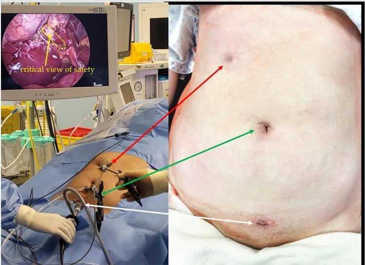
This composite clinical photograph and diagnostic display illustrate the setup and outcome of an 'Emirate' laparoscopic cholecystectomy. The image is divided into three sections: an intraoperative view, a laparoscopic monitor display, and a postoperative abdominal photograph. The laparoscopic monitor (top left) shows the 'critical view of safety' during dissection, with a yellow dashed line highlighting the hepatocystic triangle (Calot's triangle) and an arrow pointing to the cystic duct and artery area. The intraoperative photograph (bottom left) demonstrates the unique three-port placement: a 5mm intraumbilical port, a 5mm right subcostal port, and a 5mm suprapubic port. The postoperative image (right) shows the corresponding healed 5mm incisions, emphasizing the cosmetic advantage and reduced visibility of scars in the umbilical, right hypochondrium, and suprapubic regions. The content is designed for surgical education, focusing on minimally invasive techniques, anatomical landmarks for safe gallbladder removal, and port-site optimization to minimize postoperative scarring and hernia risk.
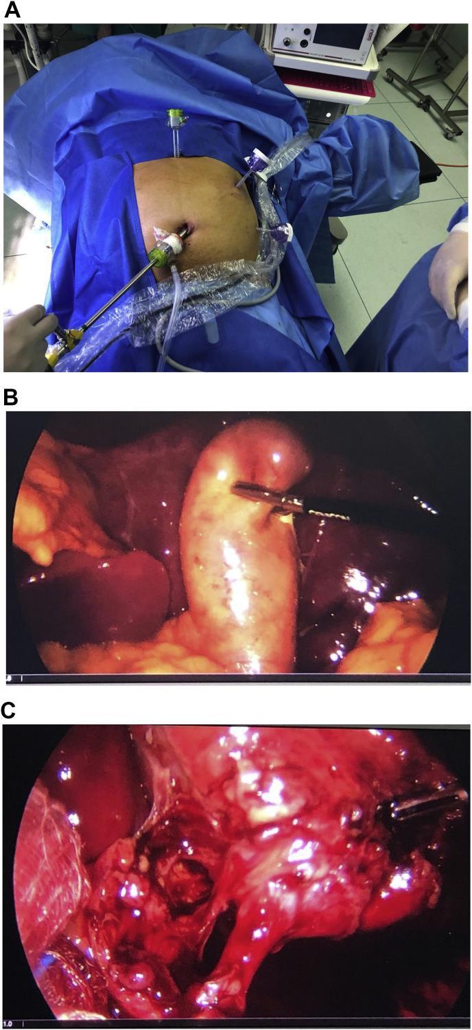
A composite medical image illustrating a laparoscopic cholecystectomy in a patient with Situs Inversus Totalis. Panel A is a clinical photograph of the patient's abdomen draped for surgery, showing a mirrored port configuration: a 10mm transumbilical port for the camera, a 10mm epigastric port, and two 5mm ports in the left upper quadrant (midclavicular and anterior axillary lines). Panels B and C are intraoperative laparoscopic views. Panel B shows the distended, inflamed gallbladder being manipulated by a grasper, located notably in the left upper quadrant due to the patient's anatomical inversion. Panel C captures the dissection of the triangle of Calot, demonstrating the 'critical view of safety' where the cystic duct and cystic artery are isolated from surrounding connective and fatty tissues before clipping. The imagery highlights the necessary technical adjustments in surgical ergonomics and port placement required when performing standard hepatobiliary procedures on patients with situs inversus.
Laparoscopic Cholecystectomy - Operative Notes Interpretation
From your handwritten notes (transcribed):
- Position: Prone + ... (likely Supine in reverse Trendelenburg - standard for lap chole; the "p & b" likely refers to positioning details)
- Incision: Port placement
- Procedure:
- GB distended, long c̄ (with) stone in Hartmann's pouch
- Dissection of Calot's triangle (△)
- Ligation of cystic duct
- Coagulation of cystic artery
- GB dissected from GB fossa
- Removal of GB
- Closure of port c̄ (with) 2-0 Ethilon
- Dressing
Step-by-Step Explanation
1. Position & Setup
- Patient is placed supine in steep reverse Trendelenburg (head-up, feet down), tilted slightly to the left.
- This displaces bowel away from the upper abdomen and improves visualization of the right upper quadrant.
- An orogastric tube decompresses the stomach for better field of view.
- General anesthesia with muscle relaxation is mandatory.
2. Port Placement (Incision)
| Port | Size | Location | Purpose |
|---|---|---|---|
| 1 | 10-12 mm | Umbilicus | Camera (laparoscope); specimen extraction |
| 2 | 5 mm | Subxiphoid / epigastric | Liver retraction |
| 3 | 5 mm | Right midclavicular line | Working port - grasp infundibulum |
| 4 | 5 mm | Right anterior axillary line | Fundus retraction |
3. Findings: GB Distended + Stone in Hartmann's Pouch
- The gallbladder was distended - consistent with obstruction (acute cholecystitis or biliary colic with impacted stone).
- A stone in Hartmann's pouch (the infundibulum, the widest part of the GB just before the cystic duct) is a common cause of obstruction.
- The fundus is grasped with the lateral port instrument and retracted cephalad toward the right shoulder.
- The infundibulum is grasped with the midclavicular port instrument and retracted inferolaterally - this opens up Calot's triangle.
4. Dissection of Calot's Triangle (△)
- Calot's triangle (hepatocystic triangle) is bounded by:
- The cystic duct (inferiorly)
- The common hepatic duct (medially)
- The inferior surface of the liver (superiorly)
- Fibrofatty tissue is cleared from this triangle until a Critical View of Safety (CVS) is achieved.
- CVS criteria (Strasberg):
- Only 2 structures are seen entering the gallbladder
- The lower 1/3 of the gallbladder is dissected free from the liver (cystic plate visible)
- The hepatocystic triangle is cleared of fat and fibrous tissue
- The Calot's node (lymph node) overlies the cystic artery and serves as a useful landmark.
- This step is the most critical for preventing common bile duct (CBD) injury - the most feared complication of lap chole.
5. Ligation of Cystic Duct
- Once CVS is confirmed, the cystic duct is clipped with metal or absorbable clips (typically 2 clips proximally, 1 distally).
- The duct is then divided between the clips.
- If stones are present in the cystic duct (as here with the Hartmann's pouch stone), gentle milking proximally or additional clips may be needed.
- An intraoperative cholangiogram (IOC) can be performed through the cystic duct at this stage if anatomy is unclear.
6. Coagulation of Cystic Artery
- The cystic artery (usually a branch of the right hepatic artery) runs in Calot's triangle.
- It is identified, clipped, and divided - often with diathermy/cautery as noted ("coagulation").
- Variant anatomy is common: the cystic artery may be duplicated or arise anomalously - careful identification before division is essential.
7. GB Dissected from GB Fossa
- Using hook diathermy or ultrasonic dissector, the gallbladder is dissected off the hepatic (gallbladder) fossa in a retrograde fashion.
- The plane is just below the GB serosa, keeping close to the gallbladder wall to avoid liver bleeding.
- Any bile leakage from the fossa (Luschka ducts) is coagulated.
8. Removal of GB
- The gallbladder is removed through the umbilical port (largest, 10-12 mm).
- If the specimen is large or stones are numerous, an endobag (retrieval bag) is used to prevent port-site contamination and stone spillage.
- The abdomen is inspected for bleeding and bile spillage, and the operative field is irrigated if needed.
9. Closure of Port with 2-0 Ethilon
- 2-0 Ethilon = non-absorbable monofilament nylon suture.
- The 10-12 mm umbilical port fascia is closed with this to prevent port-site hernia.
- 5-mm port sites typically only need skin closure.
- Skin is closed (usually with subcuticular absorbable suture or skin clips).
10. Dressing
- Standard adhesive dressings applied to all port sites.
Key Complications to Know
| Complication | Notes |
|---|---|
| CBD injury | Most serious; 0.3-0.5%; prevented by CVS |
| Bile leak | From cystic duct stump or Luschka ducts |
| Bleeding | From cystic artery or liver bed |
| Stone spillage | Can cause late intra-abdominal abscess |
| Port-site hernia | Especially at umbilical 10-mm port |
| Conversion to open | Unclear anatomy, bleeding, dense adhesions |