
5th cmc dislocation
"carpometacarpal joint" AND "dislocation" AND "fifth"
fifth carpometacarpal joint dislocation fracture-dislocation diagnosis treatment
carpometacarpal fracture dislocation fifth metacarpal base radiograph
Fifth CMC joint dislocation
Overview
Anatomy that matters
- The 4th and 5th metacarpal bases articulate with the biconcave hamate. A central ridge separates the two facets.
- The dorsal pull of the wrist extensors on the metacarpals is the deforming force. Dislocations are therefore usually dorsal.
- The ulnar motor branch lies volar to the 5th CMC joint, so check intrinsic function.
Mechanism
- The usual mechanism is an axial load on a clenched fist, such as punching.
- Higher-energy injuries can cause a reverse Bennett fracture. This is a volar fracture fragment of the 5th metacarpal base, with the shaft migrating dorsally and proximally. Hamate fractures (coronal split, dorsal) can also occur.
- Combined 4th and 5th CMC injuries are the most common pattern. Isolated 5th CMC fracture-dislocations are also common. Multiple CMC injuries are higher energy and have a worse prognosis.
Clinical assessment
- Look for loss of knuckle height, a dorsal prominence at the wrist, shortening, malrotation or scissoring, and swelling.
- Check neurovascular status, ulnar motor branch function and tendon function.
- Look for an open injury.
- Consider compartment syndrome in high-energy injuries.
- These injuries are often missed. Chronic cases have been reported (see the Kono 2024 report on chronic 4th/5th CMC dislocations).
Imaging
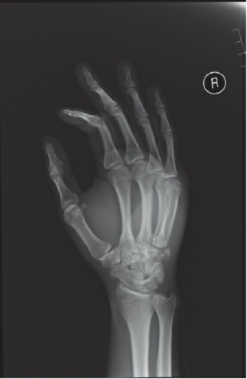
- Standard hand views: PA, lateral and oblique. The lateral view often shows the dorsal displacement best.
- Use the 30-degree pronated view to see the 4th and 5th CMC joints. The textbook calls it "crucial" for confirming the joint is reduced after reduction.
- CT is useful for occult hamate or metacarpal base fractures and for confirming reduction.
Management
- Give a hematoma block. Check neurovascular status first.
- Apply axial traction with volar pressure over the metacarpal base. A clunk is often felt.
- Splint in an ulnar gutter with the wrist in 30 degrees of extension and the MCPs in about 70 degrees of flexion.
- Confirm reduction on all views, including the 30-degree pronated view, or on CT if there is doubt.
- Follow closely, because redisplacement is common.
- Unstable or irreducible injuries
- Multiple CMC dislocations
- Intra-articular step-off
- Open injuries
- Associated metacarpal or hamate fractures
- A common construct is a 0.045 K-wire from the 5th metacarpal into the hamate, plus a wire into the 4th metacarpal base.
- For combined 4th and 5th injuries, use two wires from the 5th across the 4th into the 3rd metacarpal. Add wires across each CMC joint into the carpus.
Complications
- Post-traumatic CMC arthritis is the most common complication.
- Others are loss of reduction, stiffness, reduced grip strength, pin-tract problems and ulnar motor branch injury.
- Delayed diagnosis makes treatment harder.
Recent literature
- Volar dislocation of the 5th CMC (PMID 32612479)
- Ulnar-type dislocation of the 5th CMC (PMID 33335375)
- Isolated dorsal dislocations of the 4th and 5th CMC (PMID 33520509)
- A 2025 operative series (Berault et al.) reported that combined 4th/5th and isolated 5th CMC fracture-dislocations make up about 90% of non-thumb CMC injuries.
Walk me through the CRPP pin configuration step by step
https://surgeryreference.aofoundation.org/orthopedic-trauma/…
CRPP for fifth CMC dislocation: step by step
1. Indications
- The joint is unstable or irreducible after closed reduction, or it is a multiple CMC injury (Rockwood).
- It is acute enough to reduce closed (Rockwood). If the injury is chronic or the joint won't reduce, go to open reduction.
2. Setup (Rockwood)
- Use a hand table with the hand palm down over a large bump. The bump lets the hand sit in its natural metacarpal cascade and gives the right wire trajectory.
- For ring and small finger injuries, the surgeon sits between the patient's head and shoulder.
- Fluoroscopy comes from the end of the table or the axilla, opposite the surgeon.
- Have a lightweight drill, K-wires of various sizes, and backup screws or plates available.
- Place a tourniquet, but it is often not needed.
3. Reduce the joint (Rockwood)
- Hold the reduction with your non-dominant hand.
- Use axial traction and volar-directed pressure over the dorsally displaced 5th metacarpal base. This is the same maneuver as the closed reduction.
- Confirm the reduction on fluoroscopy: PA, true lateral, and the 30-degree pronated view. Check the joint line on each.
4. First wire across the 5th CMC joint (Rockwood)
- Use a 0.045 inch K-wire on "oscillate" mode.
- Drive it from the 5th metacarpal base across the CMC joint into the hamate.
- Common practice: start dorsal-ulnar on the metacarpal base and aim obliquely, proximal and radial, into the hamate body. Take care not to enter the joint line itself, and keep the wire clear of the volar structures.
- The ulnar motor branch and the palmar arch lie volar. Stop the wire at or just past the far hamate cortex. Do not drive it deep.
5. Second wire into the adjacent metacarpal (Rockwood)
- Place a wire from the 5th metacarpal base into the 4th metacarpal base. This is the adjacent-metacarpal stabilizer.
- If the 4th CMC is also unstable or dislocated (the combined injury), the textbook configuration changes:
- Run two wires from the small finger metacarpal, across the 4th, and into the 3rd metacarpal.
- Add wires across each CMC joint into the carpal bones. For the 4th, that means from its base into the hamate.
6. Fracture fragments (Rockwood)
- If there is a reverse Bennett fragment or another articular fragment, add supplementary K-wires into it. This is only needed when the fragment is large enough to hold a wire.
- If you can't reduce the fragment closed, convert to open reduction. Use a dorsal approach with lag screws or a mini-plate.
7. Check before finishing
- Common practice: take final fluoroscopy in AP, lateral, and oblique or pronated views. Confirm:
- The CMC joint is concentric.
- The wires sit in the hamate, not in the joint.
- There is no rotational malalignment of the digit.
- Do a clinical cascade check for scissoring.
- Stress-test the construct gently.
8. Finish (Rockwood)
- Bend and cut the K-wires to the appropriate length. Leave them outside the skin or buried, according to your preference.
- Apply a padded splint in an intrinsic-plus position with an ulnar gutter component for the 4th and 5th CMC joints.
9. Aftercare (Rockwood)
- Remove the pins at 4 to 6 weeks to prevent stiffness.
- Therapy may begin before the pins come out.
- Watch for loss of reduction, pin-tract infection, and later post-traumatic CMC arthritis. Post-traumatic arthritis is the most common complication.
Points to check before you operate
- The textbook says nothing about the number of pins, the exact entry points, or pin angles for an isolated 5th CMC injury. Two wires is the typical teaching, but it is not a fixed standard.
- I couldn't retrieve the AO Surgery Reference technique page. It would be a good next stop for illustrated pin trajectories: AO CMC dislocation page.
Find recent literature on hemi-hamate arthroplasty outcomes
hemihamate arthroplasty
"hamate" AND "arthroplasty" AND "fracture dislocation"
hemi-hamate reconstruction proximal interphalangeal joint
hemi-hamate arthroplasty outcomes PIP fracture dislocation systematic review range of motion complications
Overview
Anatomy that matters
- The 4th and 5th metacarpal bases articulate with the biconcave hamate. A central ridge separates the two facets.
- The dorsal pull of the wrist extensors on the metacarpals is the deforming force. Dislocations are therefore usually dorsal.
- The ulnar motor branch lies volar to the 5th CMC joint, so check intrinsic function.
Mechanism
- The usual mechanism is an axial load on a clenched fist, such as punching.
- Higher-energy injuries can cause a reverse Bennett fracture. This is a volar fracture fragment of the 5th metacarpal base, with the shaft migrating dorsally and proximally. Hamate fractures (coronal split, dorsal) can also occur.
- Combined 4th and 5th CMC injuries are the most common pattern. Isolated 5th CMC fracture-dislocations are also common. Multiple CMC injuries are higher energy and have a worse prognosis.
Clinical assessment
- Look for loss of knuckle height, a dorsal prominence at the wrist, shortening, malrotation or scissoring, and swelling.
- Check neurovascular status, ulnar motor branch function and tendon function.
- Look for an open injury.
- Consider compartment syndrome in high-energy injuries.
- These injuries are often missed. Chronic cases have been reported (see the Kono 2024 report on chronic 4th/5th CMC dislocations).
Imaging
- Standard hand views: PA, lateral and oblique. The lateral view often shows the dorsal displacement best.
- Use the 30-degree pronated view to see the 4th and 5th CMC joints. The textbook calls it "crucial" for confirming the joint is reduced after reduction.
- CT is useful for occult hamate or metacarpal base fractures and for confirming reduction.
Management
- Give a hematoma block. Check neurovascular status first.
- Apply axial traction with volar pressure over the metacarpal base. A clunk is often felt.
- Splint in an ulnar gutter with the wrist in 30 degrees of extension and the MCPs in about 70 degrees of flexion.
- Confirm reduction on all views, including the 30-degree pronated view, or on CT if there is doubt.
- Follow closely, because redisplacement is common.
- Unstable or irreducible injuries
- Multiple CMC dislocations
- Intra-articular step-off
- Open injuries
- Associated metacarpal or hamate fractures
- A common construct is a 0.045 K-wire from the 5th metacarpal into the hamate, plus a wire into the 4th metacarpal base.
- For combined 4th and 5th injuries, use two wires from the 5th across the 4th into the 3rd metacarpal. Add wires across each CMC joint into the carpus.
Complications
- Post-traumatic CMC arthritis is the most common complication.
- Others are loss of reduction, stiffness, reduced grip strength, pin-tract problems and ulnar motor branch injury.
- Delayed diagnosis makes treatment harder.
Recent literature
- Volar dislocation of the 5th CMC (PMID 32612479)
- Ulnar-type dislocation of the 5th CMC (PMID 33335375)
- Isolated dorsal dislocations of the 4th and 5th CMC (PMID 33520509)
- A 2025 operative series (Berault et al.) reported that combined 4th/5th and isolated 5th CMC fracture-dislocations make up about 90% of non-thumb CMC injuries.
1. Indications
- The joint is unstable or irreducible after closed reduction, or it is a multiple CMC injury (Rockwood).
- It is acute enough to reduce closed (Rockwood). If the injury is chronic or the joint won't reduce, go to open reduction.
2. Setup (Rockwood)
- Use a hand table with the hand palm down over a large bump. The bump lets the hand sit in its natural metacarpal cascade and gives the right wire trajectory.
- For ring and small finger injuries, the surgeon sits between the patient's head and shoulder.
- Fluoroscopy comes from the end of the table or the axilla, opposite the surgeon.
- Have a lightweight drill, K-wires of various sizes, and backup screws or plates available.
- Place a tourniquet, but it is often not needed.
3. Reduce the joint (Rockwood)
- Hold the reduction with your non-dominant hand.
- Use axial traction and volar-directed pressure over the dorsally displaced 5th metacarpal base. This is the same maneuver as the closed reduction.
- Confirm the reduction on fluoroscopy: PA, true lateral, and the 30-degree pronated view. Check the joint line on each.
4. First wire across the 5th CMC joint (Rockwood)
- Use a 0.045 inch K-wire on "oscillate" mode.
- Drive it from the 5th metacarpal base across the CMC joint into the hamate.
- Common practice: start dorsal-ulnar on the metacarpal base and aim obliquely, proximal and radial, into the hamate body. Take care not to enter the joint line itself, and keep the wire clear of the volar structures.
- The ulnar motor branch and the palmar arch lie volar. Stop the wire at or just past the far hamate cortex. Do not drive it deep.
5. Second wire into the adjacent metacarpal (Rockwood)
- Place a wire from the 5th metacarpal base into the 4th metacarpal base. This is the adjacent-metacarpal stabilizer.
- If the 4th CMC is also unstable or dislocated (the combined injury), the textbook configuration changes:
- Run two wires from the small finger metacarpal, across the 4th, and into the 3rd metacarpal.
- Add wires across each CMC joint into the carpal bones. For the 4th, that means from its base into the hamate.
6. Fracture fragments (Rockwood)
- If there is a reverse Bennett fragment or another articular fragment, add supplementary K-wires into it. This is only needed when the fragment is large enough to hold a wire.
- If you can't reduce the fragment closed, convert to open reduction. Use a dorsal approach with lag screws or a mini-plate.
7. Check before finishing
- Common practice: take final fluoroscopy in AP, lateral, and oblique or pronated views. Confirm:
- The CMC joint is concentric.
- The wires sit in the hamate, not in the joint.
- There is no rotational malalignment of the digit.
- Do a clinical cascade check for scissoring.
- Stress-test the construct gently.
8. Finish (Rockwood)
- Bend and cut the K-wires to the appropriate length. Leave them outside the skin or buried, according to your preference.
- Apply a padded splint in an intrinsic-plus position with an ulnar gutter component for the 4th and 5th CMC joints.
9. Aftercare (Rockwood)
- Remove the pins at 4 to 6 weeks to prevent stiffness.
- Therapy may begin before the pins come out.
- Watch for loss of reduction, pin-tract infection, and later post-traumatic CMC arthritis. Post-traumatic arthritis is the most common complication.
Points to check before you operate
- The textbook says nothing about the number of pins, the exact entry points, or pin angles for an isolated 5th CMC injury. Two wires is the typical teaching, but it is not a fixed standard.
- I couldn't retrieve the AO Surgery Reference technique page. It would be a good next stop for illustrated pin trajectories: AO CMC dislocation page.
Volar dislocation of 5th cmc Old
"carpometacarpal joint" AND "dislocation" AND (chronic OR neglected OR old)
volar dislocation fifth carpometacarpal
chronic neglected dislocation fifth carpometacarpal joint treatment open reduction arthrodesis ligament reconstruction
PMID: 39189581
PMID: 6744715
PMID: 7380863
https://jocr.co.in/wp/2021/01/operative-intervention-for-a-c…
Old (chronic) volar dislocation of the fifth CMC joint
What the literature shows
| Report | Injury | Treatment | Outcome |
|---|---|---|---|
| Boltuch and Harker 2021, J Orthop Case Rep | Chronic isolated volar-ulnar 5th CMC dislocation | Open reduction, excision of fibrous tissue, K-wire fixation for 6 weeks | Excellent. No significant loss of digital motion or grip strength. |
| Kono et al. 2024 (PMID 39189581) | Chronic 4th and 5th CMC dislocation (dorsal), missed for 1 month as a "contusion" | Closed reduction failed. Open reduction found soft tissue in the joint, a ruptured dorsal CMC ligament and hamate cartilage injury. Repaired with 2 mini-anchors plus K-wire for 4 weeks. | At 8 months, full ROM and grip, mild discomfort only |
| Papagrigorakis et al. 2024, J Wrist Surg (PMID 38505201) | Delayed diagnosis of all four ulnar CMC joints | Surgical strategy discussed. I only saw the title, not the abstract. | Not reviewed |
Practical approach (textbook and case-report synthesis, not a validated protocol)
- CT is essential. It shows the dislocation, hamate or metacarpal base fractures, articular damage, and early arthrosis. Rockwood notes that CT shows injuries not easily seen on plain films.
- Examine the ulnar motor branch. It lies volar to the 5th CMC joint, and a volar dislocation can compress it. Ulnar neuropathy with volar 5th CMC dislocation has been reported (Sreedharan and Chew, J Hand Surg Eur 2008).
- Document grip, pain, function, and the extensor tendon and flexor tendon status.
- Reduction is reasonable for a symptomatic joint with a functional deficit: pain, weak grip, a prominent metacarpal base, or ulnar nerve symptoms.
- If the patient is minimally symptomatic and the injury is very old, observation is an option. This is my judgment, not from a source I retrieved.
- Untreated dislocations risk chronic instability, early degeneration, post-traumatic arthrosis, and complex regional pain syndrome (Desai 2020).
- Closed reduction is unlikely to work. Expect to need open reduction through a dorsal approach.
- Clear the interposed fibrous or soft tissue. Kono found it blocked reduction, and Boltuch found it necessary for anatomic alignment.
- Inspect the hamate and metacarpal base cartilage.
- Hold the reduction with K-wires (4 to 6 weeks). The wire configuration is as described in my earlier CRPP answer.
- If the ligaments are ruptured and the reduction won't hold, repair or anchor the dorsal CMC ligament (Kono).
- Immobilize in an ulnar gutter splint. Rockwood's chapter covers the postoperative regimen.
- Reduction alone is unlikely to work.
- Rockwood cites 5th CMC arthroplasty for post-traumatic arthritis (J Wrist Surg 2015;4:110), based on its annotated references.
- Arthrodesis is another option. The small finger CMC joint contributes motion to grip, so I would use it as a last resort. I'm not citing a source for that.
- I did not find direct evidence comparing these options for the 5th CMC joint.
Caveats
- The two chronic-case reports I read are single patients with good outcomes. Publication bias favors good results.
- The Kono case was a dorsal dislocation, not volar, so it applies only partly to your scenario.
- I read only the abstracts, not the full texts.
Earlier request
5th cmc with motor branch of ulnar nerve compression
"carpometacarpal" AND "ulnar nerve" AND "dislocation"
"ulnar nerve" AND "deep motor branch" AND (hamate OR "fifth metacarpal") AND (fracture OR dislocation)
fifth carpometacarpal dislocation ulnar nerve motor branch compression intrinsic weakness decompression case report
PMID: 3722763
PMID: 4329822
PMID: 9764243
PMID: 8501372
PMID: 2258977
https://link.springer.com/article/10.1007/s00238-002-0440-2
Fifth CMC dislocation with deep motor branch of the ulnar nerve compression
Why it happens
- The deep motor branch passes around the hook of the hamate and then crosses the palm deep to the flexor tendons. It lies volar to the 5th CMC joint, as Rockwood notes (Rockwood and Green's 10th ed, p. 2058).
- A volar displacement of the 5th metacarpal base is the pattern that tents or compresses the nerve. Don Griot reports that nerve damage "may be expected" with this dislocation because of the close relationship.
- Volar fracture-dislocations of the base are also described with the same nerve problem (O'Rourke 1993).
Clinical picture (anatomy-based, my own synthesis)
- Motor loss only. There is no sensory loss, because the deep branch is purely motor.
- Hypothenar muscles are often spared, since their branches usually leave before the nerve reaches the injury.
- Expect weak finger abduction and adduction (interossei), weak thumb adduction (Froment sign) and first dorsal interosseous wasting, and possibly ring and little finger clawing.
- Don Griot's patient had mild first dorsal interosseous wasting and severe loss of thumb adduction strength 7 weeks after reduction.
What the case reports show
| Report | Injury | Management | Nerve outcome |
|---|---|---|---|
| Peterson and Sacks 1986 | Fracture-dislocation of the 5th metacarpal base, neuropraxia | Details not in the abstract | Complete functional recovery at 4 months, mild first dorsal interosseous wasting |
| O'Rourke and Quinlan 1993 | Volar fracture-dislocation of the 5th metacarpal base | Operative correction of the deformity | Full recovery |
| Murphy and Parkhill 1990 | Delayed diagnosis of a 5th metacarpal base fracture-dislocation | Fracture reduction plus ulnar neurolysis | Marked improvement |
| Don Griot and Hage 2003 | Volar 5th CMC dislocation | Reduction | Complete recovery at 4 months |
| Pimpalnerkar 1997 (PMID 9764243) | CMC dislocation, transient motor neurapraxia | Bone wires (per MeSH terms) | "Transient" (abstract not available) |
| Gore 1971 (PMID 4329822) | CMC dislocation with deep branch compression | Arthrodesis appears in the indexing terms. No abstract was available. | Not reviewed |
Suggested approach (synthesis, not from a protocol)
- Document the motor exam before and after reduction, and tell the patient about the risk.
- Reduce urgently, then stabilize as usual (splint or pinning, see my earlier answers).
- Observe if the nerve deficit is mild. The reported recoveries took about 4 months.
- If there is no recovery, get EMG/NCS at about 3 to 6 weeks as a baseline. Consider exploration and neurolysis if there is no improvement at around 3 months. I'm giving the timing from general nerve-injury practice, not from the cases.
- Plan open reduction plus decompression or neurolysis of the deep branch (Murphy's approach).
- Remove interposed fibrous tissue and reduce the joint, as in Boltuch's chronic volar-ulnar case.
- Protect the nerve volarly and avoid deep drilling.
- Take care with K-wire exit points. The wire should stop at the hamate's far cortex.
- Get EMG before surgery. Chronic denervation changes would affect the prognosis.
- Hook of the hamate fracture (look for it on CT)
- A Guyon canal lesion such as a ganglion
- Other compressive lesions in the palm
Caveats
- Single-patient reports tend to show good outcomes.
- I don't have data on recovery after prolonged compression or on timing of exploration.
- My earlier hemi-hamate search was cancelled. I can run it if hamate damage is part of this case.