
What is axilla and tell me its anatomy
Reading File
Now let me search the medical library for axilla anatomy content and also do a web search in parallel.
Finding Sources
Searching the Web
axilla anatomy definition contents walls
Now let me read the detailed axilla content from Gray's Anatomy for Students (the most comprehensive anatomy source):
Reading File
Reading File
Reading File
Reading File
Reading File
Reading File
Now let me fetch the key diagrams to include in the answer:
Loading Image
Loading Image
I now have comprehensive information from Gray's Anatomy for Students to provide a thorough answer. Let me compose the full response.
The Axilla - Definition and Anatomy
What is the Axilla?
The axilla (commonly called the armpit) is the gateway to the upper limb - an irregularly shaped pyramidal space located at the junction between the neck/thorax and the upper limb, inferior to the glenohumeral joint. It serves as a passageway through which all major vessels, nerves, and lymphatics pass to enter and exit the upper limb.
It has:
- 4 walls (anterior, posterior, medial, lateral)
- 1 inlet (apex, superiorly)
- 1 floor (base, inferiorly)
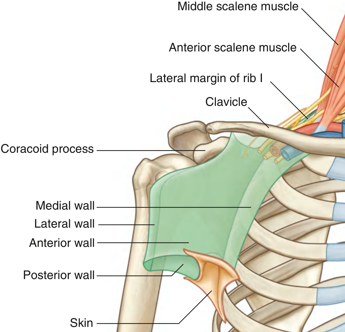
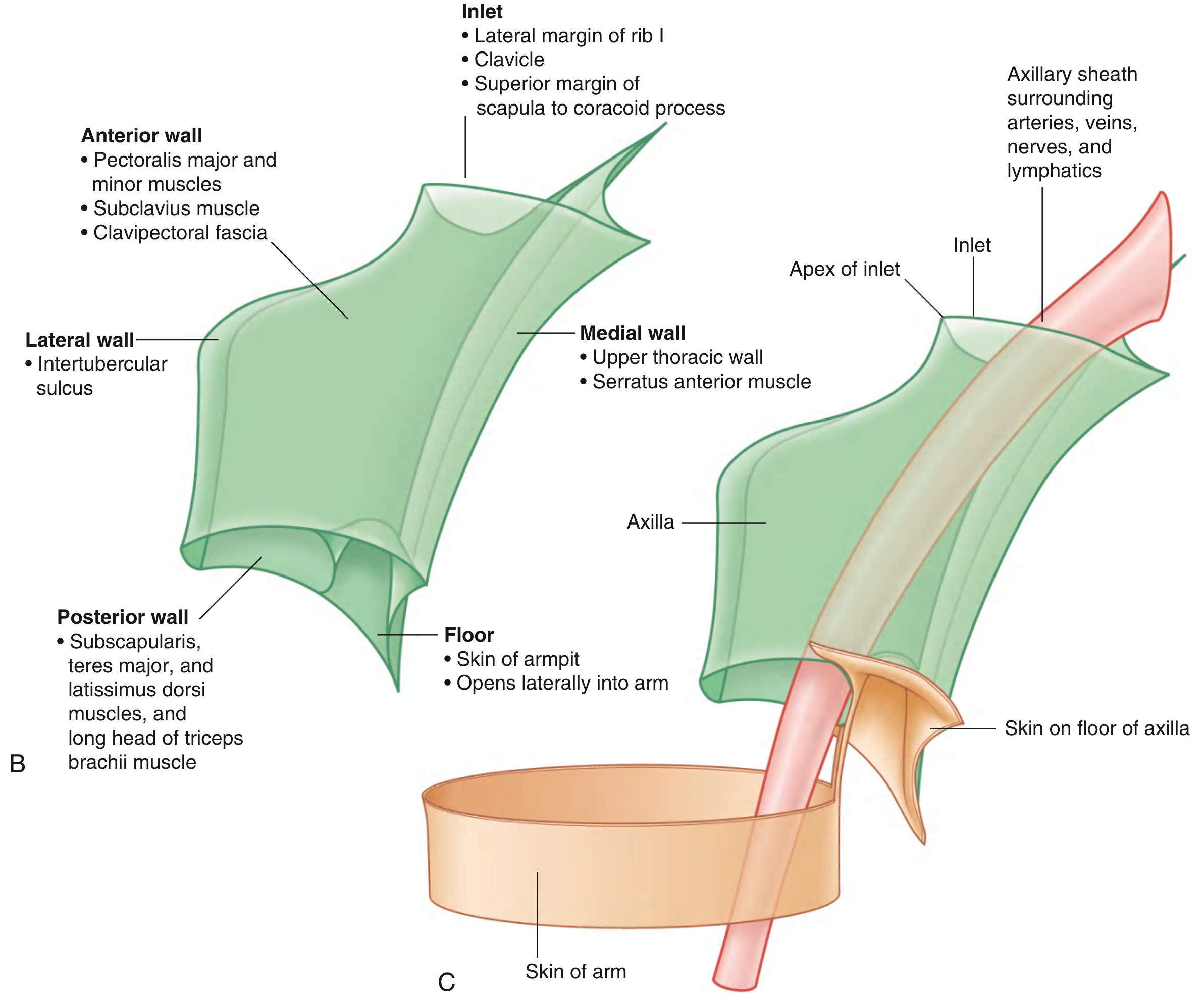
1. Axillary Inlet (Apex)
The inlet is oriented in the horizontal plane and is triangular, with the apex directed laterally. Its margins are formed entirely by bone:
| Margin | Structure |
|---|---|
| Medial | Lateral border of rib I |
| Anterior | Posterior surface of the clavicle |
| Posterior | Superior border of the scapula to the coracoid process |
| Apex (lateral) | Medial aspect of the coracoid process |
The subclavian artery becomes the axillary artery as it crosses the lateral margin of rib I into the axilla. Conversely, the axillary vein becomes the subclavian vein as it exits. At the inlet, from anterior to posterior: axillary vein → axillary artery → brachial plexus trunks.
2. Walls of the Axilla
Anterior Wall
Formed by:
- Pectoralis major - large, fan-shaped muscle forming the anterior axillary fold
- Pectoralis minor - deep to pec major, protracts/depresses the scapula
- Subclavius - small muscle between clavicle and rib I
- Clavipectoral fascia - thick connective tissue sheet connecting the clavicle to the axillary floor, enclosing subclavius and pectoralis minor
Structures traversing the clavipectoral fascia: cephalic vein, thoracoacromial artery, lateral pectoral nerve.
Posterior Wall
Formed by:
- Subscapularis (largest component) - from subscapular fossa to lesser tubercle of humerus; part of the rotator cuff; innervated by superior and inferior subscapular nerves
- Teres major - forms the inferolateral part; its inferior margin marks the inferior limit of the axilla laterally
- Latissimus dorsi - tendon curves around teres major to insert into the intertubercular sulcus
- Long head of triceps brachii - passes vertically through the posterior wall, forming three apertures:
| Aperture | Boundaries | Contents |
|---|---|---|
| Quadrangular space | Teres minor (superior), teres major (inferior), long head of triceps (medial), surgical neck of humerus (lateral) | Axillary nerve, posterior circumflex humeral artery |
| Triangular space | Teres minor/subscapularis (superior), teres major (inferior), long head of triceps (lateral) | Circumflex scapular artery |
| Triangular interval | Teres major (superior), long head of triceps (medial), humerus (lateral) | Radial nerve, profunda brachii artery |
The axillary artery becomes the brachial artery as it crosses the inferior margin of teres major.
Medial Wall
Formed by:
- Upper thoracic wall (ribs 1-4 and related intercostal muscles)
- Serratus anterior muscle - originates from ribs I-IX, inserts on the costal surface of the medial border of the scapula; pulls the scapula forward and keeps it closely apposed to the thoracic wall; innervated by the long thoracic nerve (C5, C6, C7)
Lateral Wall
The narrowest wall, formed by:
- The intertubercular sulcus (bicipital groove) of the humerus - where the long head of biceps brachii tendon runs
3. Floor (Base) of the Axilla
Formed by the skin of the armpit and the axillary fascia. Laterally, the floor opens into the arm, continuous with the skin of the medial arm. The skin over the floor is supplied by the intercostobrachial nerve (lateral cutaneous branch of T2).
4. Contents of the Axilla
A. Muscles (proximal parts)
| Muscle | Origin | Insertion | Nerve | Function |
|---|---|---|---|---|
| Biceps brachii | Short head - coracoid process; Long head - supraglenoid tubercle | Radial tuberosity | Musculocutaneous (C5, C6) | Flexion of forearm, supination |
| Coracobrachialis | Apex of coracoid process | Medial shaft of humerus | Musculocutaneous (C5, C6, C7) | Flexion and adduction of arm |
B. Axillary Artery
Divided into 3 parts by the pectoralis minor muscle:
- Part 1 (medial to pec minor): 1 branch - superior thoracic artery
- Part 2 (posterior to pec minor): 2 branches - thoracoacromial artery (pectoral, deltoid, clavicular, acromial branches) and lateral thoracic artery
- Part 3 (lateral to pec minor): 3 branches - subscapular artery (largest; divides into circumflex scapular + thoracodorsal arteries), anterior circumflex humeral artery, posterior circumflex humeral artery
C. Axillary Vein
Lies medial and anterior to the axillary artery. It becomes the subclavian vein at the lateral margin of rib I. The cephalic vein drains into it.
D. Brachial Plexus
Formed by anterior rami of C5-T1. Organized as:
- Roots: C5, C6, C7, C8, T1 - pass between anterior and middle scalene muscles
- Trunks (3): Superior (C5+C6), Middle (C7), Inferior (C8+T1) - cross rib I
- Divisions (6): Each trunk divides into anterior and posterior divisions
- Cords (3):
- Lateral cord (anterior divisions of superior + middle trunks) - gives musculocutaneous nerve, lateral root of median nerve
- Medial cord (anterior division of inferior trunk) - gives medial root of median nerve, ulnar nerve, medial cutaneous nerves
- Posterior cord (all three posterior divisions) - gives axillary nerve, radial nerve
The cords are named by their relationship to the axillary artery (lateral, medial, posterior).
E. Axillary Lymph Nodes
Five groups drain the upper limb, breast, and chest wall:
| Group | Location | Drains |
|---|---|---|
| Pectoral (anterior) | Medial wall, along lateral thoracic vessels | Anterior thoracic wall, breast |
| Subscapular (posterior) | Posterior wall, along subscapular vessels | Posterior thoracic wall, scapular region |
| Lateral (humeral) | Lateral wall, along axillary vein | Upper limb (most of it) |
| Central | Fat at center of axilla | Receives from all 3 above groups |
| Apical (infraclavicular) | Apex of axilla, along cephalic vein | Receives from all other groups; drains into subclavian lymph trunk |
All groups ultimately drain into the apical nodes, then into the subclavian lymphatic trunk, which drains into the junction of the internal jugular and subclavian veins.
5. Axillary Sheath
The major neurovascular structures (axillary artery, axillary vein, and cords of the brachial plexus) are enclosed in a fibrous axillary sheath, a continuation of the prevertebral fascia from the neck. This is clinically important for axillary nerve blocks - local anesthetic injected into this sheath can spread to block all enclosed structures.
Clinical Relevance
- Winged scapula: Injury to the long thoracic nerve (e.g., during mastectomy or axillary dissection) paralyzes serratus anterior, causing the medial border of the scapula to "wing" outward
- Axillary nerve injury: Fracture of the surgical neck of the humerus or dislocation of the glenohumeral joint can injure the axillary nerve as it passes through the quadrangular space, causing loss of deltoid function and loss of sensation over the "regimental badge" area
- Radial nerve injury: Compression in the triangular interval can cause wrist drop
- Lymph node dissection: Axillary lymph node clearance (e.g., in breast cancer surgery) can damage the long thoracic nerve, thoracodorsal nerve, and intercostobrachial nerve
Source: Gray's Anatomy for Students, Chapter 7 (Upper Limb), pp. 841-858
How to treat cobblestone appearance in throat
Reading File
"Cobblestone throat" is a classic sign of chronic pharyngitis / post-nasal drip. Let me search the library and web in parallel for the most thorough answer.
Searching sources...
Finding Sources
Finding Sources
Searching the Web
cobblestone throat treatment chronic pharyngitis post nasal drip
Reading File
Finding Sources
Reading Web Page
https://my.clevelandclinic.org/health/symptoms/cobblestone-t…
Reading File
Finding Sources
Reading File
Reading File
Reading File
Reading File
Reading File
I now have comprehensive information. Let me compose the full, well-structured answer.
Cobblestone Throat - Causes and Treatment
What Is Cobblestone Throat?
"Cobblestone throat" is a descriptive term for the bumpy, pebble-like appearance of the back of the throat (posterior pharynx). The "cobblestones" are swollen, fluid-filled lymphoid tissue (hypertrophied lymphoid follicles on the posterior pharyngeal wall) that form in response to persistent irritation, infection, or inflammation. The appearance is a sign - not a disease itself - and resolves once the underlying cause is treated.
As noted in Goldman-Cecil Medicine, oropharyngeal secretions or a cobblestone appearance to the mucosa is a hallmark physical finding of postnasal drip syndrome.
Causes (Treat the Cause to Cure the Cobblestoning)
| Cause | Key Feature |
|---|---|
| Postnasal drip (most common) | Mucus drips down the back of the throat, chronically irritating lymphoid tissue |
| Allergic rhinitis | Pollen, dust mites, pet dander trigger mucus overproduction |
| Viral pharyngitis | Rhinovirus, adenovirus, EBV, influenza |
| Bacterial pharyngitis | Group A beta-haemolytic Streptococcus (GABHS) most common bacterial cause |
| Laryngopharyngeal reflux (LPR) / GERD | Stomach acid reaching the throat inflames mucosa |
| Sinusitis | Infected sinuses drip mucus into the throat |
| Chronic irritants | Smoking, dry air, chemical fumes, excessive voice use |
| Mouth breathing | Bypasses nasal humidification, drying and irritating the pharynx |
Treatment - By Underlying Cause
1. Postnasal Drip and Allergic Rhinitis (Most Common)
This is the most frequent driver of cobblestone throat. Treatment is layered:
- Intranasal corticosteroids (e.g., fluticasone, mometasone) - first-line; topical effect with minimal systemic side effects; use in the head-down position for best delivery
- Antihistamines (e.g., cetirizine, loratadine, fexofenadine) - especially for allergic triggers; reduce mucus production
- Intranasal ipratropium bromide - anticholinergic spray to dry excessive nasal secretions; provides additional benefit on top of steroids and antihistamines
- Oral decongestants (e.g., pseudoephedrine) or topical decongestant sprays (e.g., oxymetazoline) - useful short-term (max 3-5 days) for congestion; prolonged use causes rebound nasal obstruction (rhinitis medicamentosa), so avoid long-term use
- Allergen avoidance - identify and reduce exposure to triggers (dust, pollen, pet dander, mould)
- Saline nasal irrigation - flushes mucus and irritants from nasal passages, reducing drip
The combination of topical corticosteroid + antihistamine + anticholinergic has been shown to benefit chronic cough from postnasal drip and improve nasal discharge and endoscopic appearance - Murray & Nadel's Textbook of Respiratory Medicine.
2. Viral Pharyngitis
Most cases are self-limiting in 5-7 days. Management is symptomatic:
- NSAIDs (ibuprofen 400 mg three times daily) - superior to paracetamol in adults for pain relief, fever, and headache; avoid in patients at risk of dehydration due to renal toxicity concerns
- Paracetamol (1 g four times daily) - alternative if ibuprofen is contraindicated; first choice in children
- Corticosteroids - a single oral or IM dose of corticosteroid (e.g., dexamethasone) can hasten pain resolution and achieves 3x greater complete symptom resolution at 24 hours vs. placebo; mean time to pain relief is 6.3 hours earlier; recommended as adjunct in adults alongside antibiotics when needed
- Throat lozenges / gargling with warm salt water - soothe irritation
- Adequate hydration and rest
- Antibiotics are NOT indicated for viral pharyngitis
3. Bacterial Pharyngitis (GABHS / Strep Throat)
Bacteria cause only 5-30% of pharyngitis cases. Clinical scoring (modified Centor/McIsaac score) guides testing and treatment:
- Penicillin V - first-line antibiotic; 10-day course; prevents rheumatic fever
- Amoxicillin - equally effective, better tolerated in children
- Azithromycin or clarithromycin - for penicillin-allergic patients
- Clindamycin - for recurrent streptococcal carriage or treatment failures
- Rapid antigen test or throat culture - should guide antibiotic prescribing; don't treat empirically in low-risk adults
Antibiotics reduce symptom duration by about 16 hours compared to placebo and reduce suppurative complications (e.g., peritonsillar abscess) and non-suppurative complications (e.g., rheumatic fever) - Scott-Brown's Otorhinolaryngology.
4. GERD / Laryngopharyngeal Reflux (LPR)
Acid reflux reaching the pharynx is an underrecognized cause of chronic cobblestone appearance:
- Lifestyle changes: elevate head of bed 15-20 cm, avoid eating 2-3 hours before bed, reduce alcohol and caffeine, avoid spicy/fatty foods, lose weight if overweight
- Proton pump inhibitors (e.g., omeprazole, lansoprazole) - reduce acid production; often need a longer course (2-3 months) for LPR than for typical GERD
- H2-receptor antagonists (e.g., famotidine) - milder alternative
- Antacids - short-term symptom relief
5. Sinusitis
- Acute bacterial sinusitis: antibiotic therapy (amoxicillin-clavulanate is first-line) when mucopurulent discharge has persisted for at least 10 days
- Chronic sinusitis: intranasal steroids, saline irrigation; ENT referral for surgical options (FESS) if refractory
- Viral sinusitis: supportive treatment only
6. Chronic Irritant / Non-Infectious Causes
- Smoking cessation - the single most important intervention for smoking-related cobblestone throat
- Humidification - use a room humidifier to prevent throat drying from dry air or mouth breathing
- Voice rest / vocal hygiene - for cases driven by voice overuse
- Treat mouth breathing - nasal strips, adenoidectomy if indicated
Home Remedies (Supportive Relief)
These do not treat the cause but reduce discomfort:
- Warm salt water gargling - 1/4 to 1/2 teaspoon salt in 8 oz warm water; reduces inflammation
- Honey and warm water or tea - soothing, mild antimicrobial properties
- Staying well hydrated - thins mucus and helps drainage
- Steam inhalation - loosens mucus
- Avoid irritants - smoke, alcohol, very dry or cold air
When to See a Doctor
See an ENT or GP if:
- Cobblestone throat persists beyond 2 weeks without a clear cause
- Symptoms include fever above 38.5°C, difficulty swallowing or breathing, drooling, or neck stiffness (may indicate serious infection or peritonsillar abscess)
- There is unilateral throat swelling (abscess)
- A neck lump or unexplained weight loss accompanies it (rule out malignancy)
- You are immunocompromised - HIV, diabetes, on steroids (higher risk of unusual pathogens like Candida)
For recurrent tonsillitis/pharyngitis, tonsillectomy may be considered using the SIGN/Paradise criteria: 7+ episodes in 1 year, or 5+ per year for 2 years, or 3+ per year for 3 years, provided episodes are disabling and prevent normal functioning.
Sources: Scott-Brown's Otorhinolaryngology Head & Neck Surgery; Goldman-Cecil Medicine 2-Vol Set; Murray & Nadel's Textbook of Respiratory Medicine; Cleveland Clinic
This is a shared conversation. Sign in to Orris to start your own chat.