
Difference between sciatica pain and pain due to DVT?.
sciatica DVT differential diagnosis leg pain
sciatica nerve compression L4 L5 dermatomal leg pain diagram
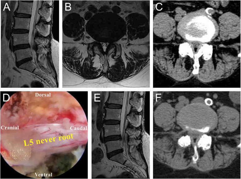
This composite figure demonstrates the preoperative, intraoperative, and postoperative findings of a 60-year-old patient with lateral recess stenosis and L4/5 disc herniation treated with visualized reamed foraminoplasty (VRF). Panels A and B (MRI) and Panel C (CT) represent preoperative imaging, showing significant narrowing of the right lateral recess and compression of the L5 nerve root at the L4/5 level. Panel D is an intraoperative endoscopic photograph showing the L5 nerve root fully released and decompressed. The nerve appears as a white, elongated structure against reddish surgical tissue; directional labels (Dorsal, Ventral, Cranial, Caudal) orient the viewer to the surgical field. Panels E (MRI) and F (CT) provide postoperative imaging, illustrating a successfully enlarged lateral recess and the resolution of compression in both the dorsal and ventral regions of the L5 nerve root. This case highlights the efficacy of the VRF technique in achieving spinal canal decompression and symptomatic relief for radiating leg pain associated with lumbar stenosis.
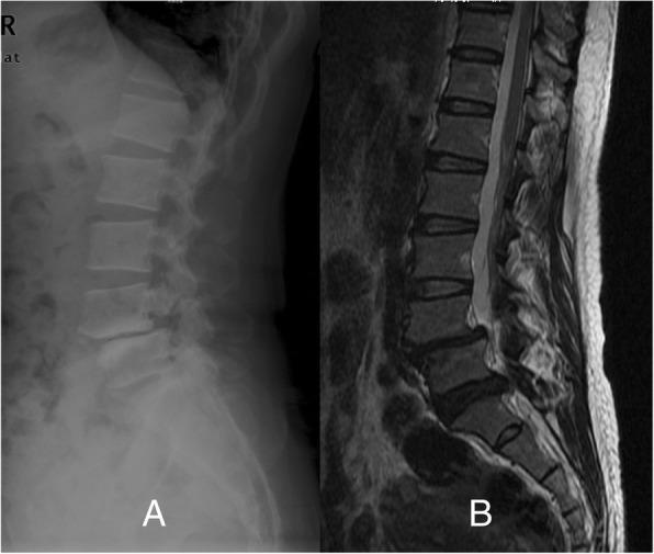
This composite diagnostic image displays a lateral X-ray (A) and a sagittal MRI (B) of the lumbar spine. The lateral radiograph demonstrates an anterior displacement of the L4 vertebral body relative to L5, characteristic of spondylolisthesis. There is evidence of spondylolysis, visible as a lucent defect in the pars interarticularis of the L4 vertebra. The sagittal MRI (B) provides a detailed view of the soft tissues and spinal canal, highlighting a significant disc protrusion at the L5/S1 level. This herniation causes mechanical compression of the thecal sac and likely impacts the descending L5 and S1 nerve roots. The imaging illustrates the mechanical instability of the mid-lumbar spine combined with secondary degenerative disc disease and neural foraminal/canal stenosis. These findings are clinically relevant for evaluating chronic low back pain, radiculopathy, and dermatomal sensory deficits in the lower extremities.
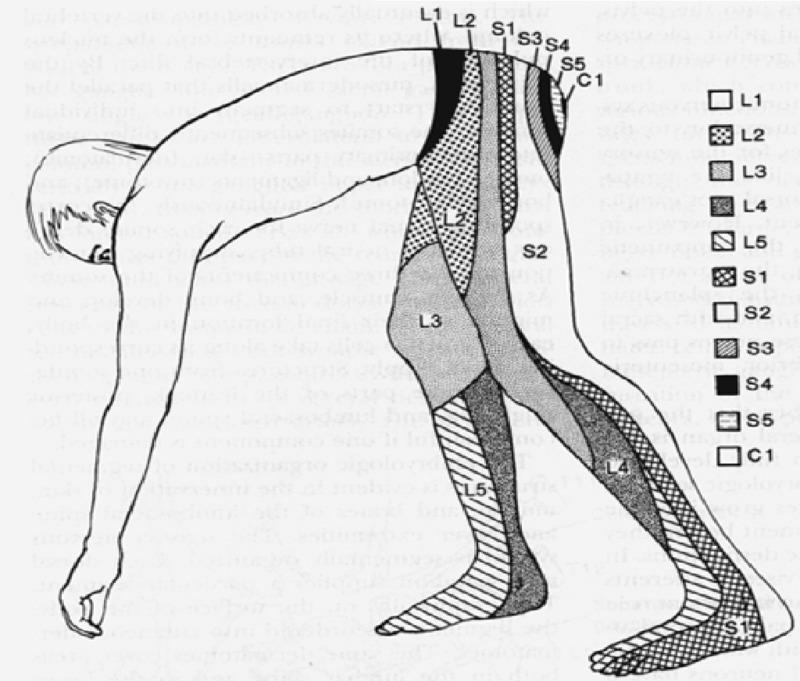
Anatomical diagram depicting a human dermatome map of the lumbosacral and coccygeal regions, shown from a lateral, bent-over perspective to highlight the sensory distribution along the lower extremities and posterior trunk. The illustration uses distinct shading patterns and alphanumeric labels to delineate specific cutaneous areas innervated by individual spinal nerve roots. Key segments include the lumbar dermatomes L1 through L5, sacral dermatomes S1 through S5, and the coccygeal dermatome C1. Visually, L1 covers the inguinal and hip region, L2-L3 transition down the anterior and lateral thigh, and L4-L5 extend down the lower leg to the dorsal foot. Sacral segments S1 and S2 cover the posterior leg and heel, while S3-S5 and C1 are concentrated in the perineal and gluteal regions. This medical illustration is a standard educational tool for neurological examinations, aiding in the localization of spinal nerve root compression or radiculopathy based on sensory loss patterns.
Sciatica vs. DVT: Differentiating Leg Pain
1. Underlying Mechanism
| Sciatica | DVT | |
|---|---|---|
| Pathology | Nerve compression/irritation | Thrombosis (blood clot) in a deep vein |
| Structure involved | Sciatic nerve or spinal nerve roots (usually L4-L5 or L5-S1) | Venous system (calf, popliteal, femoral, iliac veins) |
| Origin | Spine/neural foramen - most commonly a herniated intervertebral disc | Intravascular - Virchow's triad (stasis, hypercoagulability, endothelial injury) |
2. Pain Character
Sciatica
- Radiating, shooting, electric shock-like pain that travels from the lower back down the buttock, posterior thigh, and into the leg/foot
- Pain follows a dermatomal pattern - typically L5 (lateral leg, dorsum of foot) or S1 (posterior thigh, heel, lateral foot)
- Frequently described as burning, stabbing, or "pins and needles"
- Worse with movement, bending, coughing, sneezing (Valsalva maneuver increases intradiscal pressure)
- Associated with numbness, tingling, and weakness in the affected dermatome/myotome
- Back pain is usually also present (though patients with disk herniation often complain more about the radicular symptoms than the back pain)
- Pain extends below the knee in L4-L5 or L5-S1 involvement - this is a key differentiator
DVT
- Dull aching, cramping, heavy, or throbbing pain - localized rather than radiating
- Most common in the calf (posterior), but can affect the thigh if proximal veins are involved
- No dermatomal pattern; pain does not radiate from the spine
- Worsened by dependency (standing/dangling the leg); often relieved by elevation
- Does not follow nerve distribution - there is no numbness or tingling
- May be entirely asymptomatic in up to 50% of cases
3. Location of Pain
| Feature | Sciatica | DVT |
|---|---|---|
| Starting point | Lower back / buttock | Calf or thigh (site of clot) |
| Distribution | Radiates from back → leg → foot | Localized to affected venous segment |
| Below knee | Yes - characteristic | Yes (calf DVT), but does not radiate |
| Dermatomal | Yes | No |
4. Associated Signs
Sciatica
- Positive straight leg raise (SLR) test - pain reproduced at 30-70° of passive leg elevation (sensitivity ~80%)
- Neurological deficits: weakness (foot drop with L5), reduced reflexes (absent ankle jerk with S1), sensory loss
- Tenderness along the spine or paravertebral muscles
- No swelling, no skin color change, no warmth in the leg
DVT
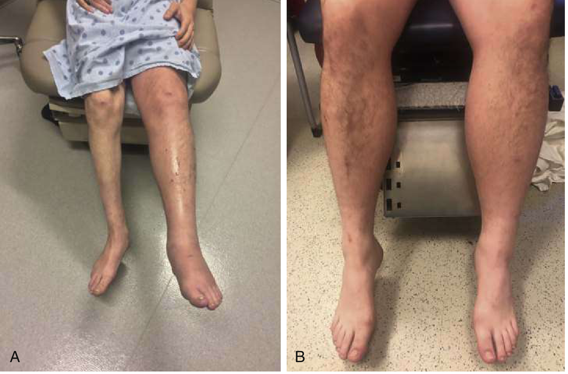
- Leg swelling - often unilateral, pitting edema (calf circumference >3 cm more than the other side is significant)
- Warmth and erythema over the affected area
- Homan's sign (pain on dorsiflexion) - historically used but poor sensitivity/specificity (not reliable)
- Palpable cord (hardened vein) in superficial thrombophlebitis; in deep veins, direct tenderness along the vein course
- No neurological deficits (no weakness, no reflex changes, no dermatomal numbness)
5. Visual Comparison - DVT
6. Dermatomal Pain Map (Sciatica)
7. Risk Factors
| Sciatica | DVT | |
|---|---|---|
| Herniated disc, degenerative spine disease | ✓ | |
| Prolonged immobility, bed rest | Indirect | ✓ (directly) |
| Recent surgery | ✓ | |
| Malignancy | Possible (vertebral metastases) | ✓ (hypercoagulability) |
| Pregnancy | ✓ (especially left leg) | |
| Oral contraceptives / hormone therapy | ✓ | |
| Obesity | ✓ | ✓ |
| Piriformis syndrome, foraminal stenosis | ✓ | |
| Prior DVT / thrombophilia | ✓ |
8. Investigations
| Test | Sciatica | DVT |
|---|---|---|
| MRI spine | Gold standard (disc herniation, cord compression) | Not indicated |
| Venous Doppler ultrasound | Not indicated | Gold standard |
| D-dimer | Not useful | High sensitivity screening test (if negative, rules out DVT in low-probability patients) |
| CT/MR venography | Not indicated | For pelvic vein DVT or equivocal US |
| Plain X-ray | May show spondylolisthesis, narrowing | Not useful |
| Wells score | Not applicable | Pretest probability scoring (clinical decision tool) |
| EMG/nerve conduction | Confirms radiculopathy | Not useful |
9. Urgency and Treatment
| Sciatica | DVT | |
|---|---|---|
| Urgency | Mostly non-urgent; urgent only with cauda equina syndrome (bowel/bladder dysfunction) | Medical emergency - risk of pulmonary embolism (PE) and death |
| Treatment | NSAIDs, activity modification, physiotherapy; epidural steroids; surgery in selected cases | Anticoagulation (DOACs like rivaroxaban/apixaban preferred; LMWH/warfarin also used) |
| Life-threatening complication | Cauda equina syndrome (rare) | Pulmonary embolism - can be fatal |
Key Clinical Differentiators at a Glance
| Feature | Sciatica | DVT |
|---|---|---|
| Pain type | Electric, shooting, radiating | Dull, aching, cramping |
| Origin | Back → buttock → leg | Calf/thigh (localized) |
| Dermatomal | Yes | No |
| Numbness/tingling | Common | Absent |
| Limb swelling | Absent | Present (often unilateral) |
| Skin warmth/redness | Absent | Present |
| SLR test | Often positive | Negative |
| Neurological deficit | Possible | Absent |
| Worse with | Coughing, bending, movement | Dependency (standing) |
| Better with | Lying flat, epidural steroids | Leg elevation |
| Emergency? | Only if cauda equina involved | Always - risk of PE |
- Tintinalli's Emergency Medicine - Low Back Pain with Sciatica and Disk Herniation section
- Rosen's Emergency Medicine - DVT Differential Diagnosis, Box 74.1
- Gray's Anatomy for Students - Deep Vein Thrombosis section
- S Das Manual on Clinical Surgery - Types of Venous Thrombosis