
Peripheral artery disease gen surgery
Peripheral vascular disease gen surgery
"peripheral artery disease"[MeSH Terms] AND management
peripheral arterial disease ankle brachial index angiography diagram
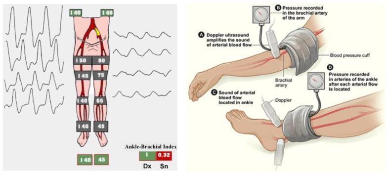
This composite educational graphic illustrates the assessment of Lower Extremity Arterial Disease (LEAD) using the Ankle-Brachial Index (ABI). The left panel presents a schematic diagram of the arterial tree in the lower limbs alongside segment-specific systolic blood pressure (SBP) measurements and arterial pulse waveforms. The right side (Dx) shows normal values (SBP 140-150 mmHg; ABI 1.0) with sharp, triphasic waveforms. In contrast, the left side (Sn) demonstrates arterial stenosis, evidenced by progressively decreasing pressures (from 80 mmHg at the thigh to 45 mmHg at the ankle), a pathologically low ABI of 0.32, and flattened, monophasic waveforms. The right panel is an anatomical illustration of the ABI measurement technique. It depicts the placement of blood pressure cuffs on the upper arm and ankle, accompanied by the use of a continuous-wave Doppler ultrasound probe to detect arterial signals in the brachial artery and the dorsalis pedis or posterior tibial arteries. The diagram highlights key steps: inflating the cuff to occlude flow and recording the SBP at the moment the Doppler signal returns during deflation.
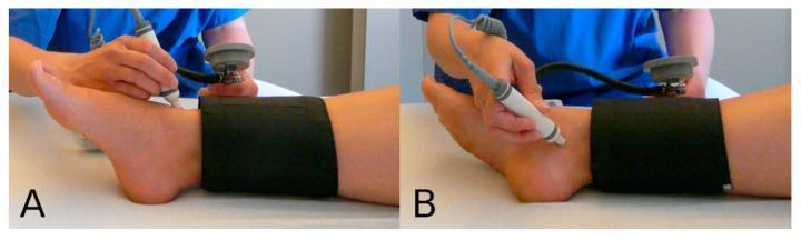
A two-panel clinical photograph demonstrating the procedural technique for measuring the Ankle-Brachial Index (ABI) using a handheld Doppler probe and a sphygmomanometer. In both images, a black pneumatic blood pressure cuff is wrapped around the distal lower leg superior to the malleoli. Panel A illustrates the assessment of the anterior tibial artery, with the Doppler probe positioned on the anterior aspect of the ankle. Panel B shows the assessment of the posterior tibial artery, with the probe positioned on the posterior-medial aspect of the ankle near the medial malleolus. An examiner is shown holding the probe at an appropriate angle to detect arterial flow while monitoring the pressure gauge attached to the cuff. This procedural visual serves as an educational guide for vascular assessment, specifically in screening for peripheral artery disease (PAD) by identifying systolic pressure at specific anatomical landmarks.
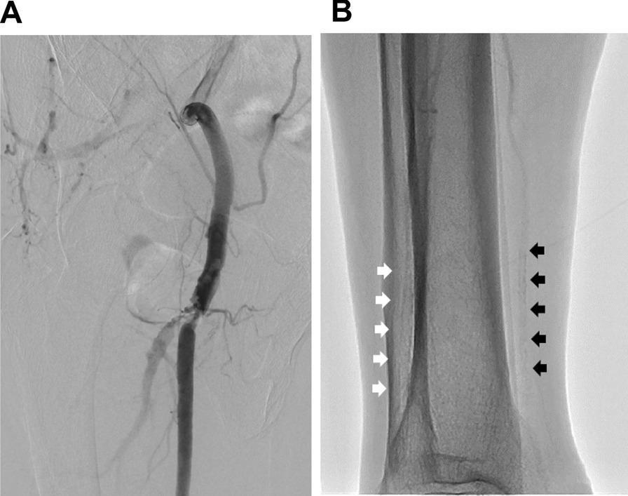
This composite of diagnostic images illustrates peripheral artery disease (PAD) in the right lower extremity. Image A is a Digital Subtraction Angiography (DSA) of the right thigh, showing the common femoral artery bifurcation. A severe focal stenotic lesion is visible at the origin of the right superficial femoral artery (SFA), characterized by a localized narrowing of the contrast column and reduced enhancement indicating restricted blood flow. Image B is a non-contrast fluoroscopic image of the right lower leg (calf) near the ankle. It demonstrates extensive medial arterial calcification (Mönckeberg sclerosis), a common finding in long-standing diabetes mellitus. White arrows indicate linear radiopaque deposits along the course of the anterior tibial artery, while black arrows highlight similar severe calcifications of the posterior tibial artery. These images demonstrate the coexistence of proximal atherosclerotic stenosis and distal infrapopliteal arterial stiffness, which can lead to falsely elevated or normal ankle-brachial index (ABI) measurements due to vessel incompressibility.
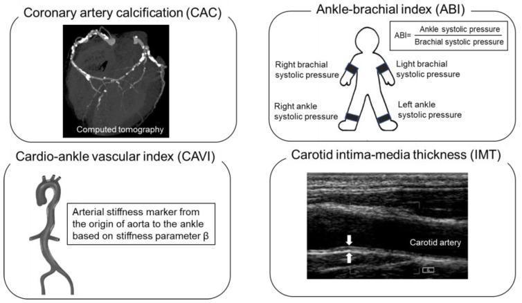
Educational infographic panel demonstrating four non-invasive modalities for atherosclerosis assessment and cardiovascular risk stratification. 1. Coronary Artery Calcification (CAC): A cross-sectional Computed Tomography (CT) scan of the heart showing hyperdense (bright) calcium deposits along the coronary arteries. 2. Ankle-Brachial Index (ABI): A schematic diagram of a human figure indicating systolic blood pressure measurement points at the brachial and ankle arteries, including the calculation formula (Ankle SP / Brachial SP) used to screen for peripheral artery disease. 3. Cardio-Ankle Vascular Index (CAVI): An anatomical diagram of the aorta and central arterial tree, labeled as an arterial stiffness marker derived from the stiffness parameter beta. 4. Carotid Intima-Media Thickness (IMT): A grayscale longitudinal ultrasound image of the carotid artery, with calipers/arrows highlighting the measurement of the vessel wall layers. This compilation serves as a clinical guide for evaluating subclinical atherosclerosis and predicting heart failure risk through diverse imaging and physiological markers.
Peripheral Vascular Disease (PVD) - General Surgery
Definition & Scope
- Peripheral arterial disease (PAD) - atherosclerotic occlusive disease of the limb arteries (most common)
- Acute limb ischemia - sudden occlusion (embolism or thrombosis)
- Venous disease - varicose veins, chronic venous insufficiency, DVT
- Aneurysmal disease - femoral, popliteal aneurysms
- Vasospastic disorders - Buerger's disease, Raynaud's
Epidemiology
- Overall prevalence 3-10%; rises to 15-20% in persons over 70 years
- Present in 50% of patients with diabetic foot ulcer
- PAD is an independent predictor of limb loss and cardiovascular death - Mulholland and Greenfield's Surgery, 7e
Chronic Arterial Ischemia - Pathophysiology & Causes
- Atherosclerosis is the dominant cause; most commonly affects the femoropopliteal segment (calf claudication) and aortoiliac segment (thigh/buttock claudication - Leriche syndrome)
- Less common: embolism from the heart or aorta causing chronic ischemia, Buerger's disease (thromboangiitis obliterans) in young male heavy smokers
- Risk factors: smoking, diabetes, hypertension, hyperlipidaemia, chronic renal failure - Pye's Surgical Handicraft, 22nd Ed
Clinical Presentation & Classification
Fontaine and Rutherford Staging
| Fontaine Stage | Clinical | Rutherford Grade | Category | Clinical |
|---|---|---|---|---|
| I | Asymptomatic | 0 | 0 | Asymptomatic |
| IIa | Mild claudication >200 m | I | 1 | Mild claudication |
| IIb | Moderate-severe claudication <200 m | I | 2-3 | Moderate-severe claudication |
| III | Ischemic rest pain | II | 4 | Ischemic rest pain |
| IV | Ulceration or gangrene | III/IV | 5-6 | Minor/major tissue loss |
Symptoms of Chronic Ischemia
- Intermittent claudication - reproducible muscle pain on walking, relieved by rest (not position)
- Rest pain - constant, worse at night, relieved by hanging the leg dependent
- Tissue loss - non-healing ulcers (punched-out, painful, on toes/pressure points), gangrene
- Signs: absent/diminished pulses, pallor on elevation, dependent rubor, hair loss on dorsum of foot, thickened toenails, muscle wasting, cold feet
Differential Diagnosis of Claudication
- Lumbosacral root irritation (spinal claudication - not relieved by standing, no pulse changes)
- Hip/knee arthritis
- Chronic venous insufficiency
Investigations
Ankle-Brachial Index (ABI)
| ABI | Clinical Status |
|---|---|
| >1.3 | Abnormal - significant arterial wall calcification |
| 1.0-1.2 | Normal |
| 0.6 ± 0.2 | Intermittent claudication |
| 0.3 ± 0.1 | Ischemic rest pain |
| 0.1 ± 0.1 | Impending tissue necrosis |
- Falsely elevated (>1.4) in calcified tibial arteries (diabetes, renal failure - Monckeberg's sclerosis); use toe pressures or plethysmographic waveforms instead
- Falsely elevated with bilateral subclavian/axillary disease
- Normal at rest in aortoiliac disease - always do post-exercise ABI
Segmental Limb Pressures
Duplex Ultrasound
Arteriography / CTA / MRA
- Digital Subtraction Angiography (DSA) remains the gold standard for planning intervention (Seldinger technique via femoral artery)
- CTA and MRA are excellent non-invasive alternatives
Management
Conservative (Medical) Management
- Smoking cessation - single most important intervention; reduces disease progression, preserves graft/stent patency, prevents amputation
- Exercise therapy - supervised walking programme; increases claudication distance
- Antiplatelet therapy - aspirin or clopidogrel (reduces cardiovascular events)
- Statins - lipid lowering and pleiotropic vascular benefits
- ACE inhibitors - ramipril significantly reduces MI, stroke, and death in PAD patients (HOPE trial)
- Antihypertensive control - target BP to reduce cerebrovascular and cardiovascular events
- Glycaemic control - HbA1c <7% in diabetics; note SGLT2-i concerns with canagliflozin and amputations (CANVAS trial) though black box warning later lifted
- Cilostazol - phosphodiesterase III inhibitor; vasodilator + antiplatelet; improves claudication walking distance; contraindicated in heart failure - Fuster and Hurst's The Heart, 15th Ed
Interventional/Surgical - Criteria for Revascularisation
- Claudication causes significant disability affecting activities of daily living
- Exercise and maximum pharmacological therapy are unsatisfactory
- Lesion anatomy is favourable with acceptable risk
- Benefit in symptoms is expected (rule out non-vascular cause)
Revascularisation Options by Anatomy
| Level of Disease | Symptoms | Treatment |
|---|---|---|
| Aortoiliac (inflow disease) | Thigh, hip, buttock claudication; Leriche syndrome | Iliac angioplasty + stenting (preferred). Aortofemoral/aortobifemoral bypass reserved for extensive disease |
| Femoropopliteal (outflow disease) | Calf claudication | Balloon angioplasty ± stenting. Femoro-popliteal (above/below knee) bypass for complex lesions |
| Infrapopliteal | Rest pain, ischemic ulcers | Angioplasty for CTLI. Tibial bypass for limb salvage |
- Type A (short segment stenosis) and Type B → endovascular first
- Type C → endovascular first with surgery backup if failure
- Type D (long occlusions, complex) → surgical bypass preferred
Specific Surgical Procedures
- For severe bilateral aortoiliac occlusive disease
- Long abdominal incision + bilateral groin incisions
- Dacron or PTFE graft from aorta to common femoral/profunda femoris arteries
- Excellent long-term limb salvage but carries significant morbidity/mortality
- Above-knee or below-knee popliteal
- Autologous long saphenous vein is the preferred conduit (reversed or in-situ)
- Synthetic (Dacron/PTFE) used when vein unavailable; worse patency below the knee
- Axillobifemoral bypass (unilateral aortoiliac disease + high operative risk)
- Femorofemoral crossover graft (contralateral iliac disease)
- Limited role; used for rest pain relief or aiding ulcer healing in patients unfit for reconstruction
- Increasingly done as chemical sympathectomy (phenol injection under image intensifier guidance) - Pye's Surgical Handicraft
Graft Conduit Choice Summary
| Situation | Preferred Conduit |
|---|---|
| Below-knee bypass | Autologous long saphenous vein |
| Above-knee femoropopliteal | Vein or synthetic (Dacron/PTFE) |
| Aortic reconstruction | Knitted Dacron or PTFE |
Acute Limb Ischemia
The 6 P's
- Paralysis and paraesthesia = impending irreversible ischemia (surgical emergency)
- Causes: cardiac embolism (AF most common), in-situ thrombosis on atherosclerotic plaque, graft thrombosis, trauma
Management
- Immediate anticoagulation - IV heparin
- Embolectomy - Fogarty balloon catheter (for embolic occlusion)
- Intra-arterial thrombolysis - catheter-directed tPA for thrombotic occlusion
- Surgical bypass - if anatomy unsuitable for catheter techniques
- Fasciotomy - for compartment syndrome post-reperfusion
- Amputation - for non-viable, irreversible ischemia (fixed mottling, rigor, muscle necrosis)
Venous Disease (PVD - Venous Component)
Varicose Veins
- Dilated, tortuous superficial veins due to valvular incompetence
- Risk factors: family history, prolonged standing, pregnancy, obesity
- Complications: aching, skin changes (lipodermatosclerosis), eczema, haemorrhage, thrombophlebitis, venous ulceration
Chronic Venous Insufficiency & Venous Ulcers
- Typically over medial gaiter area (above medial malleolus)
- Shallow, irregular edges, sloping margins, often painless
- ABI must be measured before applying compression bandaging (contraindicated if ABI <0.8)
Diabetic Foot - PVD Component
- Combined neuropathic + ischaemic ulcer = neuroischaemic foot
- Monckeberg's calcification causes falsely elevated ABI - use toe pressures or transcutaneous oxygen tension (TcPO2) instead
- Revascularisation is essential for all DFU with PVD to promote healing and prevent amputation - Mulholland and Greenfield's Surgery, 7e
Key Surgical Pearls for Exams
| Fact | Detail |
|---|---|
| Most common site of PAD | Femoropopliteal segment |
| ABI for claudication | ~0.4-0.8 |
| ABI for critical ischemia | <0.3 |
| ABI >1.4 | Abnormal - calcified (incompressible) vessels |
| Best conduit for below-knee bypass | Long saphenous vein |
| Drug of choice for claudication | Cilostazol (contraindicated in CCF) |
| Conservative treatment mainstay | Smoking cessation + exercise |
| 20% of claudicants will | Progress to rest pain/gangrene requiring surgery |
| Leriche syndrome | Aortoiliac occlusion: buttock/thigh claudication + impotence + absent femoral pulses |
| Embolectomy catheter | Fogarty balloon catheter |