
Approach to encephalopathy in infants
neonatal encephalopathy diagnosis management
neonatal encephalopathy Sarnat staging MRI brain hypoxic ischemic
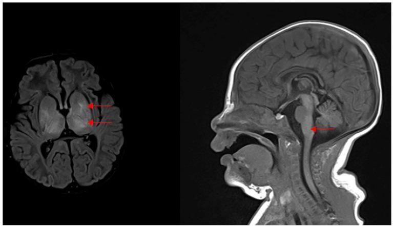
This diagnostic image consists of two brain MRI panels (axial and sagittal views) illustrating severe neonatal hypoxic-ischemic encephalopathy (HIE). The left panel is an axial T2-weighted or FLAIR sequence showing symmetric, bilateral hyperintensities (increased signal) within the basal ganglia (putamen and globus pallidus) and the thalamus, marked by red arrows. This 'basal ganglia-thalamus pattern' is characteristic of acute profound asphyxia. The right panel is a sagittal view showing an area of increased signal intensity and subtle swelling in the dorsal brainstem, specifically in the region of the pons/medulla junction near the vestibulocochlear nerve nuclei. These findings are clinically significant as they correlate with Stage III Sarnat HIE and are associated with profound sensorineural hearing impairment and poor neurological outcomes in neonates.
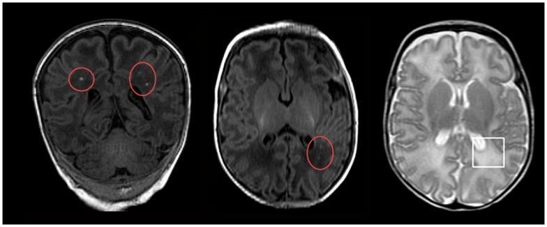
Diagnostic axial MRI images of a neonatal brain demonstrating pathological changes associated with hypoxic-ischemic encephalopathy (HIE) and white matter injury. The image panel contains two T1-weighted sequences and one T2-weighted sequence. On the T1-weighted images (left and center), red circles highlight multiple small, punctate hyperintensities located within the periventricular and subcortical white matter of the frontal and temporal lobes, characteristic of focal white matter lesions. On the T2-weighted image (right), a white square demarcates a region of diffuse excessive high signal intensity (DEHSI) in the posterior periventricular white matter, primarily involving the occipital region. This T2-hyperintensity is indicative of increased water content, such as vasogenic edema or dysmyelination. These imaging findings represent common neuroradiological markers for neonatal seizures and subsequent neurodevelopmental risks following hypoxic-ischemic events.
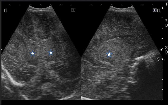
This diagnostic image displays side-by-side coronal (left) and sagittal (right) cranial ultrasound views of a neonate. The imaging shows findings characteristic of severe hypoxic-ischemic encephalopathy (HIE), specifically Sarnat stage III. Both views demonstrate diffuse, markedly increased echogenicity involving the cerebral cortex and deep gray matter structures. Specifically, white stars mark the bilateral thalamic and basal ganglia regions, which appear hyperechoic compared to normal neonatal brain tissue. Significant cerebral edema is present, resulting in a loss of normal anatomical definition and the compression of the lateral ventricles, which are slit-like or barely visible. These visual markers indicate profound global ischemic injury and are critical for the bedside evaluation of neurodevelopmental prognosis in neonatal intensive care. The image serves as a teaching tool for recognizing sonographic patterns of acute perinatal asphyxia and diffuse brain swelling.
Approach to Encephalopathy in Infants
Definition
- Bradley and Daroff's Neurology in Clinical Practice (line 1269)
- Adams and Victor's Principles of Neurology, 12th Ed. (line 1372)
Etiology (Causes by Category)
1. Hypoxic-Ischemic (Most Common)
2. Metabolic
- Hypoglycemia - most immediately treatable cause; must be excluded in all encephalopathic infants
- Hypocalcemia, hyponatremia, hypomagnesemia - electrolyte abnormalities causing seizures/encephalopathy
- Inborn errors of metabolism (IEM) - typically present 2-3 days after birth once feeding begins (organic acidemias, urea cycle defects, maple syrup urine disease, non-ketotic hyperglycinemia)
- Pyridoxine (B6) deficiency - important cause of neonatal seizures, treatable with pyridoxine
- Hyperbilirubinemia/kernicterus - prematurity, acidosis, and prior hypoxic-ischemic injury predispose to bilirubin neurotoxicity, causing staining of specific nuclei
3. Infectious
- Bacterial meningitis/sepsis - GBS, E. coli, Listeria in neonates
- Viral encephalitis - HSV (herpes simplex), enterovirus, CMV
- TORCH infections - congenital infections presenting with encephalopathy from birth
4. Structural / Vascular
- Perinatal arterial ischemic stroke (PAIS) - focal clonic seizures, often in left MCA territory
- Cerebral sinovenous thrombosis (CSVT)
- Neonatal hemorrhagic stroke
- Congenital CNS malformations
- Periventricular leukomalacia - especially in preterm infants
- Cerebellar hemorrhage - in preterm infants, from germinal matrix rupture
5. Toxic/Drug-Related
- Neonates born to mothers using cocaine, heroin, or ethanol - drug-withdrawal encephalopathy/seizures in the first few days of life
- Harrison's Principles of Internal Medicine 22E (line 1430)
Clinical Assessment
Signs of Encephalopathy in Infants
| Feature | Significance |
|---|---|
| Altered consciousness (excessive drowsiness or inability to maintain wakefulness) | Core marker - full-term infants should spend some time awake with eyes open |
| Excessive irritability | Cortical irritation |
| Poor feeding / absent suck | Encephalopathy or brainstem dysfunction |
| Hypotonia with relatively preserved power | Central (not peripheral) cause |
| Fisting (thumb trapped in closed hand) | Central dysfunction |
| Normal/brisk tendon reflexes | Favors central over peripheral cause |
| Absent/abnormal primitive reflexes (Moro, ATNR) | Degree of encephalopathy |
| Seizures (focal, multifocal, subtle) | Cortical irritation |
| Abnormal posturing (decerebrate/decorticate) | Severe encephalopathy |
- Bradley and Daroff's Neurology in Clinical Practice (lines 2133-2135)
Staging of HIE - Sarnat Classification
| Category | Mild (Stage 1) | Moderate (Stage 2) | Severe (Stage 3) |
|---|---|---|---|
| Consciousness | Hyperalert, wide-open eyes | Lethargic, obtunded | Stupor/coma |
| Spontaneous activity | Normal | Decreased | Absent |
| Tone | Mild distal flexion | Decreased, hypotonic | Flaccid |
| Muscle stretch reflexes | Increased | Decreased | Absent |
| Suck | Decreased | Decreased | Absent |
| Moro reflex | Increased | Decreased | Absent |
| Clinical seizures | None | Present (++) | May be present (+) |
| EEG background | Normal | Abnormal | Severely abnormal/suppressed |
| Autonomic | Sympathetic predominance | Parasympathetic | Both depressed |
| Prognosis | Usually full recovery | Variable | High mortality/disability |
- Bradley and Daroff's Neurology in Clinical Practice, Table 110.3 (line 1311)
Investigations
Immediate / Bedside
- Blood glucose - must be checked immediately; hypoglycemia is rapidly reversible
- ABG/lactate - acidosis, elevated lactate supports HIE or metabolic disorder
- Electrolytes (Na, Ca, Mg), renal and liver function
- Full blood count, CRP/PCT, blood cultures - sepsis screen
- Cord blood gas / Apgar scores - retrospective evidence of perinatal asphyxia
Laboratory
- Ammonia, amino acids (plasma), organic acids (urine) - IEM screen
- Urine toxicology - drug exposure
- CSF analysis (cell count, glucose, protein, culture, HSV PCR) - infection/inflammation
- Serum bilirubin - kernicterus risk
- Thyroid function, lactate/pyruvate ratio, biotinidase
- Chromosomal/genetic testing when malformation suspected
Electrophysiology
- Amplitude-integrated EEG (aEEG) / continuous EEG monitoring - essential for seizure detection (many neonatal seizures are subclinical), EEG background pattern has prognostic value. Epileptiform activity and voltage suppression are associated with unfavorable outcomes.
- Visual and auditory evoked potentials - poor prognostic signs when abnormal
Neuroimaging
-
Basal ganglia-thalamus (BGT) pattern - symmetric restriction on DWI in ventrolateral thalami and posterior putamina ± perirolandic cortex. Seen after acute profound asphyxia (e.g., cord prolapse). Correlates with severe outcome.
-
Watershed predominant pattern - restriction in anterior-MCA and posterior-MCA watershed zones (cortical and subcortical white matter). Seen with prolonged partial asphyxia.
- DWI restriction: begins soon after injury, peaks within a few days, "normalizes" by end of first week
- T1/T2 changes: begin around day 3-4 (absence of normal high T1 signal in posterior limb of internal capsule is a key finding)
- MR spectroscopy: elevated lactate:NAA ratio in deep gray nuclei has prognostic value
- Bradley and Daroff's Neurology in Clinical Practice (lines 1305-1320)
Management
1. Immediate Stabilization (ABC)
- Secure airway; support breathing (many severe cases require mechanical ventilation)
- Correct hypoglycemia immediately (IV dextrose)
- Correct electrolyte abnormalities
- Treat sepsis empirically (ampicillin + gentamicin ± acyclovir if HSV suspected) pending cultures
- Avoid hyperthermia (worsens neuronal injury)
2. Therapeutic Hypothermia (HIE-specific, EVIDENCE-BASED)
-
Indication: Moderate or severe HIE (Sarnat stage 2 or 3), gestational age ≥36 weeks
-
Window: Must be initiated within 6 hours of birth
-
Target temperature: 33.5°C (whole-body cooling) for 72 hours, then slow rewarming
-
NNT = 7 to prevent one death or moderate-severe impairment
-
Despite hypothermia, mortality + morbidity in moderate-severe HIE remains nearly 50%
-
Adjuncts under study: erythropoietin (EPO), melatonin, allopurinol
-
Bradley and Daroff's Neurology in Clinical Practice (line 1326)
3. Seizure Management
- First-line: Phenobarbital (20 mg/kg IV loading dose)
- Second-line: Levetiracetam (gaining use as alternative/adjunct), phenytoin/fosphenytoin
- Pyridoxine 100 mg IV if seizures unresponsive - trial for pyridoxine-dependent epilepsy
- Continuous EEG monitoring - subclinical seizures are common and must be detected
- Avoid prolonged seizures - they compound neuronal injury
4. Metabolic/IEM Management
- Restrict protein if urea cycle defect suspected
- Thiamine, biotin, cofactor trials in refractory neonatal seizures
- Specific dietary/enzyme replacement therapies once IEM confirmed
5. Neuroprotective Supportive Care
- Maintain normoglycemia (glucose 50-100 mg/dL)
- Normotension (avoid hypotension)
- Normocapnia (avoid hypo/hypercarbia - both worsen outcome)
- Avoid hyperthermia
- Treat hyperbilirubinemia promptly (phototherapy, exchange transfusion)
Prognosis
| Stage | Outcome |
|---|---|
| Mild (Sarnat 1) | Full recovery in most |
| Moderate (Sarnat 2) | Variable - may improve by 48-72h or deteriorate with cerebral edema and seizures |
| Severe (Sarnat 3) | High mortality; survivors often have severe neurodevelopmental disability |
- Seizures with EEG burst-suppression or flat background
- Absent brainstem reflexes
- Severe BGT pattern on MRI
- Abnormal visual/auditory evoked potentials
- Absent posterior limb of internal capsule signal on T1 MRI
Summary Algorithm
Infant with encephalopathy
    │
    ├── STABILIZE: Airway, breathing, circulation
    │
    ├── IMMEDIATE INVESTIGATIONS
    │       Blood glucose → treat hypoglycemia NOW
    │       ABG, electrolytes, ammonia, LFTs, CBC, cultures
    │       CSF (LP when stable)
    │
    ├── DETERMINE CAUSE
    │       Perinatal history → HIE?
    │       Fever/maternal illness → Infection?
    │       Onset after feeds → IEM?
    │       Drug exposure → Withdrawal?
    │       Dysmorphism/malformation → Structural/genetic?
    │
    ├── STAGE ENCEPHALOPATHY (Sarnat)
    │       EEG / aEEG monitoring
    │       MRI brain (preferably day 3-5)
    │
    └── TREAT
            HIE moderate/severe → Therapeutic hypothermia (within 6h)
            Seizures → Phenobarbital ± levetiracetam ± pyridoxine
            Infection → Antibiotics ± acyclovir
            IEM → Dietary restriction, cofactors
            Electrolyte → Correction
- Bradley and Daroff's Neurology in Clinical Practice (lines 1267-1332, 2131-2148)
- Adams and Victor's Principles of Neurology, 12th Edition (lines 1372-1400)
- Harrison's Principles of Internal Medicine 22E (line 1430)