
Anatomy of pelvic autonomic nervous system. Detailed overview using tables and flowchart if required.
Reading File
Now let me search the library for relevant sections on pelvic autonomic nervous system anatomy.
Finding Sources
Finding Sources
Finding Sources
Finding Sources
Finding Sources
Finding Sources
Excellent - found several key sources. Let me now read the most relevant sections in parallel.
Reading File
Reading File
Reading File
Reading File
Reading File
Reading File
I now have comprehensive information. Let me also fetch the key diagram from Berek & Novak's and check the Campbell Walsh section on pelvic plexus.
Loading Image
Reading File
I now have rich content from multiple authoritative sources. Let me compile the comprehensive answer.
Anatomy of the Pelvic Autonomic Nervous System
Overview
The pelvic autonomic nervous system controls the bladder, rectum, internal anal and urethral sphincters, reproductive organs, and erectile/clitoral tissues. It is composed of two divisions - sympathetic (thoracolumbar) and parasympathetic (sacral craniosacral) - organized into a hierarchy of plexuses that converge into a "final common pathway" at the inferior hypogastric plexus.
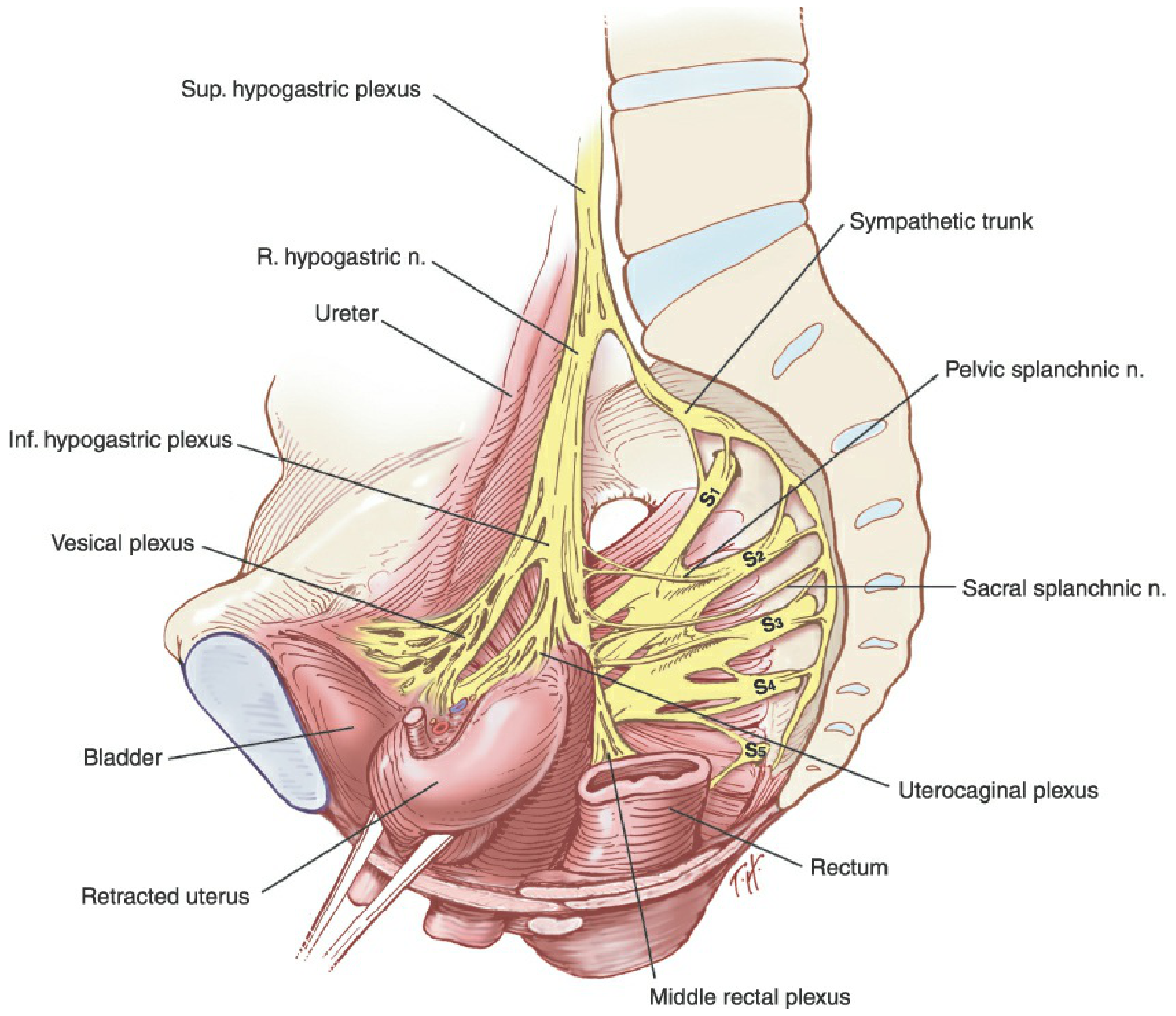
1. Structural Hierarchy
Thoracic/Lumbar Spinal Cord (T10-L2)
         │ (Preganglionic sympathetic fibers)
         ▼
Lumbar Sympathetic Trunks
         │
         ▼
Inferior Mesenteric Plexus ──→ (Aortic Plexus continuation)
         │
         ▼
Superior Hypogastric Plexus (Presacral Nerve)
    [Anterior to L5/Sacral promontory, between bifurcation of aorta]
         │
    Divides into
    ┌────┴────┐
    ▼         ▼
Left          Right
Hypogastric   Hypogastric
Nerve         Nerve
    └────┬────┘
         │ + Pelvic Splanchnic Nerves (S2-S4) [parasympathetic]
           + Sacral Splanchnic Nerves [sympathetic, from sacral trunk]
         ▼
Inferior Hypogastric Plexus (Pelvic Plexus)
    [Lateral pelvic wall, beside rectum/lateral to pelvic viscera]
         │
    ┌────┼────┬────┐
    ▼    ▼    ▼    ▼
Vesical  Rectal  Prostatic/  Uterovaginal
Plexus  Plexus  Uterovaginal  (Frankenhäuser)
                Plexus         Plexus
2. Components: Sympathetic Division
Origin and Course
| Feature | Detail |
|---|---|
| Spinal cord levels | T10 - L2 (thoracolumbar) |
| Preganglionic neurons | Intermediolateral cell column of spinal cord |
| Pre-ganglionic fibers exit via | Anterior (ventral) roots → white rami communicantes → sympathetic trunks |
| Synapse location | Paravertebral ganglia OR prevertebral ganglia (distant from target organ) |
| Route into pelvis | Via lumbar splanchnic nerves → aortic plexus → inferior mesenteric plexus → superior hypogastric plexus |
| Pelvic entry | Superior hypogastric plexus crosses pelvic inlet anterior to L5/sacral promontory, medial to ureters |
| Pelvic structure | Divides below sacral promontory into left and right hypogastric nerves |
| Additional pelvic input | Sacral splanchnic nerves from sacral sympathetic trunks join inferior hypogastric plexus directly |
Superior Hypogastric Plexus (Presacral Nerve)
- Located anterior to the terminal aorta, fifth lumbar vertebra, and sacral promontory
- Embedded in loose areolar tissue, overlies middle sacral vessels
- Usually composed of two or three incompletely fused trunks
- Contains: preganglionic fibers from lumbar nerves, postganglionic fibers from higher sympathetic ganglia and sacral sympathetic trunks, and visceral afferent fibers
- Medial to the ureters on each side
Hypogastric Nerves
- Paired nerves formed by the split of the superior hypogastric plexus just below the sacral promontory
- Course inferiorly and laterally to connect with the inferior hypogastric plexuses
- Often appear as intermingling strings rather than a single defined nerve
- Intimately associated with the presacral fascia
- Cross the pelvic inlet medially to the internal iliac vessels
- Pass through the peritoneum of the posterior cul-de-sac and uterosacral ligaments
Functions of Sympathetic Fibers in Pelvis
| Function | Target |
|---|---|
| Innervate blood vessels (vasoconstriction) | Pelvic vasculature |
| Contract smooth muscle | Internal urethral sphincter |
| Contract smooth muscle | Internal anal sphincter |
| Smooth muscle contraction | Reproductive tract and accessory glands |
| Move secretions into urethra | Epididymis, seminal vesicles, prostate (ejaculation) |
| Bladder filling | Detrusor relaxation, sphincter contraction |
3. Components: Parasympathetic Division
Origin and Course
| Feature | Detail |
|---|---|
| Spinal cord levels | S2, S3, S4 (sacral - craniosacral division) |
| Preganglionic neurons | Sacral parasympathetic nucleus (intermediolateral cell column, sacral cord) |
| Preganglionic fibers exit via | Ventral (anterior) sacral nerve roots → sacral plexus → pelvic splanchnic nerves |
| Alternative name | Nervi erigentes (erigent nerves) - named for function |
| Route | Pelvic splanchnic nerves pierce the presacral fascia (left and right sides) |
| Synapse location | On or near the target organ (ganglia in the inferior hypogastric plexus or organ wall) |
| Junction point | Join hypogastric nerves to form the inferior hypogastric plexus (pelvic plexus) |
Pelvic Splanchnic Nerves (Nervi Erigentes)
- Arise from S2-S4 sacral anterior roots
- Pierce the presacral fascia bilaterally and join the inferior hypogastric plexus
- Some fibers ascend superiorly in the prevertebral plexus to innervate the descending colon and sigmoid (i.e., parasympathetic supply to hindgut extends retrograde from pelvis)
Functions of Parasympathetic Fibers in Pelvis
| Function | Target |
|---|---|
| Vasodilation | Erectile tissues (penis, clitoris), pelvic vasculature |
| Stimulate detrusor contraction | Urinary bladder (micturition) |
| Promote erection | Cavernous bodies (penis, clitoris), vestibular bulbs |
| Lubrication of external genitalia | Vestibular/Bartholin glands |
| Modulate enteric nervous system | Colon distal to left colic flexure, sigmoid, rectum |
| Transmit rectal distension | Via S2-S4 splanchnic nerves (defecation reflex) |
4. Inferior Hypogastric Plexus (Pelvic Plexus) - The Final Common Pathway
Structure
- Paired - one on each side
- Retroperitoneal, lateral pelvic sidewall, medially to major vessels and somatic nerves
- In men: beside the rectum, 5-11 cm from the anal verge, forms a fenestrated rectangular plate in the sagittal plane with its midpoint at the level of the tip of the seminal vesicle
- In women: extends anteriorly in a fan-like fashion along the lower lateral posterior wall of the vagina
- Branches of the inferior vesical artery and vein perforate the pelvic plexus
Fiber Composition
| Fiber Type | Source | Function |
|---|---|---|
| Preganglionic parasympathetic | Pelvic splanchnic nerves (S2-S4) | Vasodilation, bladder contraction, erection |
| Postganglionic sympathetic | Hypogastric nerves (from superior hypogastric plexus) | Ejaculation, sphincter tone, vasoconstriction |
| Postganglionic sympathetic | Sacral splanchnic nerves (sacral trunk) | Additional sympathetic supply |
| Visceral afferent (sensory) | Travel with both sympathetic and parasympathetic | Pain, distension, reflex arcs |
Subsidiary Plexuses
| Subsidiary Plexus | Target Organs | Course |
|---|---|---|
| Vesical plexus | Bladder and urethra | Along vesical vessels |
| Rectal (middle rectal) plexus | Rectum, internal anal sphincter | Along middle rectal vessels |
| Prostatic plexus (men) | Prostate, seminal vesicles, urethra, corpora cavernosa | Lateral to prostate, gives off cavernous nerves |
| Uterovaginal plexus / Frankenhäuser ganglion (women) | Uterus, vagina, clitoris, vestibular bulbs | Along uterine vessels, through cardinal and uterosacral ligaments |
5. Cavernous Nerves and Neurovascular Bundle (NVB)
- Terminal branches of the prostatic plexus (men) or uterovaginal plexus (women)
- In men: travel outside the prostatic capsule and Denonvilliers fascia in the lateral pelvic fascia dorsolaterally between prostate and rectum
- The neurovascular bundle of Walsh = cavernous nerves + capsular arteries and veins, located between prostatic fascia and levator fascia
- Cavernous branches join capsular vessels in a spray-like distribution 20-30 mm distal to the bladder-prostate junction
- After piercing the urogenital diaphragm, enter the corpora cavernosa
- Clinically critical: preservation during radical prostatectomy prevents erectile dysfunction
6. Visceral Afferent (Sensory) Fibers
| Fiber Path | Spinal Level | Sensation Carried |
|---|---|---|
| Via sympathetic (thoracolumbar) path | Lower thoracic/lumbar | Pain from most pelvic viscera |
| Via parasympathetic (sacral) path | S2-S4 | Pain from cervix, bladder, urethra; rectal distension |
| Ovarian plexus (with ovarian vessels) | Aortic/renal plexus origin | Pain from ovary/fallopian tube (NOT relieved by presacral neurectomy) |
7. Additional Pelvic Plexuses
| Plexus | Origin | Structures Innervated |
|---|---|---|
| Ovarian plexus | Aortic and renal plexuses (accompanies ovarian vessels) | Ovaries, fallopian tubes, broad ligament |
| Inferior mesenteric plexus | Celiac/aortic plexus continuation | Left colon, sigmoid, rectum (sympathetic) |
| Sacral sympathetic trunks | Continuation of lumbar sympathetic trunks | Pass behind common iliac vessels, anterior to sacrum |
8. Summary Comparison: Sympathetic vs. Parasympathetic
| Feature | Sympathetic | Parasympathetic |
|---|---|---|
| Origin | T10-L2 spinal cord | S2-S4 sacral cord |
| Preganglionic fiber | Short | Long |
| Ganglia location | Paravertebral / prevertebral (distant from organ) | On or near target organ |
| Route into pelvis | Lumbar splanchnic → SHP → hypogastric nerves | Pelvic splanchnic nerves (nervi erigentes) |
| Key plexus | Superior hypogastric plexus → hypogastric nerves | Pelvic splanchnic nerves → IHP |
| Bladder effect | Detrusor relaxation + sphincter contraction (filling) | Detrusor contraction + sphincter relaxation (micturition) |
| Genital effect | Ejaculation, smooth muscle of genital tract | Erection, vasodilation, lubrication |
| Anal sphincter | Internal sphincter contraction | Facilitates defecation |
| Neurotransmitter | Noradrenaline (postganglionic) | Acetylcholine (+VIP as cotransmitter for vasodilation) |
9. Clinical Correlates
| Clinical Situation | Nerve at Risk | Consequence |
|---|---|---|
| Low anterior rectal resection / TME | Hypogastric nerves, pelvic splanchnic nerves, pelvic plexus | Bladder, bowel, and sexual dysfunction |
| Radical prostatectomy | NVB of Walsh (cavernous nerves) | Erectile dysfunction (impotence) |
| Radical hysterectomy | Inferior hypogastric plexus, uterovaginal plexus | Bladder, bowel, sexual dysfunction |
| Sacrocolpopexy mesh fixation (anterior S1) | Sacral hypogastric plexus | Hypogastric nerve injury |
| Uterosacral ligament transection | Hypogastric nerves (course through uterosacral ligaments) | Urinary retention, bowel/sexual dysfunction |
| Presacral neurectomy | Superior hypogastric plexus | Dysmenorrhea relief (~50-75% cases) - but ovarian pain NOT relieved |
| Posterior rectal dissection | Presacral fascia + adherent hypogastric nerves | Sympathetic denervation |
Sources
- Fischer's Mastery of Surgery, 8th ed. - "Autonomic Nervous System" (p. 4002-4003)
- Gray's Anatomy for Students - "Pelvic extensions of the prevertebral plexus" (p. 570-572)
- Berek & Novak's Gynecology - "Efferent Innervation / Afferent Innervation" (p. 173-175)
- Campbell-Walsh-Wein Urology - "Pelvic Plexus" (p. 4649)
This is a shared conversation. Sign in to Orris to start your own chat.