
Varicose vein
Reading File
Now let me search the medical library for varicose veins, running parallel queries:
Finding Sources
Searching PubMed
"varicose veins"[MeSH Terms] AND treatment
Finding Sources
Good. Now let me read the main Bailey & Love section on varicose veins and the S. Das section in parallel:
Reading File
Reading File
Reading File
Reading File
Reading File
Reading File
Reading File
I now have excellent content. Let me also fetch the clinical image of varicose veins:
Loading Image
I now have comprehensive information. Here is a complete, well-structured answer on varicose veins:
Varicose Veins
Definition
Varicose veins are tortuous, dilated, elongated subcutaneous veins resulting from sustained venous hypertension and valvular incompetence, predominantly affecting the superficial venous system of the lower limbs. They are distinguished from telangiectasia (thread veins) and reticular veins, which are smaller and more superficial.
Clinical Photograph
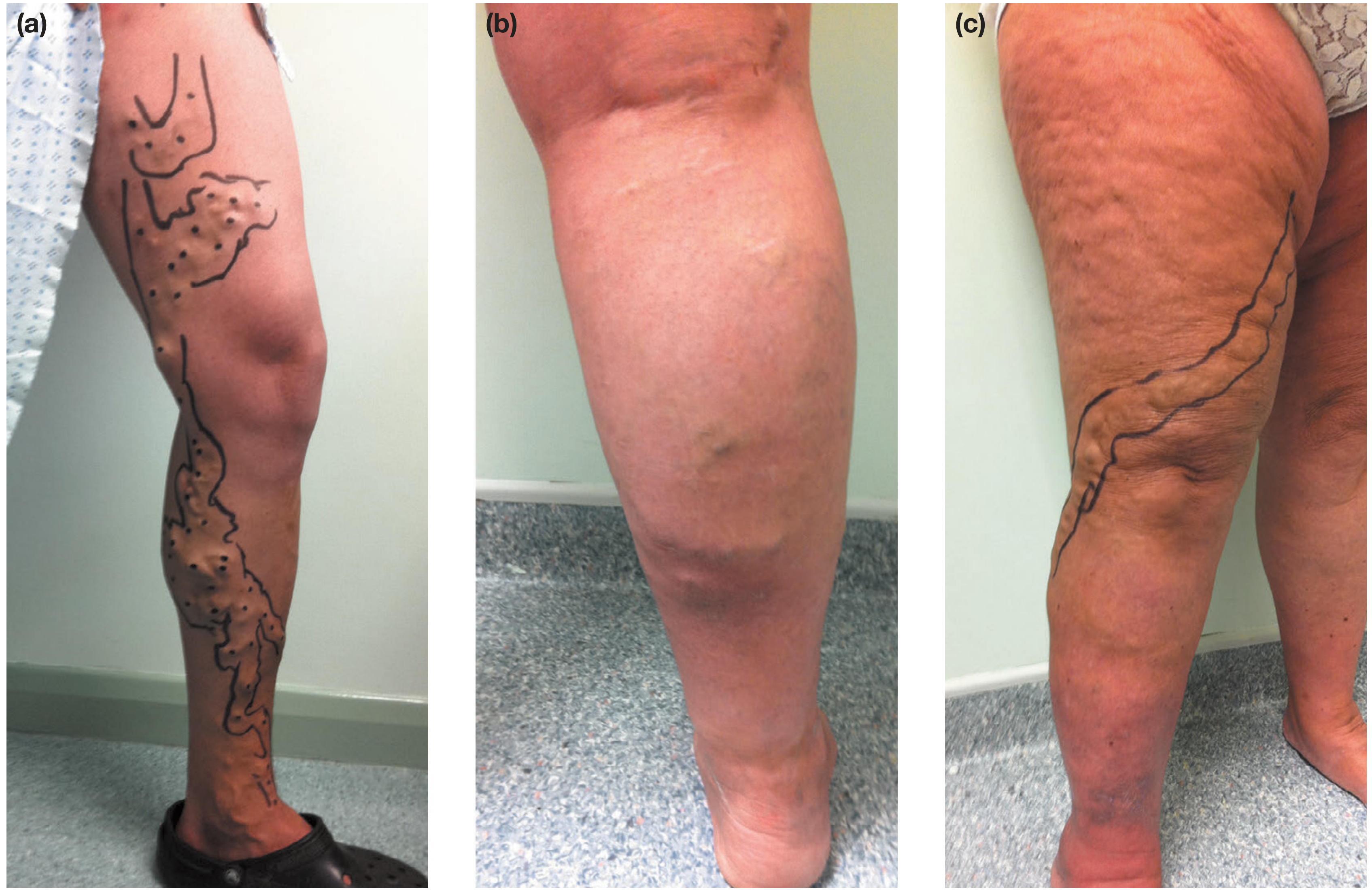
Figure: (a) Left leg GSV varicosities marked for intervention; (b) Right leg SSV distribution with recent phlebitis; (c) Anterolateral AAGSV distribution with gaiter area skin changes - Bailey & Love, p.1049
Epidemiology
- Adult prevalence: 30-50% of the general population
- More common in women than men
- Prevalence increases with age - from 11.5% (age 18-24) to 55.7% (age 55-64) per the Edinburgh Vein Study
- Risk factors: pregnancy, family history, increasing BMI and height, possibly prolonged standing
- Less common in primitive/rural communities (Africa, Far East)
Pathophysiology
The primary problem lies in the vein wall rather than mechanical valve failure alone. Vein wall changes include:
- Inflammatory cell infiltration and activation
- Dysfunctional smooth muscle cell proliferation
- Collagen deposition, decreased elastin content
- Increased matrix metalloproteinases
- Net result: loss of compliance → dilatation → elongation (tortuosity) → secondary valvular dysfunction
This creates retrograde flow with gravity (venous incompetence). Secondary varicosities can follow deep vein thrombosis (post-thrombotic) or occur with congenital arteriovenous fistulae.
Causes of predisposition:
- Primary: valvular incompetence, vein wall weakness
- Secondary: obstruction to venous return - pregnancy, pelvic tumours (fibroid, ovarian cyst, carcinoma cervix/rectum), ascites, abdominal lymphadenopathy, retroperitoneal fibrosis
- Congenital: arteriovenous fistula (in younger patients)
CEAP Classification
The CEAP (Clinical-Etiology-Anatomy-Pathophysiology) system is used internationally:
Clinical categories (C):
| Grade | Description |
|---|---|
| C0 | No visible or palpable signs of venous disease |
| C1 | Telangiectasia/reticular veins |
| C2 | Varicose veins |
| C3 | Oedema |
| C4a | Pigmentation/eczema |
| C4b | Lipodermatosclerosis/atrophie blanche |
| C5 | Healed venous ulcer |
| C6 | Active venous ulcer |
Anatomical (A): Superficial (As), Perforator (Ap), Deep (Ad)
Pathophysiological (P): Reflux (Pr), Obstruction (Po), Both (Pr,o)
Symptoms
Patients describe:
- Aching, heaviness, throbbing, burning, or bursting sensation in the legs
- Symptoms worsen throughout the day or with prolonged standing
- Relieved by elevation and compression hosiery
- Ankle swelling (especially by end of day)
- Pruritus (itching), especially with complications
- Night cramps
- Bursting pain on walking (suggests associated deep vein thrombosis)
Note: symptoms are independent of the degree of venous incompetence - asymptomatic varicosities can coexist with severe symptoms from minimal veins on the other side. - S. Das, p.105
Clinical Signs
Inspection:
- Tortuous dilated subcutaneous veins
- Distribution gives clue to axis involved:
- Medial thigh/calf → GSV incompetence
- Posterolateral calf → SSV incompetence
- Anterolateral thigh/calf → AAGSV incompetence
- Saphena varix: dilated vein at the SFJ appearing as a groin lump, disappears when recumbent (can mimic a femoral hernia - impulse on coughing)
- Skin changes: pigmentation, eczema, lipodermatosclerosis, venous ulcer (medial malleolus)
Key clinical tests:
| Test | Method | What it detects |
|---|---|---|
| Trendelenburg test | Tourniquet applied at SFJ after leg elevation; observe vein filling on standing | SFJ incompetence |
| Schwartz test (Percussion) | Tap prominent varicosity; impulse felt at saphenous opening | Continuous column of blood (incompetent valves) |
| Cough impulse (Morrissey) | Leg elevated, then patient coughs; expansile impulse at saphenous opening | SFJ valve incompetence |
| Fegan's method | Mark varicosity bulges; elevate limb; palpate for fascial gaps | Sites of incompetent perforators |
| Auscultation | Continuous machinery murmur | Arteriovenous fistula (secondary varicosities) |
Investigation
Duplex ultrasound is now the gold standard - tourniquet tests and hand-held Doppler have been abandoned. Duplex establishes:
- Presence and distribution of reflux in deep and superficial systems
- Anatomy of the SFJ and SPJ
- Presence of deep vein thrombosis or obstruction
- Location of incompetent perforators
A 7.5-13 MHz linear array transducer is standard. Blue = antegrade flow toward heart; red = reverse (reflux).
Treatment
Conservative
- Compression hosiery (first-line for symptom relief)
- Leg elevation, weight loss, exercise
- Useful as trial to confirm symptoms are venous before intervention
Interventional (current evidence)
1. Endovenous Thermal Ablation
- Endovenous Laser Ablation (EVLA): Laser fibre inserted under ultrasound guidance; perivenous tumescent anaesthesia used; energy delivered as fibre is withdrawn. Highest technical efficacy.
- Radiofrequency Ablation (RFA): Uses radiofrequency energy via catheter. Comparable efficacy to EVLA with possibly less post-procedural pain.
- A 2024 meta-analysis (Jiang et al., J Vasc Surg Venous Lymphat Disord, PMID 38316290) found RFA and EVLA have comparable clinical outcomes; RFA may have slight advantage in post-operative comfort.
2. Foam Sclerotherapy
- Sclerosant (e.g., sodium tetradecyl sulphate or polidocanol) mixed with air/gas to create foam
- Ultrasound-guided for truncal veins
- Can be used alongside thermal ablation for tributaries
3. Ambulatory Phlebectomy (Stab Avulsion)
- Micro-incisions (2-3 mm) to remove varicosities
- Often combined with truncal ablation in same sitting
4. Open Surgery (Trendelenburg + Stripping)
- Flush ligation of SFJ + stripping of GSV
- Now less commonly performed; reserved for recurrent disease, complex anatomy, or where endovenous techniques are unavailable
For recurrent varicose veins from the SFJ/GSV, a 2024 systematic review (Bontinis et al., Int Angiol, PMID 39559844) found endovenous techniques are effective for recurrence.
Complications
| Type | Examples |
|---|---|
| Chronic | Pigmentation, eczema, lipodermatosclerosis, atrophie blanche, venous ulcer |
| Acute | Superficial vein thrombosis (thrombophlebitis), haemorrhage (from rupture) |
| Post-operative | DVT, nerve injury (saphenous/sural nerve), wound haematoma, recurrence |
Differential Diagnosis of Varicose Veins (causes of dilated leg veins)
- Primary (idiopathic) varicose veins
- Secondary to DVT (post-thrombotic syndrome)
- Secondary to pelvic obstruction (pregnancy, tumour)
- Congenital AV fistula (Klippel-Trenaunay syndrome - port wine stain + limb hypertrophy + varicosities)
- May-Thurner syndrome (iliac vein compression)
Sources:
- Bailey and Love's Short Practice of Surgery, 28th Edition, pp. 1047-1060
- S. Das: A Manual on Clinical Surgery, 13th Edition, pp. 105-109
- Pfenninger and Fowler's Procedures for Primary Care, 3rd Edition
Venoux system of lower limb
Reading File
This is a broad anatomical topic (Visual + Broad). Searching in parallel:
Finding Sources
Finding Sources
Excellent - found a dedicated anatomy section in Bailey & Love, plus anatomy atlases. Let me read all key sources in parallel:
Reading File
Reading File
Reading File
Reading File
Reading File
Excellent. Now let me fetch all the key anatomical diagrams in parallel:
Loading Image
Loading Image
Loading Image
Loading Image
Reading File
Reading File
Loading Image
Loading Image
I now have comprehensive material with excellent diagrams. Here is the complete answer:
Venous System of the Lower Limb
The venous system of the lower limb is divided into three interconnected compartments: the deep venous system, the superficial venous system, and the perforating (communicating) veins that link them. Blood flows from superficial → deep → central circulation.
1. Overview
The venous system contains approximately 60% of total blood volume at a mean pressure of 5-10 mmHg. Return of blood from the lower limb to the right atrium depends on:
- Venous valves (preventing reflux)
- Calf muscle pump (augmenting flow during exercise)
- Respiratory/thoracic pressure changes
2. Deep Venous System
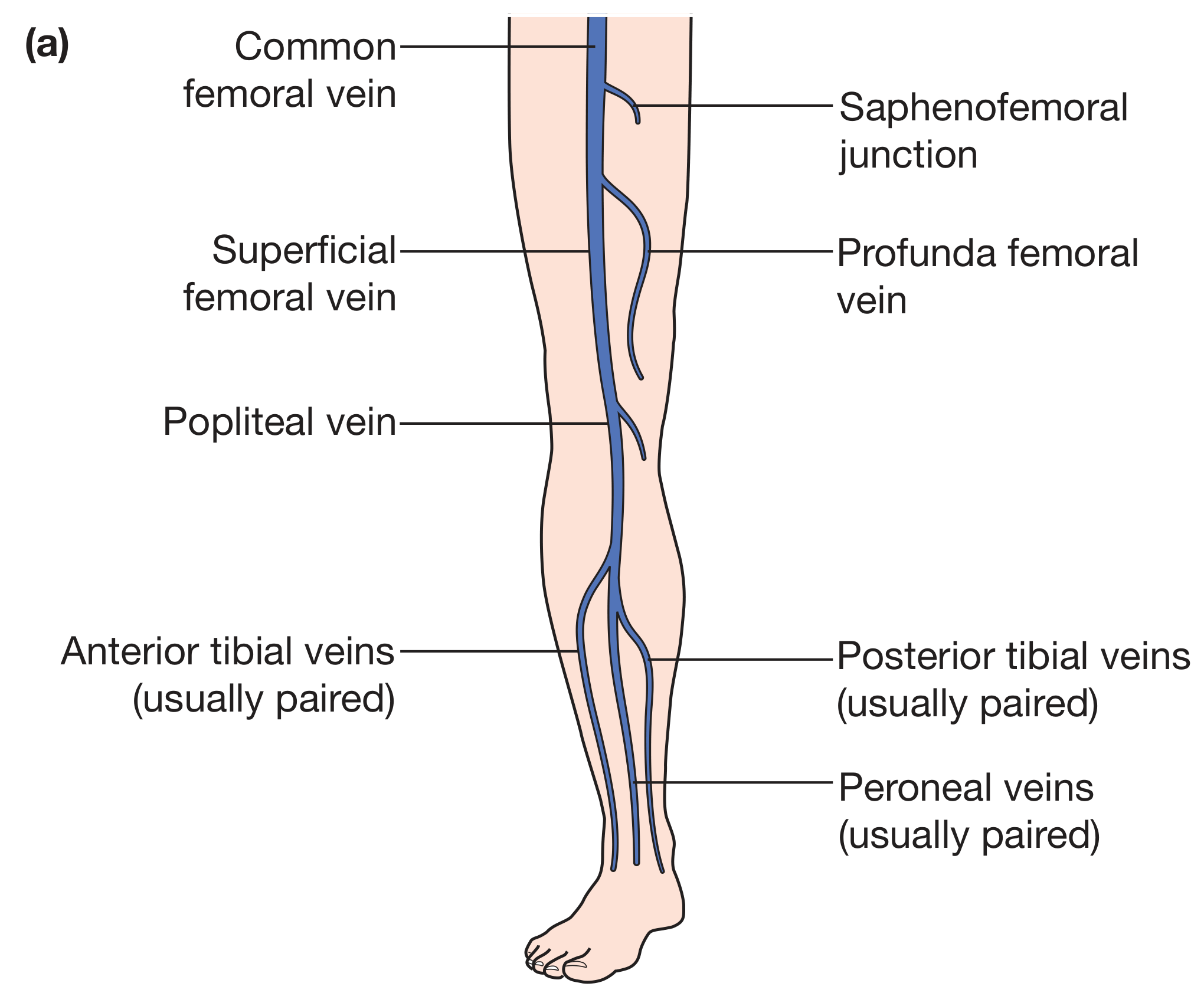
Figure: Anatomy of the deep veins of the lower limb - Bailey & Love, p.1047
The deep veins lie beneath the deep fascia and accompany the arterial tree:
Leg (Crural Veins)
Three pairs of venae comitantes (paired veins running alongside each artery):
- Anterior tibial veins (paired) - accompany anterior tibial artery
- Posterior tibial veins (paired) - accompany posterior tibial artery
- Peroneal (fibular) veins (paired) - accompany peroneal artery
These six veins intercommunicate freely and converge in the popliteal fossa.
They also receive the muscular veins of the calf:
- Soleal veins - drain the soleus muscle (form a sinus/plexus - a common site for DVT initiation)
- Gastrocnemius veins - drain both heads of gastrocnemius
Popliteal Fossa
All six crural veins + soleal + gastrocnemius veins join to form the popliteal vein.
Thigh
The popliteal vein passes through the adductor hiatus → enters the subsartorial canal as the femoral vein (previously called "superficial femoral vein" - a misnomer, it is a deep vein).
In the femoral triangle, it receives the profunda femoris (deep femoral) vein before passing behind the inguinal ligament to become the external iliac vein.
Pelvis and Abdomen
- External iliac vein + Internal iliac vein → Common iliac vein
- Left common iliac vein passes behind the right common iliac artery (clinically important - site of May-Thurner compression syndrome)
- Right and left common iliac veins join on the right side of the aorta → Inferior vena cava → Right atrium
3. Superficial Venous System
The superficial veins lie in the subcutaneous fat, superficial to the deep fascia (fascia lata) but deep to the saphenous fascia - within what is called the saphenous envelope. There are two main axes.
3A. Great Saphenous Vein (GSV)
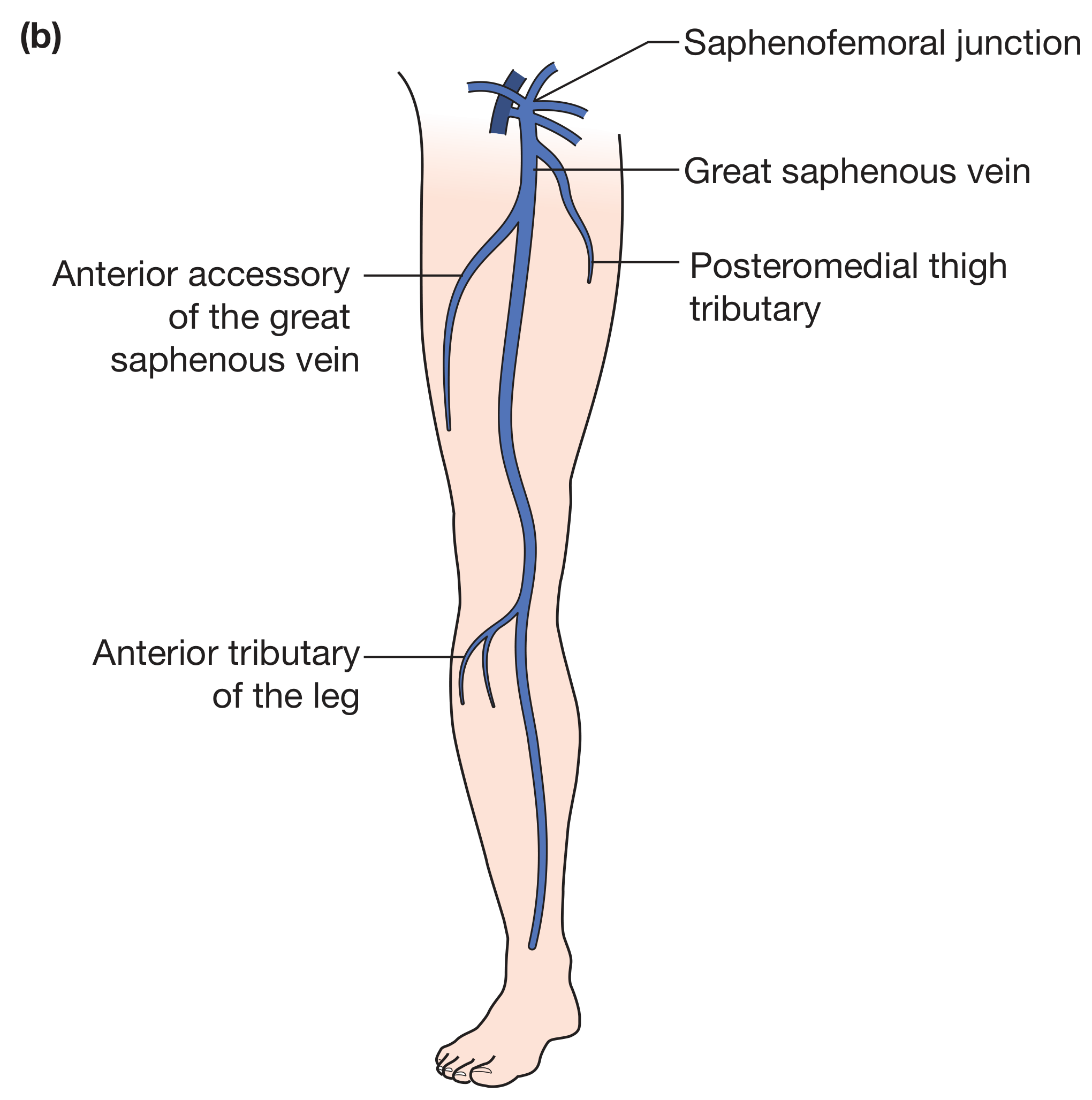
Figure: Great saphenous vein axis with AAGSV and tributaries - Bailey & Love, p.1047
| Feature | Detail |
|---|---|
| Origin | Medial side of the dorsal venous arch of the foot |
| Ankle | Passes anterior to the medial malleolus |
| Leg | Ascends medially with the saphenous nerve; medial to tibia |
| Knee | Loops posteriorly around the medial condyle of femur |
| Thigh | Continues in the medial thigh |
| Termination | Pierces the cribriform fascia at the saphenous opening (~2.5 cm below and lateral to the pubic tubercle) → drains into the common femoral vein at the saphenofemoral junction (SFJ) |
| Clinical significance | Longest vein in the body; most frequently affected by incompetence |
Tributaries at the SFJ (the "bouquet"):
- Superficial external pudendal vein
- Superficial circumflex iliac vein
- Superficial epigastric vein
- Anterior femoral cutaneous vein
- Posteromedial thigh tributary
- Anterolateral thigh tributary
Anterior Accessory of the Great Saphenous Vein (AAGSV):
- One of the most common tributaries
- Originates around the lateral border of the knee (sometimes from the lateral end of the dorsal venous arch)
- Courses anterolateral to the GSV
- Drains into the GSV at or near the SFJ
- Commonly mistaken for the GSV on duplex scan ("duplex GSV" - a true duplicated GSV is rare)
3B. Small Saphenous Vein (SSV)
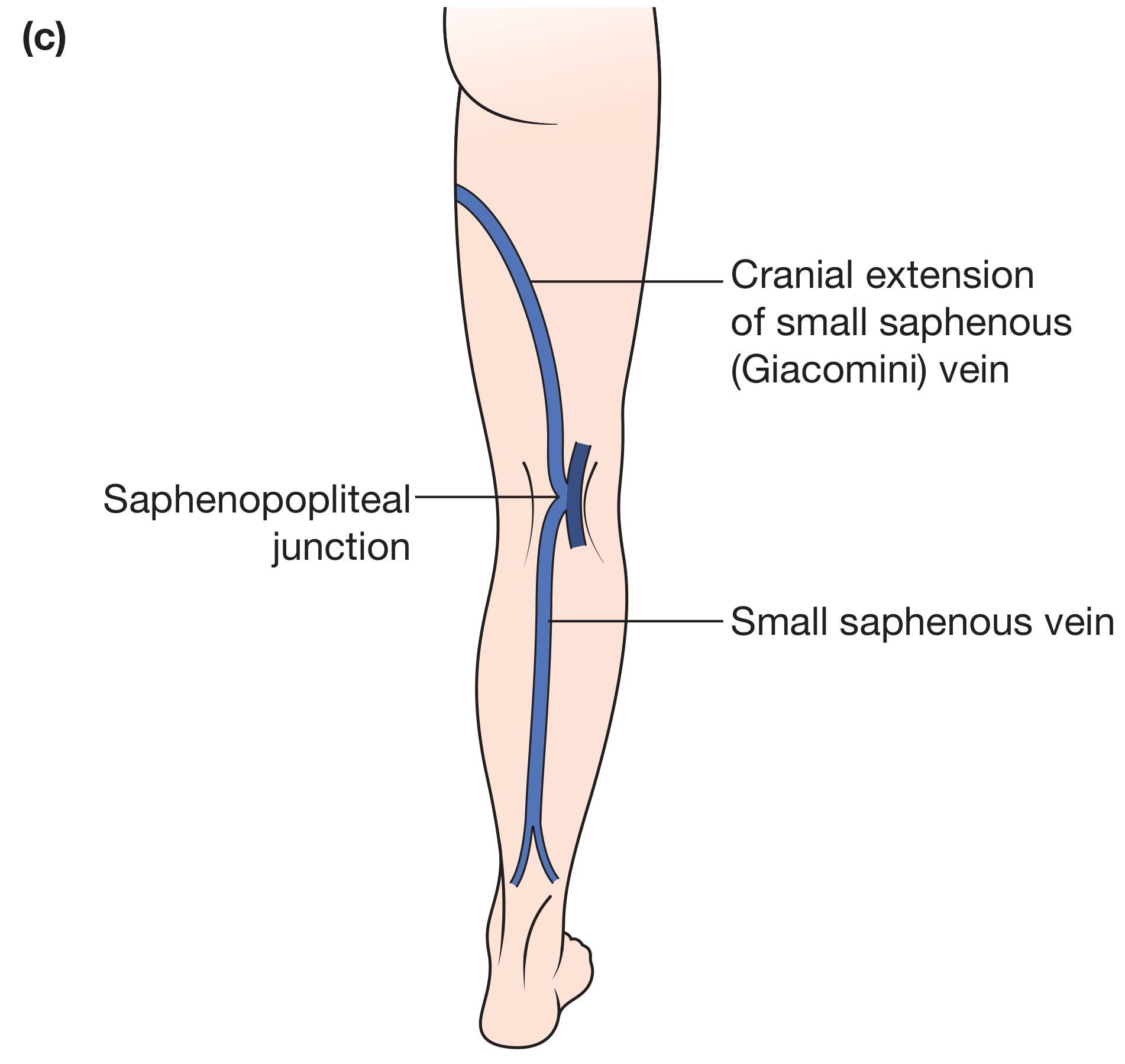
Figure: Small saphenous vein, saphenopopliteal junction, and Giacomini vein - Bailey & Love, p.1047
| Feature | Detail |
|---|---|
| Origin | Lateral side of the dorsal venous arch |
| Ankle | Posterior to the lateral malleolus (with the sural nerve) |
| Leg | Ascends in the posterior midline of the calf; sits in the groove between the two heads of gastrocnemius |
| Termination | Pierces the fascia of the popliteal fossa → drains into the popliteal vein at the saphenopopliteal junction (SPJ) - highly variable, may be as low as mid-calf |
| Extension | May continue cranially as the Giacomini vein - communicates with the GSV system, sometimes joining the GSV at/near the SFJ |
3C. Veins of the Foot
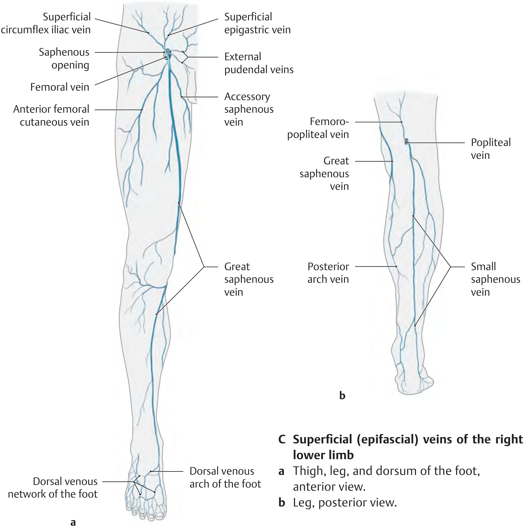
Figure: Superficial (epifascial) veins of the right lower limb - THIEME Atlas
| Structure | Location | Drains into |
|---|---|---|
| Dorsal venous arch | Over metatarsal heads | Medial end → GSV; lateral end → SSV |
| Medial marginal vein | Medial border of foot | GSV |
| Lateral marginal vein | Lateral border of foot | SSV |
| Dorsal venous network | Dorsum of foot | Dorsal arch, then GSV/SSV |
| Plantar venous arch | Plantar surface | Posterior tibial veins (deep) via plantar digital/metatarsal veins |
| Plantar venous network | Plantar surface | Plantar arch → posterior tibial veins |
| Posterior venous arch of foot | Dorsum, connects arches | Empties into SSV/GSV |
The dorsal and plantar arches are connected by intercapitular veins.
Because the sole bears significant weight and pressure, most venous drainage of the foot is via the dorsal venous arch rather than the plantar surface.
4. Perforating (Communicating) Veins
Perforating veins are valved vessels that pierce the deep fascia, connecting superficial to deep veins. Valves normally allow flow only from superficial → deep. When valves become incompetent, reversal of flow causes superficial hypertension and varicosities.
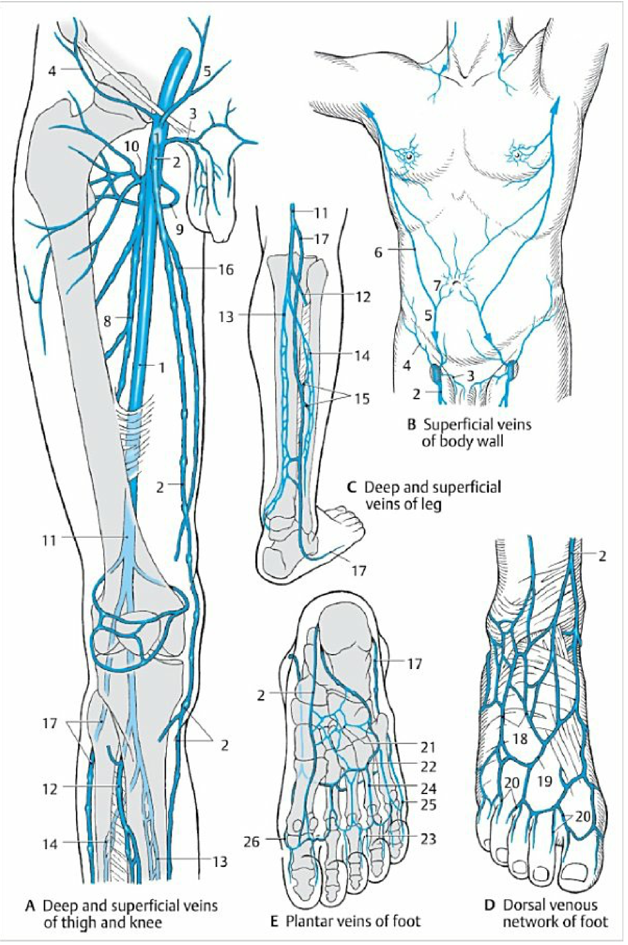
Figure: Deep and superficial veins with perforators, plantar and dorsal venous networks - Color Atlas of Human Anatomy
The three clinically important groups of perforators (THIEME Atlas, p.551):
| Group | Location | Clinical relevance |
|---|---|---|
| Dodd group | Medial thigh, middle third | Incompetence → medial thigh varicosities |
| Boyd group | Medial leg, just below knee | Common site of incompetence |
| Cockett group | Medial distal leg (ankle region) | Most important clinically; incompetence causes gaiter area pigmentation, lipodermatosclerosis, venous ulcers |
The Cockett perforators are located at approximately 7.5, 12.5, and 18.5 cm above the medial malleolus.
5. Venous Physiology - Calf Muscle Pump
At rest (standing), superficial venous pressure in the foot is ~80-90 mmHg (hydrostatic). During walking:
- Calf muscle contraction compresses the deep veins
- Valves direct flow upward (antegrade)
- Superficial pressure falls to ~25 mmHg
- On stopping exercise, pressure rises slowly back to resting level (refilling time)
In venous incompetence (valve failure), pressure does not fall adequately during exercise, producing venous hypertension - the root cause of all complications (oedema, skin changes, ulceration).
6. Summary: Flow Pathway
Foot veins (dorsal venous arch / plantar network)
↓
Great saphenous vein Small saphenous vein
(medial) (posterior)
↓ ↓
Saphenofemoral junction Saphenopopliteal junction
(common femoral vein) (popliteal vein)
↓
Femoral vein
↓
+ Profunda femoris vein
↓
External iliac vein
↓
Common iliac vein
↓
Inferior vena cava
↓
Right atrium
All along this pathway: perforating veins connect superficial → deep systems at the leg and thigh.
Sources:
- Bailey and Love's Short Practice of Surgery, 28th Edition, pp. 1046-1049
- Gray's Anatomy for Students, p. 671
- THIEME Atlas of Anatomy: General Anatomy and Musculoskeletal System, p. 551
- Color Atlas of Human Anatomy, Vol. 2 (Internal Organs), pp. 144-146
Operation and treatment of vericose vein in detail
Finding Sources
Finding Sources
Finding Sources
Finding Sources
Reading File
Reading File
Reading File
Reading File
Reading File
Reading File
Reading File
Reading File
Reading File
Reading File
Reading File
Reading File
Reading File
Now let me fetch the key operative diagrams:
Loading Image
Loading Image
Loading Image
Reading File
Reading File
I now have exhaustive content from four textbooks. Let me compile the complete, detailed answer:
Treatment and Operation of Varicose Veins - Detailed
Indications for Treatment
Treatment is considered in patients with:
- Symptomatic varicose veins (aching, heaviness, swelling, itching) not responding to conservative measures
- Complications: superficial thrombophlebitis, haemorrhage, chronic skin changes (pigmentation, eczema, lipodermatosclerosis)
- Active or healed venous ulceration
- Cosmetic concerns (selected cases)
- Evidence of GSV/SSV incompetence on duplex ultrasound
Pre-treatment Assessment
Duplex ultrasound is mandatory before any intervention. It establishes:
- Presence and location of reflux in the deep and superficial systems
- Diameter and tortuosity of incompetent truncal veins
- Patency of the deep venous system (DVT must be excluded)
- Location of the SFJ/SPJ
- Presence of incompetent perforators
- Pelvic source of reflux
Reflux is defined as retrograde flow lasting ≥0.5 seconds in superficial/crural veins, and ≥1 second in proximal deep veins.
Overview of Treatment Options
┌────────────────────────────────────────────────────────────┐
│ TREATMENT OF VARICOSE VEINS │
├──────────────────┬─────────────────────────────────────────┤
│ Conservative │ Compression, elevation, lifestyle │
├──────────────────┼─────────────────────────────────────────┤
│ Interventional │ Thermal ablation (EVLA, RFA) │
│ │ Non-thermal ablation (MOCA, CAC, PEM) │
│ │ Sclerotherapy (liquid/foam/UGFS) │
│ │ Open surgery (ligation + stripping) │
│ │ Phlebectomy (ambulatory / TriVex) │
│ │ Perforator ligation (SEPS) │
└──────────────────┴─────────────────────────────────────────┘
A. Conservative Treatment
1. Compression Therapy
- Elastic compression stockings (class I: 14-17 mmHg; class II: 18-24 mmHg; class III: 25-35 mmHg)
- Mechanism: reduces superficial venous pressure, augments calf muscle pump, reduces oedema
- First-line for symptom relief and in patients unsuitable for intervention
- Also used as a trial to confirm symptoms are venous before committing to surgery
- Worn after all interventional procedures to improve results
2. Lifestyle Modifications
- Leg elevation (above heart level when resting)
- Regular walking (activates calf muscle pump)
- Weight loss (reduces venous pressure)
- Avoid prolonged standing
3. Pharmacotherapy
- Venoactive drugs (phlebotonic agents): horse chestnut seed extract (aescin), ruscus extract, micronized purified flavonoid fraction (diosmin + hesperidin), pentoxifylline
- Some evidence for reduction in leg heaviness and oedema; insufficient evidence for routine recommendation
- No FDA-approved drugs specifically for chronic venous insufficiency
- Diuretics: may reduce oedema but risk volume depletion and renal compromise
- Topical steroids: short-term use for stasis dermatitis only
B. Endovenous Thermal Ablation
The current gold-standard first-line interventional treatment. Two energy modalities are available.
General Procedure (Common to both EVLA and RFA)
Step-by-step technique:
- Pre-procedure: Patient stands; duplex scan marks the truncal vein and varicosities
- Positioning:
- GSV treatment: supine, hip externally rotated, slightly flexed
- SSV treatment: prone position
- Percutaneous access: Ultrasound-guided cannulation at the lowest point of reflux
- Catheter/fibre advancement: Tip positioned under ultrasound guidance just below the SFJ (position is controversial - some place several cm distal to junction, others aim for flush occlusion)
- Perivenous tumescent anaesthesia (PVTA): Critical step - dilute local anaesthetic (e.g. lignocaine + adrenaline in saline) injected around the vein under ultrasound guidance, creating a "halo" around the vein. Purposes:
- Anaesthesia
- Compresses vein around catheter (reduces diameter, improves contact)
- Heat sink (protects surrounding tissues)
- Energy delivery during catheter withdrawal
- Compression applied immediately after
1. Endovenous Laser Ablation (EVLA)
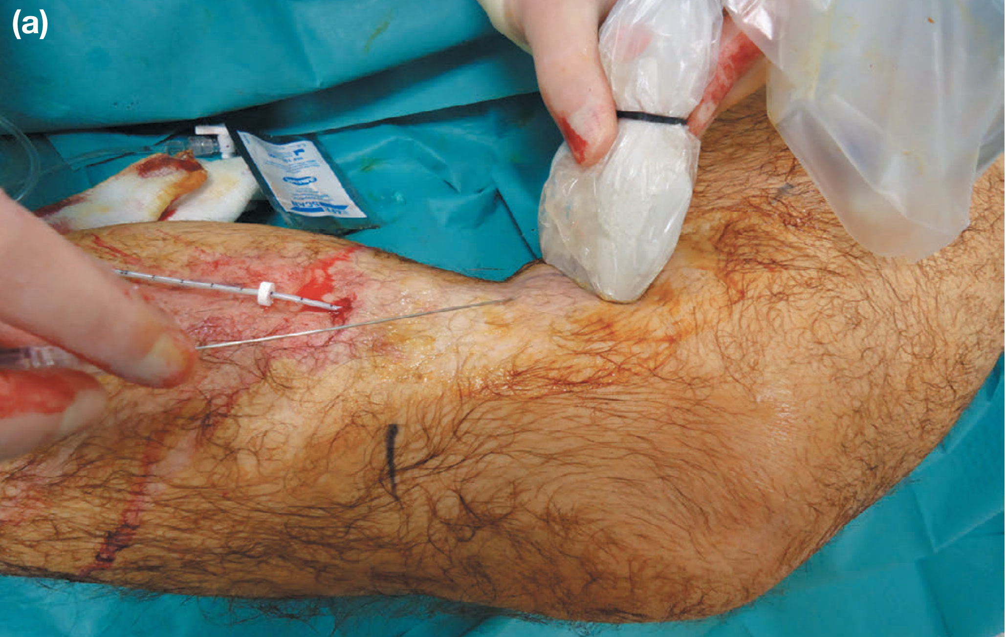
Figure: Ultrasound-guided perivenous tumescent anaesthesia infiltration - Bailey & Love, p.1055
Mechanism: Laser energy (wavelengths 810-1500 nm) delivered via optical fibre converts to heat, causing direct thermal injury to the vein wall endothelium and media → thrombosis → fibrosis → permanent occlusion.
Fibre tip designs:
- Bare-tip (forward firing): concentrates energy in a small area
- Divergent forward-firing: spreads energy over larger area
- Divergent side/radial firing: even distribution, postulated to reduce vein wall perforations, pain and bruising
Energy delivery: approximately 60-80 J/cm of treated vein. Power and pullback speed are operator-adjusted.
Efficacy: Average 1-year occlusion rate >90%; 5-year occlusion >90%. The highest technical efficacy of any technique.
Advantages over RFA:
- Better for very large diameter veins (>15 mm) - can increase energy delivery
- Lower consumable costs
- Catheter can be used to treat perforators without additional devices
Disadvantages:
- Requires specific laser safety protocols for room design and staff training
- More operator-dependent (power settings and pullback speed must be manually controlled)
- Marginally more peri-procedural pain and bruising
2. Radiofrequency Ablation (RFA)
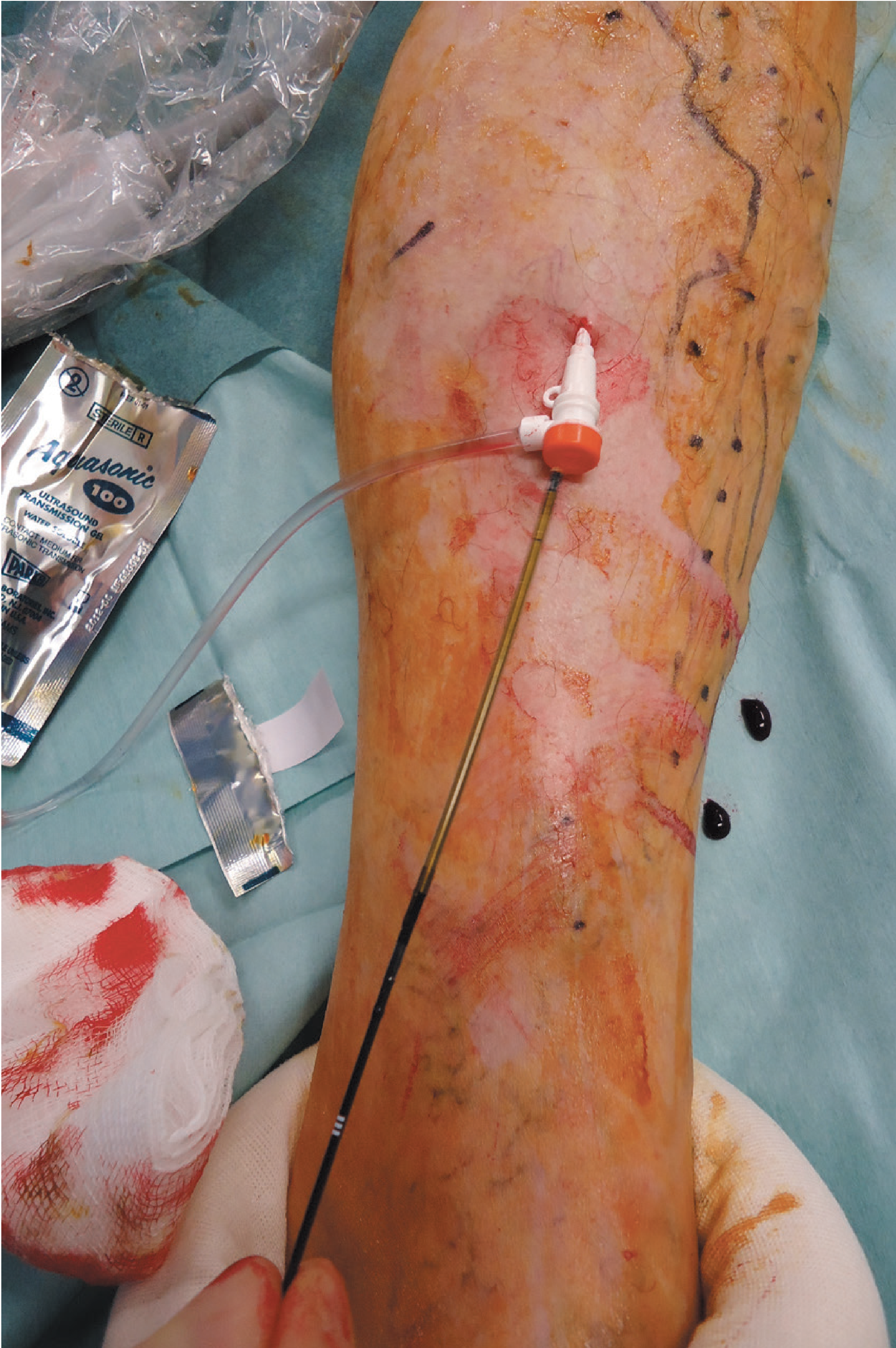
Figure: RFA with ClosureFast™ device - Bailey & Love, p.1056
Mechanism: Electromagnetic current passes through a metal coil at the catheter tip → generates heat to 120°C → thermal injury → fibrosis → occlusion.
Most popular device: ClosureFast™ (Medtronic)
- Metal coil at catheter tip (3 cm and 7 cm coil options)
- Generator maintains 120°C for a 20-second treatment cycle
- Catheter withdrawn a set distance → next cycle commenced
- Single button press per cycle - automated, frees operator's attention
Efficacy: Average 1-year occlusion rate >90%, slightly less than EVLA at 5 years.
Advantages over EVLA:
- Shorter learning curve - automated treatment cycle, less chance of energy delivery error
- Marginally less post-procedural pain and bruising
- Frees operator to communicate with patient and perform concurrent phlebectomy
- No laser safety protocols required
Disadvantages:
- Less suitable for very large diameter veins
- Higher consumable cost than EVLA (device-specific catheter)
- Specific additional device needed for perforator treatment
EVLA vs RFA Comparison
| Feature | EVLA | RFA |
|---|---|---|
| Mechanism | Laser energy | Electromagnetic heat |
| Target temp | Variable | 120°C (controlled) |
| 1-year occlusion | >90% | >90% |
| Learning curve | Longer | Shorter (automated) |
| Post-op pain | Marginally more | Marginally less |
| Large veins (>15mm) | Better | Less suitable |
| Consumable cost | Lower | Higher |
| Laser safety | Required | Not required |
| Perforator treatment | Same fibre | Additional device needed |
Bottom line: Both techniques have equivalent clinical outcomes. Choice is largely personal preference.
C. Non-thermal, Non-tumescent Ablation (NTNT)
These newer techniques avoid tumescent anaesthesia entirely - important for patients with needle phobia or when treating near nerves.
1. Mechanochemical Ablation (MOCA) - ClariVein®
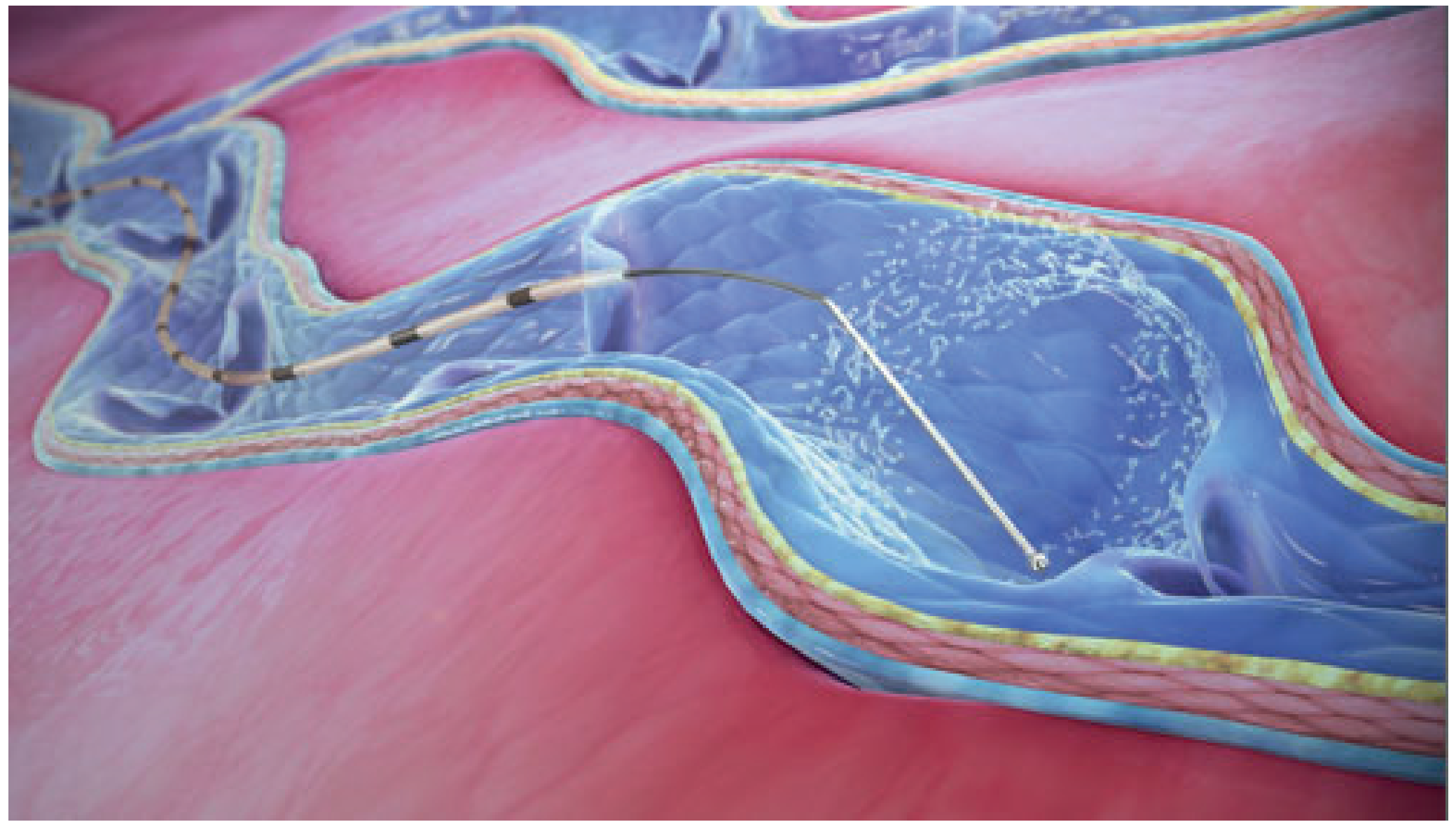
Figure: Mechanochemical ablation device - Bailey & Love, p.1058
Mechanism: A catheter deploys an angled rotating wire from its tip. The spinning wire:
- Mechanically abrades the venous endothelium
- Simultaneously infuses liquid sclerosant via catheter → deeper penetration into wall
- Combined mechanical + chemical injury causes fibrosis and occlusion
Procedure: Catheter placed in vein lumen; trigger activates spinning wire; catheter withdrawn while sclerosant is infused.
Advantages:
- No tumescent anaesthesia needed
- Less painful than thermal ablation (for axial treatment alone)
- Suitable for patients with needle phobia
Disadvantages:
- Some patients find it uncomfortable
- Device can "snag" on tortuous veins, occasionally tearing or stripping the vein
- Similar early efficacy to thermal ablation but increased medium/long-term recanalisation rates
- Catheter length limits treatment of long veins
- When combined with phlebectomy, the pain advantage over thermal ablation is lost
2. Cyanoacrylate Adhesive (CAC) - VenaSeal™
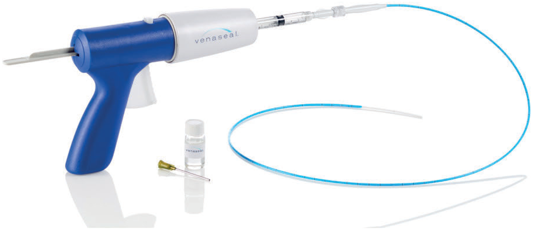
Figure: Endovenous glue device (VenaSeal) - Bailey & Love, p.1058
Mechanism: Cyanoacrylate (tissue glue) is delivered in 0.1 mL aliquots via catheter. The vein is compressed after each application, sealing the lumen. Vein is initially closed by adhesive, then undergoes progressive fibrosis.
Procedure:
- Ultrasound-guided catheter access (micropuncture system)
- Catheter advanced to SFJ; pulled back 3 cm distal to junction
- Glue applied at 2-3 cm intervals during catheter withdrawal
- Continuous manual compression with ultrasound probe at SFJ during injection (to prevent glue embolisation to deep system)
- Catheter captured within sheath before removal (prevents glue extravasation into subcutaneous tissue)
1-year occlusion rate: ~90%
Advantages:
- No tumescent anaesthesia
- Minimal intra-procedural pain
- Can access distally (near ankle) without nerve injury concern
Disadvantages:
- Highest consumable cost of any venous ablative technique
- Allergic reactions to cyanoacrylate (ranging from mild inflammation to tissue necrosis) - screen for prior adhesive allergy
- Long-term data still limited
3. Polidocanol Endovenous Microfoam (PEM) - Varithena®
Mechanism: FDA-approved (2013) proprietary microfoam sclerosant delivered endovenously via catheter under ultrasound guidance.
Procedure:
- Ultrasound-guided access (micropuncture system) placed as distally as possible
- PEM injected 1-2 mL/second through sheath
- Limb elevated >45° during injection to reduce DVT risk
- Digital occlusion of perforator veins during injection
- Compression at SFJ during treatment
- Post-procedure: 20-30 mmHg compression stocking; ambulate within the hour
- Duplex scan at 2-5 days post-procedure
1-year occlusion rate: ~90%
DVT risk: 1.5-3%; mitigation strategies include limb elevation, reducing total volume used, digital occlusion of perforators, and proximal compression.
D. Sclerotherapy
1. Liquid Sclerotherapy
Mechanism: Sclerosant injected directly into vein → chemical injury to endothelium → fibrosis and obliteration.
Approved sclerosants (US/UK):
- Sodium tetradecyl sulphate (STS / Fibrovein)
- Polidocanol (Aethoxysclerol)
- Sodium morrhuate
- Glycerin (for telangiectasia)
Best used for:
- Telangiectasia and reticular veins
- Small varicose tributaries
- Residual veins after truncal ablation
Technique: Injection with fine needle, patient supine/seated; compression applied after.
2. Ultrasound-Guided Foam Sclerotherapy (UGFS)
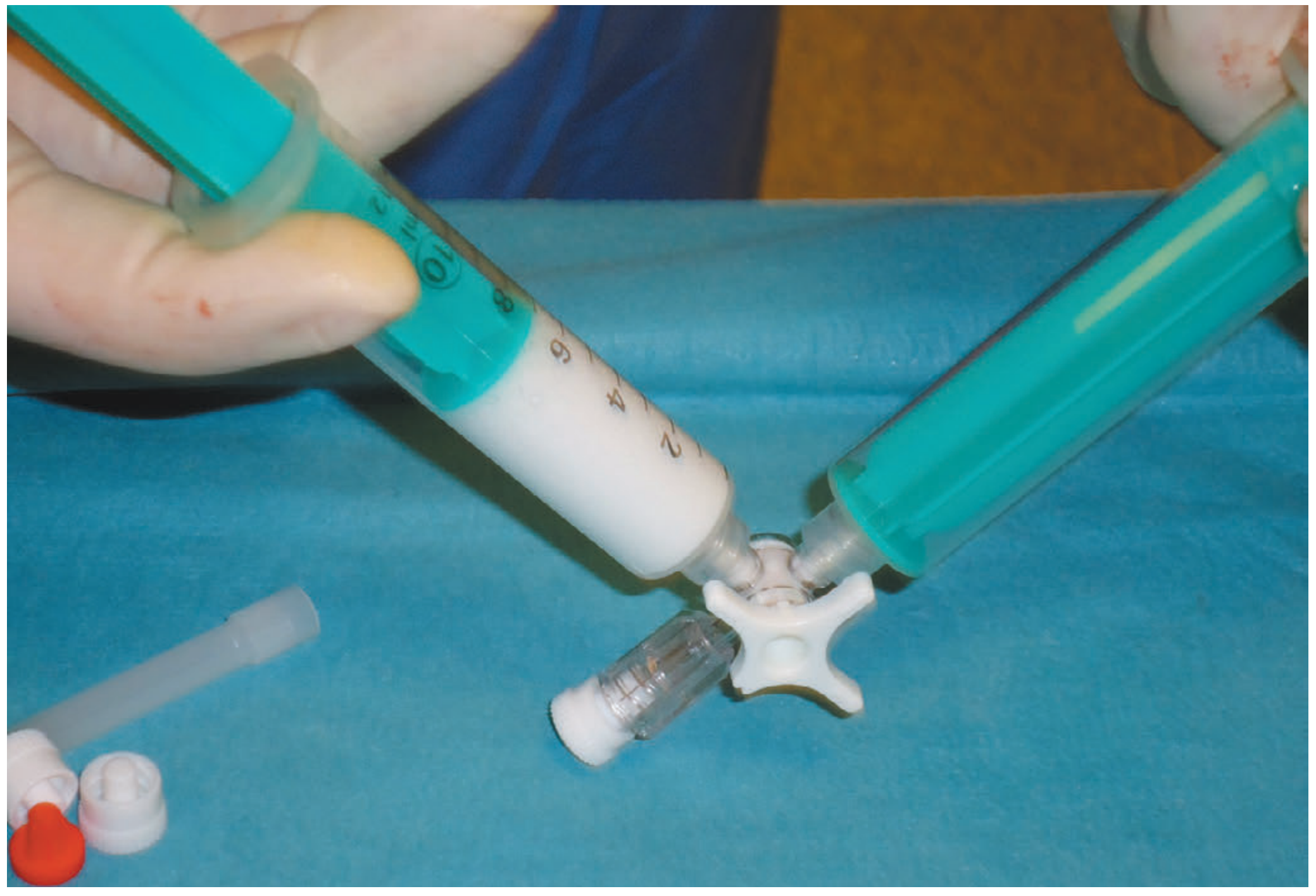
Figure: Tessari method of foam preparation with two syringes and a three-way tap - Bailey & Love, p.1057
Mechanism: Sclerosant mixed with air/CO₂ to create foam (ratio 1:3 or 1:4 sclerosant:gas). Foam is more effective than liquid because:
- Displaces blood from vein (maximises endothelial contact)
- Larger volume effect with smaller drug dose
- Echogenic under ultrasound - visible during injection
Tessari method (most common foam preparation):
- Two syringes connected by a three-way tap
- 1:3 or 1:4 sclerosant:air drawn into one syringe
- Vigorously oscillated between syringes 10-20 times
- Foam stable for ~2 minutes - inject immediately
Procedure:
- Leg elevated to empty veins
- Cannulate vein under ultrasound guidance
- Inject foam into superficial varicosities first, then the GSV/SSV
- Maximum 1-2 mL per injection; distribution monitored with ultrasound probe
- Stop when foam visualised at junctional incompetence
- Maximum total dose: 10-12 mL per session (complication risk rises with larger volumes)
- Apply compression after
Efficacy: 1-year occlusion ~81%; 5-year ~74% - lower than thermal ablation.
Advantages of UGFS over thermal ablation:
- No tumescent anaesthesia (less painful procedure)
- All veins suitable regardless of tortuosity
- Can treat calf veins beneath damaged/ulcerated skin without needle puncture through the damage
- Very low consumable cost
- Useful adjunct for treating neovascularisation, residual tributaries
Disadvantages:
- Significantly lower long-term efficacy → higher re-intervention rates
- Higher rates of phlebitis and post-procedural pigmentation
- Complications: DVT, haematoma, nerve injury (saphenous/sural), hyperpigmentation, skin necrosis (if inadvertent subcutaneous injection), anaphylaxis (rare but severe)
E. Open Surgery
Now largely superseded by endovenous techniques, but still used for complex, recurrent cases or where endovenous techniques are unavailable.
1. Saphenofemoral Junction (SFJ) Ligation and GSV Stripping (Trendelenburg Operation)
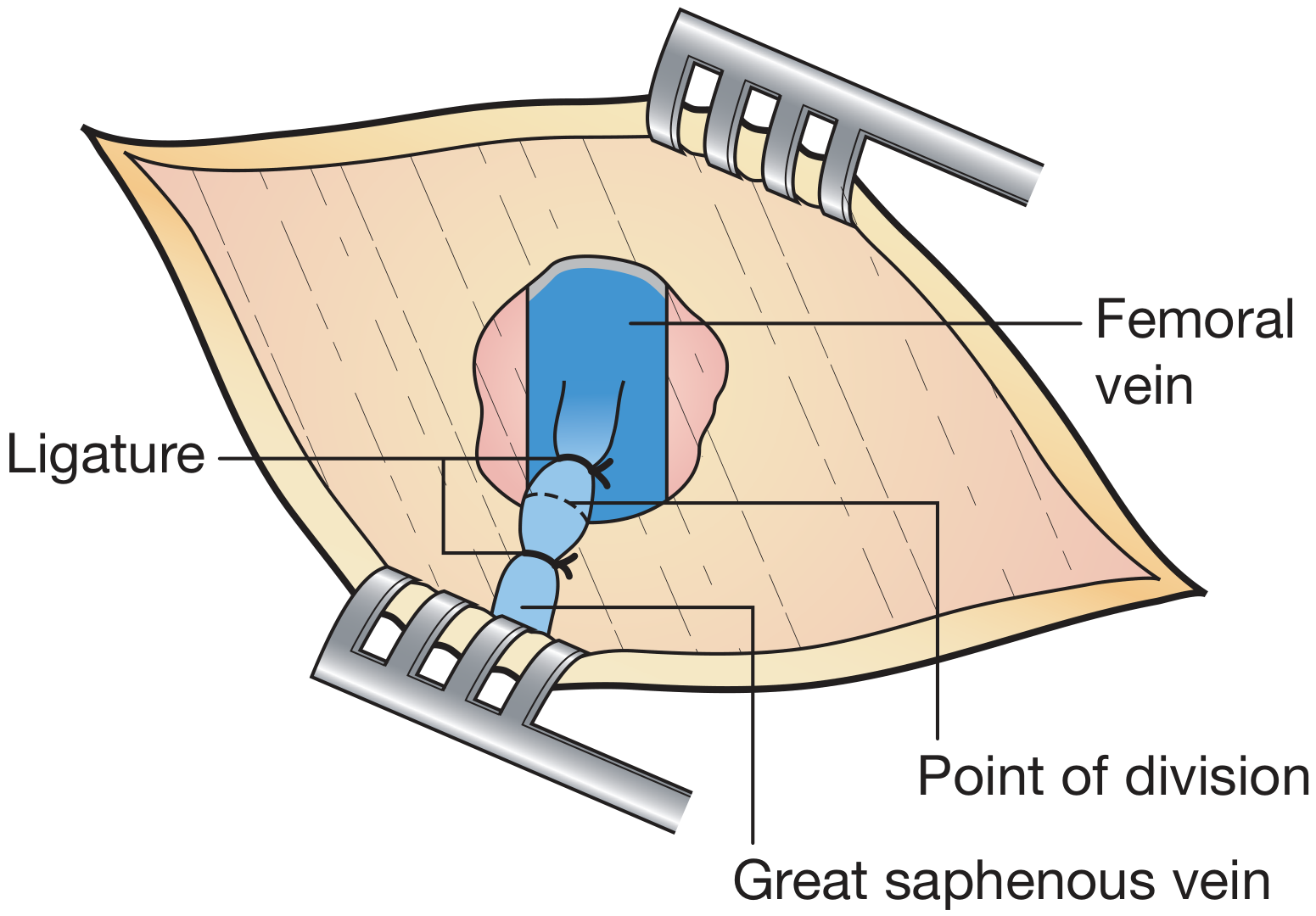
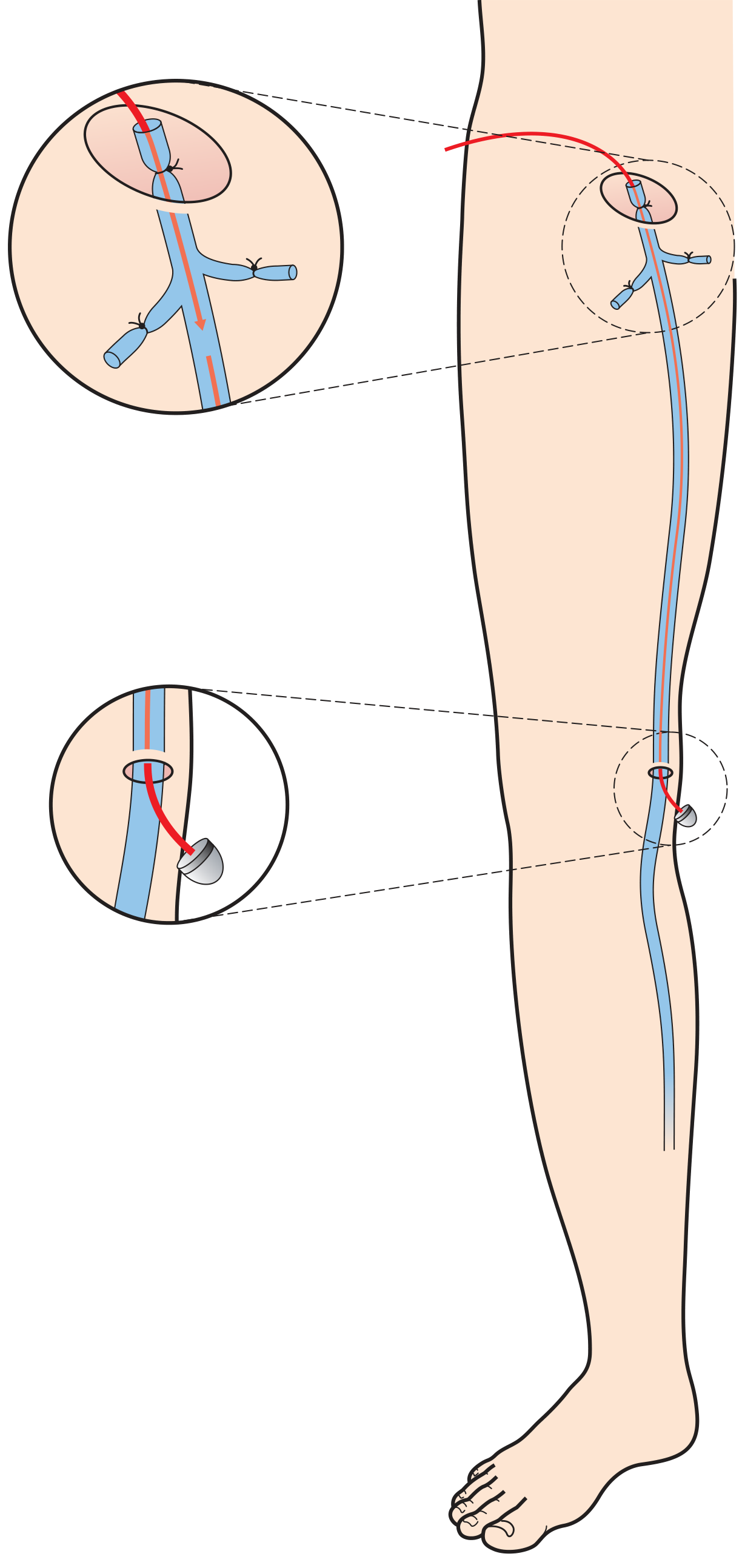
Step-by-step technique:
- Anaesthesia: General or spinal (rarely local alone)
- Patient position: Supine; varicosities marked preoperatively while standing
- Groin incision: Oblique incision lateral to pubic tubercle, above the groin crease
- Dissection: GSV identified and traced to SFJ; anatomy confirmed before any division
- Tributaries ligated at SFJ (the 6 tributaries encountered):
- Laterally: superficial inferior epigastric vein, superficial circumflex iliac vein
- Medially: superficial external pudendal vein, deep external pudendal vein
- Distally: anterior accessory GSV, posteromedial thigh vein
- Flush SFJ ligation: GSV ligated flush with the common femoral vein
- Retrograde stripping: A stripper passed from the groin down to approximately the knee (stripping to ankle increases saphenous nerve injury risk)
- Phlebectomy of remaining varicosities via stab incisions
- Closure: Cribriform fascia closure does not reduce groin recurrence
Note on neovascularisation: Surgical trauma in the groin triggers formation of new, valve-less veins that may bridge the ligated junction to tributaries - a major cause of recurrence. This theoretical concern has led some surgeons to avoid flush junction ligation, though there is no clear clinical evidence to support this.
2. Saphenopopliteal Junction (SPJ) Ligation and SSV Stripping
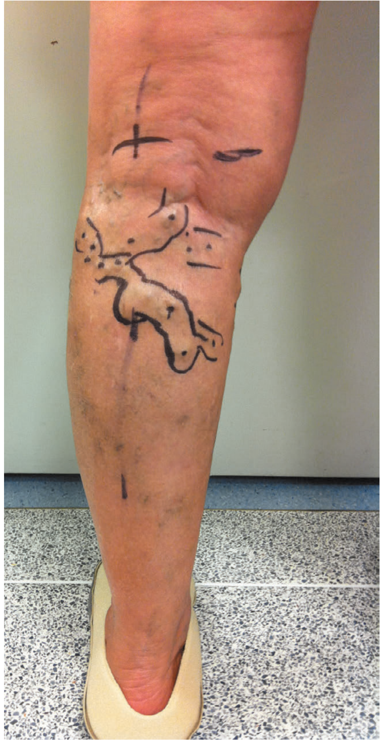
Key points:
- Duplex mapping of the SPJ is mandatory before surgery (highly variable anatomy)
- Position: Prone
- Incision: Transverse over pre-marked SPJ
- Dissection: Fascia divided, SSV exposed; SPJ dissected and ligated (flush or proximal to SSV)
- Flush ligation: avoids leaving a stump (common recurrence site), but risks popliteal vein and nerve injury
- Simple SSV ligation: safer for nerves and popliteal vein, but higher recurrence from residual stump
- SSV stripping: reduces recurrence but increases sural nerve injury risk
- Phlebectomy performed after SPJ ligation
F. Phlebectomy (Ambulatory / Stab Avulsion)
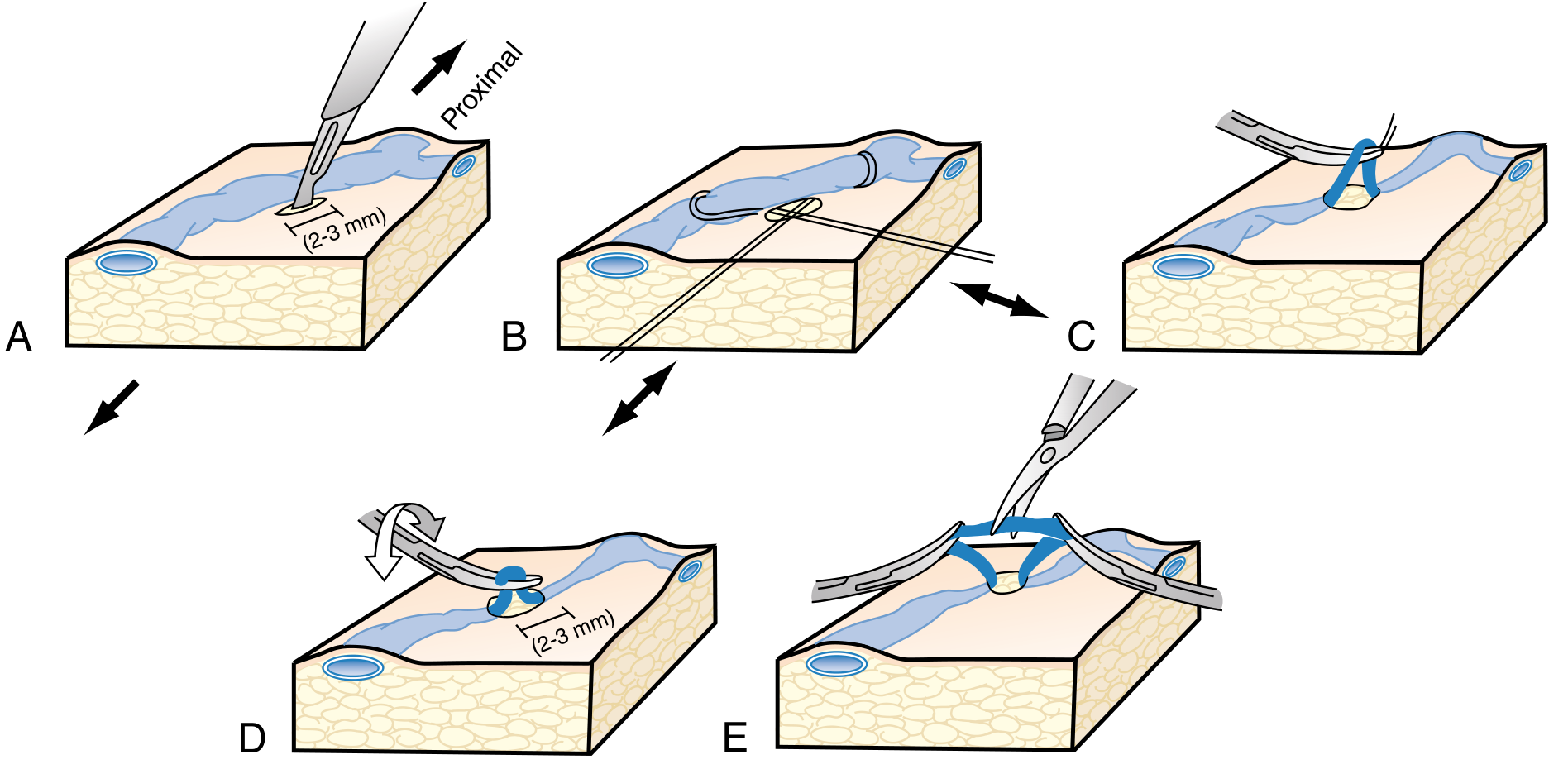
Figure: Stab avulsion phlebectomy technique - A: 2-3mm incision; B: hook under vein; C: vein exteriorised; D: rotation; E: avulsion with forceps - Sabiston Textbook of Surgery
Indications:
- Adjunct to truncal ablation for removal of varicose tributaries
- Sole treatment for isolated tributary incompetence
Technique (step-by-step):
- Varicosities marked preoperatively while standing
- Local tumescent anaesthesia injected
- 2-3 mm stab incisions along Langer's lines at 2 cm intervals over varicosities
- Vein retrieved through incision with mosquito forceps or phlebectomy hook
- Continuous traction to maximise vein removal
- Direct pressure after avulsion
- Compression dressing applied; patient walks same day
- Compression stockings worn for 2 weeks
Post-operative course: Minimal; paracetamol/NSAIDs usually sufficient.
Complications: Bleeding, infection, temporary/permanent paresthesia, phlebitis from retained segments, recurrence.
TriVex (Transilluminated Powered Phlebectomy)
- Useful for extensive branch varicosities
- 2 mm incisions at varicosity boundaries
- Transilluminator: illuminates veins from below AND delivers tumescent anaesthesia
- Resector: rotating blade transects and aspirates veins
- Requires fewer incisions than standard phlebectomy but associated with more bruising and haematoma
G. Perforator Ligation
Subfascial Endoscopic Perforator Surgery (SEPS):
- Endoscope introduced subfascially via two small ports remote from damaged skin
- Incompetent perforators identified and clipped/divided
- Mainly used in patients with venous ulcers or significant skin changes
- Evidence for benefit is limited - most randomised data are lacking
- Perforators can also be ablated with endovenous laser (EVLA through a stab incision)
H. EHIT (Endovenous Heat-Induced Thrombus)
A specific complication of thermal ablation coined by Dr Lowell Kabnick:
| Class | Description | Management |
|---|---|---|
| 1 | Thrombus within GSV, not at junction | Observe; no treatment |
| 2 | Non-occlusive thrombus at SFJ/CFV | Observe; no treatment usually |
| 3 | Partial non-occlusive extension into deep vein | Anticoagulation (physician discretion) |
| 4 | Occlusive DVT | 3-month anticoagulation course |
EHIT becomes echogenic within <24 hours (unlike acute DVT which remains hypoechoic for days). DVT rate following thermal ablation: 0-4% (RFA) and 0-3% (EVLA).
Complications of Surgery
| Complication | Incidence/Notes |
|---|---|
| Wound infection (most common) | Reduced by prophylactic antibiotics |
| Saphenous nerve neuralgia | Up to 7% after GSV stripping to knee; higher to ankle |
| Sural nerve neuropraxia | Up to 20% after SSV surgery |
| Common peroneal nerve injury | Up to 4% after SSV surgery |
| VTE (DVT/PE) | ~0.5% - patient risk assessment and prophylaxis mandatory |
| Lymph leak / lymphocoele | More common with re-do surgery (40% complication rate overall in recurrent cases) |
| Haematoma | More common with tumescent techniques |
| Recurrence | 10-35% clinically at 5-10 years; duplex-detected ~70% |
Recurrent Varicose Veins
Approximately 10-20% of patients presenting with varicose veins have had previous intervention. Causes of recurrence:
- Neovascularisation - new valve-less veins bridging ligated junction to tributaries
- Reflux in residual axial vein (inadequate initial treatment)
- Inadequate initial surgery
- New junctional reflux
Higher risk of recurrence in: SSV surgery (vs GSV), high BMI, axial vein not stripped.
Limited data suggest endovenous thermal ablation has lower recurrence rates than open surgery.
Re-do open surgery has a 40% complication rate (mostly lymph leak and wound infection) - endovenous techniques preferred for recurrence where anatomically feasible.
Treatment Algorithm (Current Best Practice)
Symptomatic varicose veins
↓
Duplex ultrasound assessment
↓
Truncal incompetence?
/ \
YES NO (isolated tributaries)
↓ ↓
First line: Phlebectomy or
Endovenous sclerotherapy
thermal ablation
(EVLA or RFA)
+
Concurrent/staged phlebectomy
or foam sclerotherapy for tributaries
↓
Unsuitable for thermal ablation
(very tortuous, needle phobic)?
↓
MOCA or CAC (NTNT) or
UGFS or open surgery
Sources:
- Bailey and Love's Short Practice of Surgery, 28th Edition, pp. 1046-1060
- Sabiston Textbook of Surgery, 21st Edition, pp. 2410-2412
- Harrison's Principles of Internal Medicine, 22nd Edition, pp. 2225-2226
- S. Das: A Manual on Clinical Surgery, 13th Edition, pp. 105-109
This is a shared conversation. Sign in to Orris to start your own chat.