
Present me a case on direct inguinal hernia
"Hernia, Inguinal"[MeSH Terms]
direct inguinal hernia Hesselbach triangle
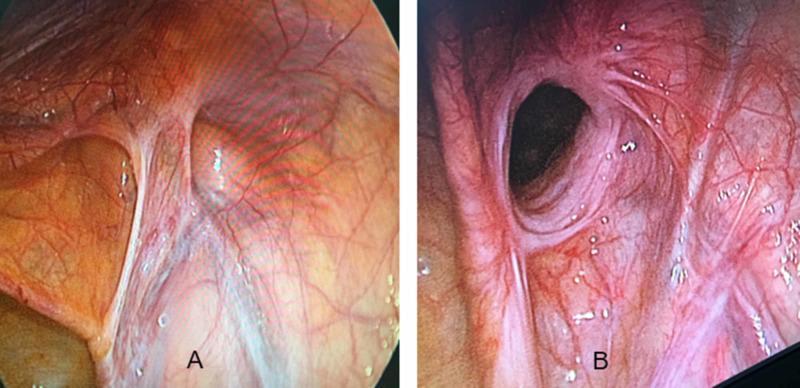
This diagnostic comparison uses laparoscopic (endoscopic) imaging to demonstrate the internal anatomical differences between an indirect and a direct inguinal hernia. Image A displays an indirect inguinal hernia, characterized by a rounded protrusion located lateral to the inferior epigastric vessels. The defect is clearly defined with a localized network of fine vasculature concentrated near the base of the herniated sac. Image B depicts a direct inguinal hernia, presenting as a more diffuse, wide-based bulge through the abdominal wall, located medial to the epigastric vessels in Hesselbach's triangle. The borders of the direct hernia are less distinct than the indirect type, and the overlying peritoneum shows a more prominent, dense, and tortuous vascular network. These images illustrate the surgical perspective during a Transabdominal Preperitoneal (TAPP) repair, emphasizing the importance of anatomical landmarks, specifically the inferior epigastric vessels, in differentiating hernia types based on their exit point relative to the deep inguinal ring.
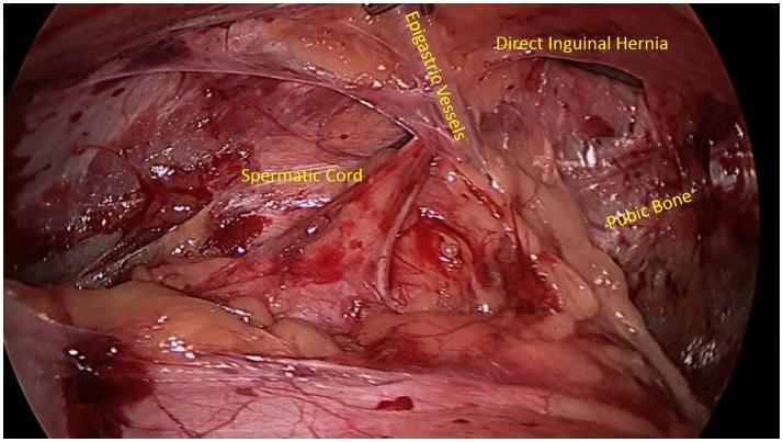
A laparoscopic (endoscopic) surgical view of the posterior inguinal abdominal wall, highlighting key anatomical landmarks for hernia identification. Centrally, the inferior epigastric vessels are visualized, serving as the critical anatomical boundary between direct and indirect inguinal spaces. Medial to these vessels (Hesselbach's triangle), a distinct bulge or protrusion is visible, identified as a direct inguinal hernia. Inferiorly, the whitish, firm structure of the pubic bone is seen, providing a caudal reference point. The spermatic cord is visible laterally, coursing toward the deep inguinal ring. The image demonstrates the laparoscopic perspective of the preperitoneal space, often utilized in TEP (Total Extraperitoneal) or TAPP (Transabdominal Preperitoneal) hernia repair procedures. This clinical photograph provides an educational example of inguinal floor weakness and the topographical relationship between the abdominal wall musculature, vasculature, and pelvic osteology in the context of general surgery and sports medicine.
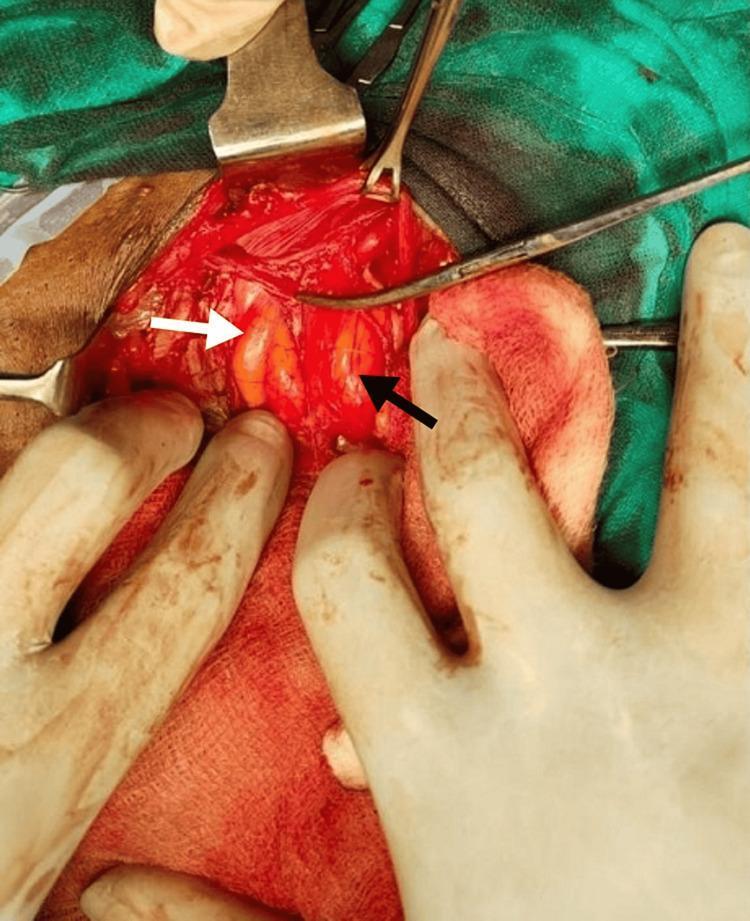
This clinical photograph provides an intra-operative view of a surgical field during an inguinal hernia repair, specifically demonstrating a Pantaloon hernia. The image illustrates the anatomical relationship between the inferior epigastric vessels and two distinct hernial protrusions. The tip of an artery forceps is used to identify and isolate the inferior epigastric vessels. Medial to these vessels, a white arrow indicates a direct inguinal hernia, presenting as a localized bulge through Hesselbach’s triangle. Lateral to the same vessels, a black arrow indicates an indirect inguinal hernia, where the herniated contents protrude through the deep inguinal ring. Surgical retractors and gloved hands are shown maintaining exposure of the inguinal canal. The educational focus of this image is to show the simultaneous presence of both direct and indirect hernia sacs on the same side, straddling the epigastric vessels like a pair of pants. This is a critical finding in general surgery for choosing the appropriate repair technique, such as the Lichtenstein procedure shown here.
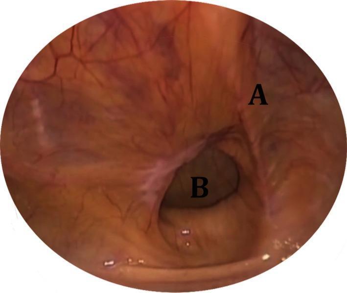
This diagnostic laparoscopic image displays the internal view of the right inguinal region during a surgical procedure. Label 'A' identifies the right medial umbilical ligament, appearing as a prominent, pinkish-tan vertical fold of peritoneum that serves as a key anatomical landmark in the abdominal wall. Label 'B' highlights a right direct inguinal hernia, visualized as a distinct, dark, circular defect or bulging within Hesselbach's triangle, medial to the inferior epigastric vessels. The surrounding parietal peritoneum is smooth and glistening with visible fine vasculature. This image is used in medical education to illustrate the intra-abdominal appearance of direct hernias and to identify relevant laparoscopic landmarks—such as the medial umbilical fold and the inguinal floor—required for TAPP (Transabdominal Preperitoneal) or TEP (Totally Extraperitoneal) hernia repair techniques.
Case: Direct inguinal hernia
Presentation
- Risk factors: a lifetime of heavy lifting, a 40-year smoking history, and chronic cough (COPD).
- Urinary symptoms: a weak stream and nocturia, so he may have prostatic outflow obstruction, which raises intra-abdominal pressure.
- Past history: no previous hernia surgery.
Examination
- He is afebrile and haemodynamically stable. BMI is 27.
- He has a mild chronic cough.
- There is a 5 cm hemispherical bulge above the medial half of the right inguinal ligament.
- It does not extend into the scrotum.
- The skin is normal and the lump is non-tender.
- The lump has an expansile cough impulse.
- It reduces easily and spontaneously on lying down.
- After reduction, the examining finger feels a wide defect in the posterior wall of the canal.
- Deep ring occlusion test: with pressure over the deep ring (mid-inguinal point), the swelling still appears on coughing. This favours a direct hernia.
- The left groin, both testes, and the femoral orifices are normal.
- Abdomen and rectum: the abdomen is soft with no mass. Digital rectal examination shows a mildly enlarged smooth prostate.
Anatomy behind the diagnosis
- Laterally: the inferior epigastric artery.
- Medially: the rectus abdominis.
- Inferiorly: the inguinal ligament.
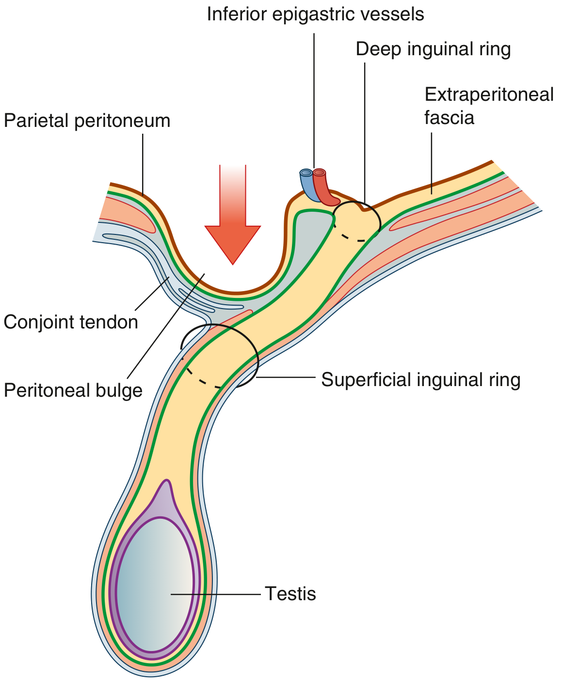
Direct versus indirect hernia
| Feature | Direct | Indirect |
|---|---|---|
| Origin | Acquired | Congenital (patent processus vaginalis) or acquired |
| Age | Older | Young and middle-aged |
| Exit point | Medial to the inferior epigastric vessels (Hesselbach's triangle) | Lateral to the inferior epigastric vessels, through the deep ring |
| Shape | Hemispherical | Pyriform |
| Course | Usually does not enter the scrotum | Often enters the scrotum |
| On lying down | Reduces spontaneously | Needs manual reduction |
| Deep ring occlusion | Bulges on coughing | Controlled |
| Neck | Wide | Narrow |
| Strangulation | Rare | More common |
Differential diagnosis
- Indirect inguinal hernia
- Femoral hernia (below and lateral to the pubic tubercle)
- Pantaloon hernia (direct and indirect together, straddling the epigastric vessels)
- Lipoma of the cord
- Inguinal lymphadenopathy
- Hydrocele of the cord
- Undescended or ectopic testis
- Psoas abscess or saphena varix (less likely here)
Investigations
- Ultrasound (dynamic): use it if the diagnosis is uncertain, in obese patients, or to check for an occult contralateral hernia.
- CT or MRI: use these when there is diagnostic doubt, or for suspected obstruction or strangulation.
- Pre-operative work-up: FBC, urea and electrolytes, ECG, and spirometry or anaesthetic review because of the COPD.
- Bladder and prostate: because of the voiding symptoms, check urinalysis, PSA after counselling, and a post-void residual. A bladder or prostate cause should be treated, because it raises the risk of recurrence.
Diagnosis
Management
- Stop smoking.
- Optimise the COPD.
- Assess and treat the prostatism.
- Avoid heavy straining.
- A symptomatic hernia in a fit patient should be repaired.
- Watchful waiting is reasonable for minimally symptomatic hernias, but he has progressive symptoms and an occupation that strains the canal.
- The main urgent risk is incarceration or strangulation. Direct hernias are less prone to it because of their wide neck.
- Open Lichtenstein tension-free mesh repair (Bailey & Love, "Open flat mesh repair"). It can be done under local, regional, or general anaesthesia. A flat polypropylene mesh reinforces the posterior wall.
- Mesh repair has fewer recurrences than open tissue repair (OR 0.37, CI 0.26-0.51) (Schwartz's Principles of Surgery, 11th Ed, Outcomes).
- Failure to achieve a tension-free repair is a key technical cause of recurrence. Mesh is considered the gold standard (Current Surgical Therapy, 14e).
- Shouldice, a tissue repair, had higher recurrence than Lichtenstein in a Cochrane review. The reviewers raised doubts about how uniformly the Shouldice technique was done across the trials (Current Surgical Therapy, 14e).
- Laparoscopic TEP or TAPP repair. This is a good choice for bilateral or recurrent hernias and for patients who need an early return to work. It needs general anaesthesia, which matters given his COPD.
- Open Lichtenstein repair under local anaesthesia with sedation. This avoids general anaesthesia in a COPD patient.
- Day-case surgery.
- Single-dose antibiotic prophylaxis is optional.
- Return to light activity within days, and avoid heavy lifting for about 4 to 6 weeks.
- Early: haematoma or seroma, urinary retention (relevant with his prostate), and wound infection.
- Late: chronic postoperative groin pain (nerve injury to the ilioinguinal, iliohypogastric, or genital branch of the genitofemoral), testicular complications, and recurrence.
- Recurrence rates of 2% or less are now routinely reported from specialist centres, for both laparoscopic and tension-free open repair (Mulholland & Greenfield's Surgery, 7e).
Viva questions
- What are the boundaries of Hesselbach's triangle, and what is the "Hesselbach's triangle" versus the "triangle of doom"?
- How does a direct hernia differ from an indirect hernia clinically and anatomically?
- What is a pantaloon hernia, and how does it affect the repair?
- Why is the Lichtenstein repair called "tension-free"?
- What are the layers encountered in an open inguinal hernia repair?
- What do you do for a patient with a hernia and bladder outlet obstruction?
Recent evidence check
- Herniotomy alone in adolescents and young adults (PMID 42397477).
- Repair timing in bladder exstrophy (PMID 40494704).
- Early versus delayed repair in preterm neonates (PMID 40494567).