
Management of acute pancreatic
I'll read the medical library skill first before answering this clinical question.
Reading File
Now I'll run parallel searches across the medical library and PubMed simultaneously.
Finding Sources
Searching PubMed
"acute pancreatitis"[MeSH Terms] AND management
Excellent - found great sources. Now I'll read all the key sections in parallel.
Reading File
Reading File
Reading File
Searching PubMed
"acute pancreatitis" AND treatment
Now I'll read more from Harrison's (which was cut off) and fetch the CT scan image, plus read more from Schwartz's for nutritional/surgical details.
Reading File
Reading File
Loading Image
Excellent - I have rich content from multiple authoritative sources. Now let me read more from the Schwartz's section on nutrition, antibiotics, and surgical management, and the Harrison's continuation on complications.
Reading File
Reading File
I now have comprehensive content from multiple authoritative sources. Let me compile a thorough, well-organized response.
Management of Acute Pancreatitis
Acute pancreatitis (AP) is an inflammatory condition of the pancreas that ranges from a mild, self-limiting illness to a life-threatening emergency with multi-organ failure. About 85-90% of cases are mild and resolve spontaneously within 3-7 days. The remaining 10-15% develop moderate-to-severe or critical disease requiring intensive care.
- Harrison's Principles of Internal Medicine, 22nd ed., p. 2791
- Schwartz's Principles of Surgery, 11th ed., p. 1471
1. Severity Classification
Revised Atlanta Classification (RAC)
| Category | Features | Mortality |
|---|---|---|
| Mild | No organ failure (OF), no local complications | <1% |
| Moderately Severe | Transient OF (<48 h) and/or local complications | ~10% |
| Severe | Persistent OF (>48 h) | 20-40% |
| Critical | Persistent OF + infected necrosis | >50% |
Severity Scoring Systems
BISAP Score (score ≥3 indicates high risk):
- B - BUN >25 mg/dL
- I - Impaired mental status
- S - SIRS (≥2 of 4 criteria)
- A - Age >60 years
- P - Pleural effusion
SIRS Criteria (≥2 required):
- Core temp <36°C or >38°C
- Heart rate >90 bpm
- Respirations >20/min or PCO2 <32 mmHg
- WBC >12,000/µL, <4,000/µL, or >10% bands
Other markers: APACHE II ≥8 at 24 h, hematocrit >44%, admission BUN >20 mg/dL, CRP >100 mg/L.
2. Initial Assessment and Monitoring
All patients with suspected AP should be admitted. High-risk patients (failure to respond to initial resuscitation) require HDU/ICU admission with:
- Invasive monitoring of vital signs, CVP, urine output, blood gases
- Frequent labs: LFTs, renal function, clotting, serum calcium, blood glucose, hematocrit, BUN every 8-12 h
- CECT abdomen if organ failure, clinical deterioration, or signs of sepsis develop
CT in AP:
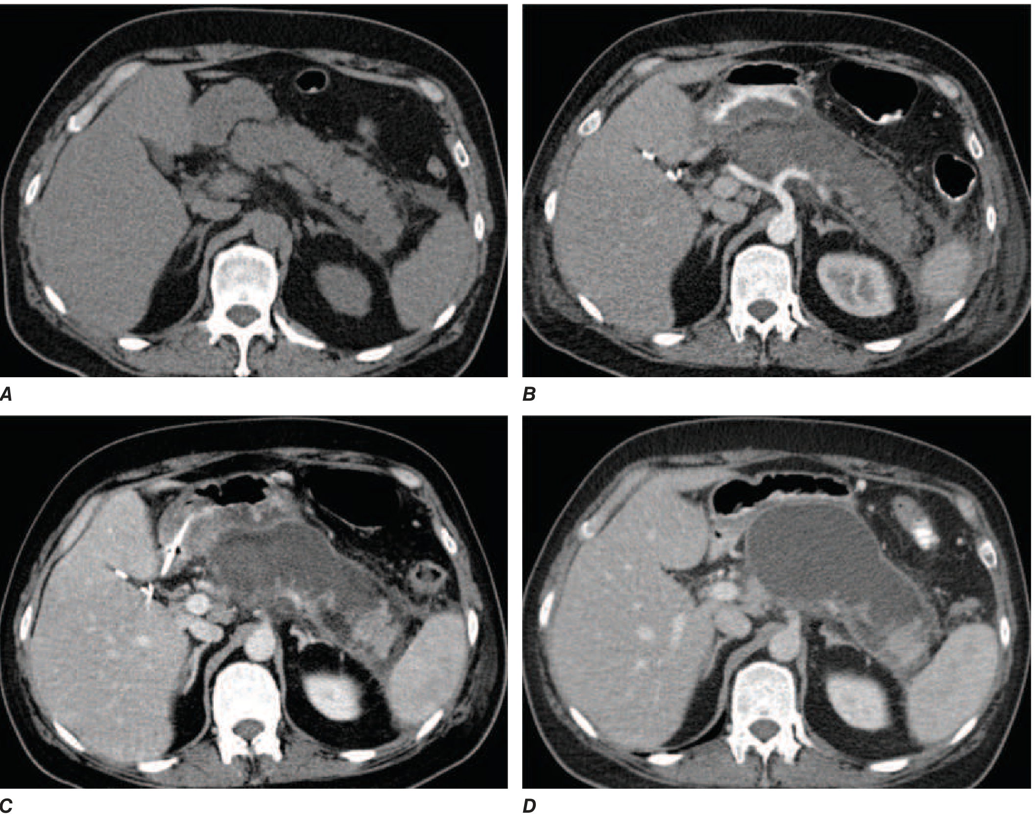
FIGURE: Evolution of acute necrotizing pancreatitis on CT. A - mild peripancreatic stranding on admission; B - extensive intrapancreatic necrosis at 1 week (lack of contrast enhancement); C - acute necrotic collection (ANC) at 2 weeks; D - walled-off necrosis (WON) replacing the pancreas at several weeks. - Harrison's Principles, p. 2790
3. Fluid Resuscitation
The single most important early intervention. Goals: restore circulating volume, normalize BP, and maintain urine output >0.5 mL/kg/h.
- Preferred fluid: Lactated Ringer's (LR) - shown to lower CRP and reduce systemic inflammation vs. normal saline
- Regimen: Initial bolus ~10-15 mL/kg, then 1.5-2 mL/kg/h
- A recent RCT showed aggressive resuscitation (15-20 mL/kg bolus + 2-3 mL/kg/h) increased fluid overload risk without improving outcomes vs. the less aggressive strategy
- Re-assess every 6-8 h; measure hematocrit + BUN every 8-12 h to guide adequacy
- Caution in elderly and those with cardiac/renal disease - over-resuscitation risk is higher
4. Analgesia
- IV narcotic analgesics (opioids) are the standard for pain control
- There is no evidence that morphine worsens AP (the old concern about sphincter of Oddi spasm has not been proven clinically significant)
- Adequate analgesia is a key pillar of early management
5. Nutritional Support
Major paradigm shift - "resting the pancreas" by prolonged NPO is no longer supported.
- Enteral nutrition (EN) is the preferred route - oral/nasogastric or nasojejunal feeding
- EN maintains gut mucosal integrity, prevents bacterial translocation, and reduces infectious complications
- Nasogastric (NG) feeding is equally effective as nasojejunal (NJ) and much easier to place
- Parenteral nutrition (PN) is reserved for cases where EN is not tolerated or not possible
- For mild AP, refeeding orally can begin once nausea/vomiting and pain have subsided (often within 24-72 h)
- For severe AP requiring nutritional support, EN should be started within 24-48 h of admission
6. Antibiotics
Prophylactic antibiotics are NOT routinely recommended for AP - multiple RCTs have failed to show benefit.
Antibiotics are indicated for:
- Proven or suspected infected pancreatic necrosis (fine-needle aspiration guided by CT, or clinical signs of infection - fever, rising WBC, deterioration at 2-4 weeks)
- Cholangitis - treat promptly
- Concurrent infections - urinary tract infection, pneumonia, line infection
When required, regimens include:
- IV imipenem-cilastatin (penetrates pancreatic tissue well)
- Ciprofloxacin + metronidazole
- Cefuroxime (IV)
- Duration: guided by cultures; prophylactic use should not exceed 14 days
7. Managing the Etiology
Gallstone Pancreatitis
- ERCP within 72 hours is indicated for severe gallstone pancreatitis or when cholangitis or biliary obstruction is present
- Cholecystectomy should be performed during the same admission for mild gallstone pancreatitis (prevents recurrence)
- For severe/necrotizing gallstone pancreatitis, timing of cholecystectomy must be individualized (usually deferred until resolution of acute inflammation)
Hypertriglyceridemia
- Aggressively lower triglycerides; insulin infusion and/or plasmapheresis may be needed
Alcohol-related AP
- Treat withdrawal prophylactically; counsel regarding cessation
8. Local Complications
Classification of Fluid Collections
| Timing | Content | Sterile | Infected |
|---|---|---|---|
| <4 weeks (no wall) | Fluid | Acute pancreatic fluid collection (APFC) | Infected APFC |
| <4 weeks (no wall) | Solid ± fluid | Acute necrotic collection (ANC) | Infected ANC |
| >4 weeks (defined wall) | Fluid | Pseudocyst | Infected pseudocyst |
| >4 weeks (defined wall) | Solid ± fluid | Walled-off necrosis (WON) | Infected WON |
Schwartz's Principles of Surgery, p. 1471
Management of Necrotizing Pancreatitis
- Sterile necrosis: conservative management; most resolves without intervention
- Infected necrosis (peaks at 3-4 weeks): step-up approach
- Percutaneous or endoscopic drainage first
- If drainage fails: minimally invasive necrosectomy (video-assisted retroperitoneal debridement - VARD, or endoscopic transluminal necrosectomy)
- Open necrosectomy reserved for failure of minimally invasive approaches
- Necrotizing AP carries >50% median rate of organ failure; mortality rises to ~50% with multi-organ failure
Pseudocyst
- Most resolve spontaneously
- Drainage indicated if: symptomatic, enlarging, infected, causing biliary/gastric obstruction
- Preferred: endoscopic transgastric or transduodenal drainage (EUS-guided)
Pancreatic Duct Disruption
- Presents as pancreatic ascites (high amylase in ascitic fluid) or enlarging fluid collection
- Confirmed by MRCP or ERCP
- Bridging pancreatic stent placement >90% effective; non-bridging stents less effective (25-50%)
9. Systemic Complications and Organ Support
| System | Complication | Management |
|---|---|---|
| Respiratory | ARDS, pleural effusion, atelectasis | O2, mechanical ventilation if needed |
| Cardiovascular | Hypotension, hypovolemia | Aggressive fluids, vasopressors/inotropes |
| Renal | Oliguria, AKI | Fluids, hemofiltration/dialysis |
| Hematologic | DIC | Supportive, FFP/platelets |
| Metabolic | Hyperglycemia, hypocalcemia | Insulin, IV calcium gluconate |
| Vascular | Splenic vein thrombosis, pseudoaneurysm | Angiography + embolization for bleeding |
10. Summary: Early Management of Severe AP (Bailey & Love, Table 72.4)
- Admit to HDU/ICU
- IV analgesics
- Aggressive fluid resuscitation with LR
- Supplemental oxygen
- Invasive monitoring (vital signs, CVP, urine output, blood gases)
- Frequent lab monitoring (LFTs, renal, clotting, Ca2+, glucose)
- Nasogastric tube only if vomiting
- Antibiotics if cholangitis or proven infection
- CECT if organ failure, deterioration, or sepsis signs
- ERCP within 72 h for severe gallstone pancreatitis or cholangitis
- Enteral (nasogastric) nutrition when nutritional support required
- Organ-specific supportive therapy (inotropes, ventilation, hemofiltration)
11. Follow-Up and Discharge Planning
- Screen for diabetes mellitus (endocrine insufficiency)
- Screen for exocrine pancreatic insufficiency (steatorrhea, weight loss)
- Assess for recurrent cholangitis or infected fluid collections
- Identify and address the underlying etiology to prevent recurrence (25% of patients experience at least one recurrence)
- Alcohol cessation counseling for alcohol-related AP
- Interval cholecystectomy for gallstone-related AP if not done during admission
Key references:
- Harrison's Principles of Internal Medicine, 22nd ed. (2025) - Chapter 359, pp. 2790-2793
- Schwartz's Principles of Surgery, 11th ed. - Chapter 33, pp. 1471-1472
- Bailey and Love's Short Practice of Surgery, 28th ed. - Table 72.4
- iLATAM-AP 2026 Guidelines - Ibero-Latin American Acute Pancreatitis Guidelines (PMID 42216617, June 2026)
This is a shared conversation. Sign in to Orris to start your own chat.