
Clinical guidelines on Hypothyroidism in pregnancy
"hypothyroidism" AND "pregnancy" AND "guidelines"
hypothyroidism pregnancy clinical guidelines 2024 2025 ATA endocrine society
PMID: 42219800
https://www.guidelinecentral.com/insights/jun-2026-ata-thyro…
hypothyroidism pregnancy TSH management algorithm
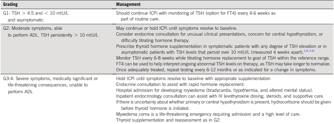
<table><thead><tr><th>Grading</th><th>Management</th></tr></thead><tbody><tr><td>G1: TSH > 4.5 and < 10 mIU/L and asymptomatic</td><td>Should continue ICPi with monitoring of TSH (option for FT4) every 4-6 weeks as part of routine care.</td></tr><tr><td>G2: Moderate symptoms, able to perform ADL. TSH persistently > 10 mIU/L</td><td>May continue or hold ICPi until symptoms resolve to baseline. Consider endocrine consultation for unusual clinical presentations, concern for central hypothyroidism, or difficulty titrating hormone therapy. Prescribe thyroid hormone supplementation in symptomatic patients with any degree of TSH elevation or in asymptomatic patients with TSH levels that persist over 10 mIU/L (measured 4 weeks apart).131,132 Monitor TSH every 6-8 weeks while titrating hormone replacement to goal of TSH within the reference range. FT4 can be used to help interpret ongoing abnormal TSH levels on therapy, as TSH may take longer to normalize. Once adequately treated, repeat testing every 6-12 months or as indicated for a change in symptoms.</td></tr><tr><td>G3-4: Severe symptoms, medically significant or life-threatening consequences, unable to perform ADL</td><td>Hold ICPi until symptoms resolve to baseline with appropriate supplementation Endocrine consultation to assist with rapid hormone replacement. Hospital admission for developing myxedema (bradycardia, hypothermia, and altered mental status). Inpatient endocrinology consultation can assist with IV levothyroxine dosing, steroids, and supportive care. If there is uncertainty about whether primary or central hypothyroidism is present, hydrocortisone should be given before thyroid hormone is initiated. Myxedema coma is a life-threatening emergency requiring admission and a high level of care. Thyroid supplementation and reassessment as in G2.</td></tr></tbody></table>
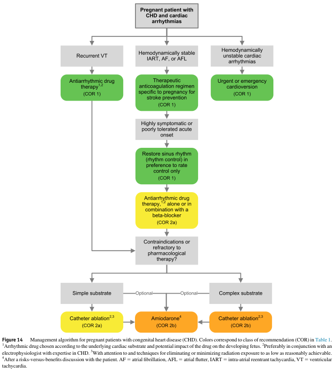
Summary : This flowchart presents a management algorithm for pregnant patients with congenital heart disease (CHD) and cardiac arrhythmias, outlining decision pathways based on arrhythmia type and hemodynamic stability, with recommendations for drug therapy, cardioversion, and ablation. flowchart: # Nodes : • Start (rectangle): "Pregnant patient with CHD and cardiac arrhythmias" • Decision (rectangle): "Recurrent VT" • Decision (rectangle): "Hemodynamically stable IART, AF, or AFL" • Decision (rectangle): "Hemodynamically unstable cardiac arrhythmias" • Action (rounded rectangle, green): "Antiarrhythmic drug therapy¹,² (COR 1)" • Action (rounded rectangle, green): "Therapeutic anticoagulation regimen specific to pregnancy for stroke prevention (COR 1)" • Action (rounded rectangle, green): "Urgent or emergency cardioversion (COR 1)" • Decision (rectangle): "Highly symptomatic or poorly tolerated acute onset" • Action (rounded rectangle, green): "Restore sinus rhythm (rhythm control) in preference to rate control only (COR 1)" • Action (rounded rectangle, yellow): "Antiarrhythmic drug therapy¹,² alone or in combination with a beta-blocker (COR 2a)" • Decision (rectangle): "Contraindications or refractory to pharmacological therapy?" • Substrate type (rectangle): "Simple substrate" • Substrate type (rectangle): "Complex substrate" • Optional (rectangle): "Optional" • Action (rounded rectangle, orange): "Catheter ablation²,³ (COR 2a)" • Action (rounded rectangle, orange): "Amiodarone⁴ (COR 2b)" • Action (rounded rectangle, orange): "Catheter ablation²,³ (COR 2b)" # Connectors : • Downward arrows connect each node in sequence, with branches for arrhythmia type and stability. • "Pregnant patient with CHD and cardiac arrhythmias" splits into three branches: "Recurrent VT", "Hemodynamically stable IART, AF, or AFL", and "Hemodynamically unstable cardiac arrhythmias". • "Recurrent VT" leads to "Antiarrhythmic drug therapy¹,² (COR 1)". • "Hemodynamically stable IART, AF, or AFL" leads to "Therapeutic anticoagulation regimen specific to pregnancy for stroke prevention (COR 1)", then to "Highly symptomatic or poorly tolerated acute onset", then to "Restore sinus rhythm (rhythm control) in preference to rate control only (COR 1)", then to "Antiarrhythmic drug therapy¹,² alone or in combination with a beta-blocker (COR 2a)", then to "Contraindications or refractory to pharmacological therapy?". • "Hemodynamically unstable cardiac arrhythmias" leads directly to "Urgent or emergency cardioversion (COR 1)". • "Contraindications or refractory to pharmacological therapy?" splits into "Simple substrate" and "Complex substrate". • "Simple substrate" leads to "Catheter ablation²,³ (COR 2a)" and optionally to "Amiodarone⁴ (COR 2b)". • "Complex substrate" leads to "Catheter ablation²,³ (COR 2b)". # Layout : • Top-down hierarchical flowchart with three main branches from the starting node. • Colour coding: green (COR 1), yellow (COR 2a), orange (COR 2a/2b), grey for decision nodes and substrate types. • Optional steps indicated by "Optional" nodes. • Explanatory footnotes and abbreviations provided below the chart. # Analysis : • The flowchart prioritises antiarrhythmic drug therapy and stroke prevention for stable arrhythmias, with urgent cardioversion for unstable cases. • Rhythm control is preferred over rate control in symptomatic cases. • Catheter ablation is considered for refractory cases, with amiodarone as an option after risk-benefit discussion. • The algorithm is structured to minimise fetal risk and tailor therapy to arrhythmia type and patient stability. • Colour coding visually distinguishes recommendation strength (COR 1, 2a, 2b).
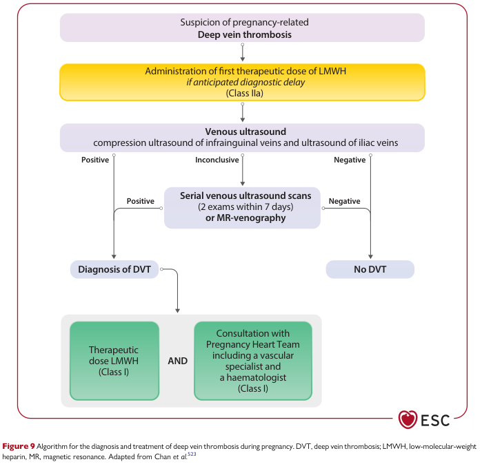
Summary : This flowchart presents an algorithm for the diagnosis and treatment of deep vein thrombosis (DVT) during pregnancy, as recommended by the European Society of Cardiology (ESC). It outlines the stepwise approach from initial suspicion to diagnosis and management, including the use of low-molecular-weight heparin (LMWH), ultrasound imaging, and specialist consultation. flowchart: # Nodes : • Start (rounded rectangle): "Suspicion of pregnancy-related Deep vein thrombosis" • Decision/Action (rounded rectangle, yellow): "Administration of first therapeutic dose of LMWH if anticipated diagnostic delay (Class IIa)" • Process (rounded rectangle): "Venous ultrasound compression ultrasound of infrainguinal veins and ultrasound of iliac veins" • Decision (split into three branches): "Positive", "Inconclusive", "Negative" • Process (rounded rectangle): "Serial venous ultrasound scans (2 exams within 7 days) or MR-venography" (for inconclusive results) • Decision (split from serial scans): "Positive", "Negative" • Terminal (rectangle): "Diagnosis of DVT" • Terminal (rectangle): "No DVT" • Management (rectangle): "Therapeutic dose LMWH (Class I)" • Management (rectangle): "Consultation with Pregnancy Heart Team including a vascular specialist and a haematologist (Class I)" # Connectors : • Downward arrows connect each step in sequence. • From "Venous ultrasound", three branches: – Positive → "Diagnosis of DVT" – Inconclusive → "Serial venous ultrasound scans or MR-venography" – Negative → "No DVT" • From "Serial venous ultrasound scans or MR-venography": – Positive → "Diagnosis of DVT" – Negative → "No DVT" • From "Diagnosis of DVT", two parallel arrows to: – "Therapeutic dose LMWH (Class I)" – "Consultation with Pregnancy Heart Team including a vascular specialist and a haematologist (Class I)" • Both management steps are joined by "AND". # Layout : • Top-down, single entry point. • Branching at ultrasound result (positive/inconclusive/negative). • Re-merging after serial scans. • Parallel management steps after DVT diagnosis. • Color coding: yellow for urgent action, purple for diagnostic steps, green for management. # Analysis : • The algorithm prioritizes early treatment with LMWH if diagnostic delay is expected. • Ultrasound is the primary diagnostic tool, with serial imaging or MR-venography for inconclusive cases. • Negative imaging at any stage rules out DVT. • Confirmed DVT leads to both therapeutic anticoagulation and multidisciplinary specialist consultation. • The flowchart emphasizes safety, rapid diagnosis, and coordinated care for pregnant patients with suspected DVT.
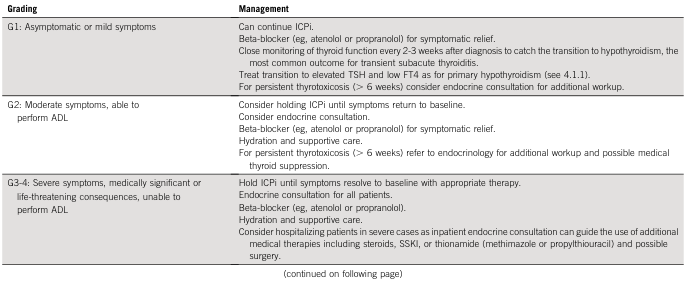
<table> <tr> <th>Grading</th> <th>Management</th> </tr> <tr> <td>G1: Asymptomatic or mild symptoms</td> <td> Can continue ICPI.<br> Beta-blocker (eg, atenolol or propranolol) for symptomatic relief.<br> Close monitoring of thyroid function every 2-3 weeks after diagnosis to catch the transition to hypothyroidism, the most common outcome for transient subacute thyroiditis.<br> Treat transition to elevated TSH and low FT4 as for primary hypothyroidism (see 4.1.1).<br> For persistent thyrotoxicosis (> 6 weeks) consider endocrine consultation for additional workup. </td> </tr> <tr> <td>G2: Moderate symptoms, able to perform ADL</td> <td> Consider holding ICPI until symptoms return to baseline.<br> Consider endocrine consultation.<br> Beta-blocker (eg, atenolol or propranolol) for symptomatic relief.<br> Hydration and supportive care.<br> For persistent thyrotoxicosis (> 6 weeks) refer to endocrinology for additional workup and possible medical thyroid suppression. </td> </tr> <tr> <td>G3-4: Severe symptoms, medically significant or life-threatening consequences, unable to perform ADL</td> <td> Hold ICPI until symptoms resolve to baseline with appropriate therapy.<br> Endocrine consultation for all patients.<br> Beta-blocker (eg, atenolol or propranolol).<br> Hydration and supportive care.<br> Consider hospitalizing patients in severe cases as inpatient endocrine consultation can guide the use of additional medical therapies including steroids, SSKI, or thionamide (methimazole or propylthiouracil) and possible surgery. </td> </tr> </table> <br> (continued on following page)
Clinical Guidelines on Hypothyroidism in Pregnancy
Important update: The American Thyroid Association released updated guidelines in May 2026, replacing the 2017 version. Key changes include revised TSH thresholds, a new approach to subclinical hypothyroidism treatment (no longer based on TPO antibody status), and greater emphasis on timing of diagnosis.
1. Physiological Changes Affecting Thyroid Function in Pregnancy
- hCG stimulation - Rising hCG in the first trimester weakly stimulates TSH receptors, causing a reciprocal fall in TSH
- Estrogen-induced rise in TBG - Increases total T4 and T3 (~1.5x), but free T4 is the same or slightly higher only at end of first trimester, then progressively decreases
- Immune modulation - Can trigger onset, exacerbation, or amelioration of autoimmune thyroid disease
- Placental type III deiodinase - Increases thyroid hormone metabolism
- Increased urinary iodide excretion - Impairs hormone production in iodine-deficient areas
2. TSH Reference Ranges in Pregnancy
| Trimester | TSH Range (mIU/L) |
|---|---|
| First | 0.1 - 2.5 |
| Second | 0.2 - 3.0 |
| Third | 0.3 - 3.0 |
"When possible, population-based, trimester-specific reference ranges for serum TSH should be defined through assessment of local population data representative of a healthcare provider's practice."
3. Epidemiology
- Overt hypothyroidism: ~0.5% of pregnant women; worldwide, iodine deficiency is the most common cause
- Subclinical hypothyroidism (SCH): ~2-3% of iodine-sufficient pregnant women
- Isolated hypothyroxinemia: ~2% (low fT4 with normal TSH, no autoantibodies)
- TPO antibody positivity: 10-20% of women in childbearing years (even if euthyroid, increased risk of miscarriage, preterm delivery, and postpartum thyroiditis)
4. Adverse Outcomes of Untreated Hypothyroidism
- Miscarriage
- Preterm delivery
- Pre-eclampsia
- Neonatal mortality (from preterm delivery)
- Low birthweight for gestational age
- Impaired neurocognitive development (decreased IQ)
- Subclinical complications occur at lower frequency than overt hypothyroidism but follow the same pattern
5. Diagnosis
- TSH is the best single test during pregnancy
- Free T4 analogue assays may give falsely low values in the second and third trimester (due to elevated binding proteins); total T4 combined with T4 uptake may more accurately reflect true free T4
- TPO and Tg autoantibodies should be checked in relevant clinical contexts
6. Treatment: Overt Hypothyroidism
Indications for LT4 (Tietz/ATA):
- TSH above trimester-specific reference interval + low fT4
- TSH > 10.0 mIU/L irrespective of fT4 level
- TSH above reference + positive TPO antibodies
Dosing:
- Thyroid hormone requirements increase by up to 45% during pregnancy
- A practical approach: increase the existing LT4 dose by ~30% as soon as pregnancy is confirmed (i.e., take two extra tablets per week for daily dosing)
- Preconception LT4 dose should be adjusted to keep TSH in the lower portion of the non-pregnant reference range before conception; this reduces the risk of first-trimester TSH elevation
Treatment Goal:
- TSH within trimester-specific reference intervals
- Total T4 at or above the upper normal range (as estrogen-elevated TBG increases total T4)
Administration Precaution:
- Administer LT4 at least 4 hours apart from prenatal vitamins and calcium supplements (which reduce T4 absorption)
7. Treatment: Subclinical Hypothyroidism (SCH)
Old approach (2017 ATA): Based on TPO antibody status
- TPO-positive + SCH → treat
- TPO-negative + SCH → insufficient evidence
New approach (2026 ATA): Based on TIMING of diagnosis
- SCH diagnosed in the first trimester: Consider treatment with LT4
- SCH diagnosed in the second or third trimester: Treatment is generally NOT recommended
- TPO antibody status no longer drives the treatment decision
- Repeated thyroid function testing to verify that mild overt or subclinical hypothyroidism is persistent before initiating treatment
- Fetal brain development is most vulnerable early in pregnancy
- SCH in the 2nd/3rd trimester may represent physiological variation
- Benefits of treatment are not proven in mild late-diagnosed SCH
Monitoring Untreated SCH:
- TSH + fT4 every 4 weeks until 16-20 weeks of gestation
- At least once between 26-32 weeks
8. Treatment: Isolated Hypothyroxinemia
- Defined as low fT4 with normal TSH and no autoantibodies
- Not recommended for treatment - insufficient evidence
- Complicated by: analogue fT4 assay inaccuracies in later pregnancy; unclear etiology in many cases
9. Monitoring During Pregnancy
| Situation | TSH Monitoring Schedule |
|---|---|
| Known hypothyroid on LT4 | Every 4 weeks until 20 weeks, then at least once in second half |
| Untreated SCH | Every 4 weeks until 16-20 weeks; once at 26-32 weeks |
| Euthyroid but TPO antibody positive (not on LT4) | Every 4 weeks in first half; once at 26-32 weeks |
10. Screening for Thyroid Disease in Pregnancy
- Family history of autoimmune thyroid disease
- Personal history of autoimmune disease (e.g., Type 1 diabetes)
- Infertility
- Prior preterm delivery or recurrent miscarriage
- Signs or symptoms of thyroid disease
- Age > 30 years
11. Euthyroid TPO Antibody-Positive Women
- Euthyroid TPO antibody-positive women with infertility
- Those planning fertility treatment
- Those with a history of recurrent miscarriage
12. Postpartum Management
- Revert LT4 dose to pre-pregnancy level immediately on the day of delivery (not tapered gradually)
- Check TSH and thyroid hormones 6 weeks postpartum (by this point, pregnancy effects on TFTs have resolved)
- Monitor TPO antibody-positive women for postpartum thyroiditis
13. Iodine Supplementation
- WHO recommendation: 250 mcg iodine/day during pregnancy and lactation
- Prenatal vitamins should contain 150 mcg iodine per tablet
- Women with iodine intake < 50 mcg/day are at highest risk for goiter and neonatal hypothyroidism
Key 2026 ATA Guideline Changes - Summary
| Topic | 2017 Position | 2026 Update |
|---|---|---|
| TSH upper limit (first trimester) | 2.5 mIU/L | ~4.0 mIU/L (population-based preferred) |
| SCH treatment basis | TPO antibody status | Timing of diagnosis (first trimester = consider; 2nd/3rd = generally do not treat) |
| Euthyroid TPO+ with infertility/recurrent miscarriage | LT4 sometimes offered | Do NOT offer LT4 |
| Confirmation of SCH | Not emphasized | Repeat TFTs to confirm persistence before treating |
- ATA 2026 Guidelines (Korevaar et al., Thyroid 2026 - PMID 42219800)
- Tietz Textbook of Laboratory Medicine, 7th Ed. - pp. 4478-4493
- Harrison's Principles of Internal Medicine, 22nd Ed. (2025) - pp. 2975-2978
- Creasy & Resnik's Maternal-Fetal Medicine - pp. 1641-1651
- Goodman & Gilman's Pharmacological Basis of Therapeutics - pp. 2460-2464
- Katzung's Basic and Clinical Pharmacology, 16th Ed. - pp. 1163-1169