
Immunostaining profiles for cytokeratin in diagnostic Pathology
Reading File
Reading File
Finding Sources
Finding Sources
Searching PubMed
cytokeratin immunohistochemistry diagnostic pathology
Searching the Web
cytokeratin 7 20 immunohistochemistry diagnostic pathology profiles review
Reading File
Reading File
Finding Sources
Finding Sources
Searching Images
cytokeratin CK7 CK20 immunohistochemistry primary site diagnostic algorithm adenocarcinoma unknown primary
Reading File
Reading File
Finding Sources
Finding Sources
Reading Web Page
https://pmc.ncbi.nlm.nih.gov/articles/PMC11987302
Cytokeratin immunostaining in diagnostic pathology
Cytokeratins (CKs) are intermediate-filament proteins of epithelial cells. In tumor pathology, pancytokeratin positivity supports epithelial differentiation and therefore favors carcinoma, but it does not by itself identify the primary site. It must be interpreted with morphology, clinical-radiologic findings, and a targeted panel of lineage/site-specific markers.
Bailey and Love's Short Practice of Surgery, 28th ed., p. 211.
Bailey and Love's Short Practice of Surgery, 28th ed., p. 211.
1. Broad-spectrum cytokeratin markers
| Marker/panel | Main diagnostic use | Important comments |
|---|---|---|
| AE1/AE3 | Broad-spectrum pancytokeratin | Initial screen for epithelial differentiation; includes many low- and high-molecular-weight CKs. |
| MNF116 | Broad epithelial marker | Useful for many carcinomas; staining spectrum differs from AE1/AE3. |
| CAM5.2 | Low-molecular-weight CKs, mainly CK8/18 | Often positive in adenocarcinomas and many neuroendocrine carcinomas. |
| OSCAR | Broad epithelial marker | Often useful in poorly differentiated carcinoma. |
| CK5/6 | Basal/squamous and mesothelial differentiation | Helps support squamous carcinoma or mesothelioma in the appropriate panel. |
| 34βE12 / HMWCK | Basal, squamous, urothelial epithelium | Supports squamous or basal-type differentiation; also used in prostate basal-cell assessment. |
Caveat: PanCK positivity is not synonymous with carcinoma. It can occur in mesothelioma, synovial sarcoma, epithelioid sarcoma, desmoplastic small round cell tumor, chordoma, and occasionally focal areas of other tumors. Conversely, some poorly differentiated carcinomas may stain weakly or lose CK expression. A broad panel is required when morphology is atypical.
2. Useful individual CK profiles
| Cytokeratin | Common positive tumors/tissues | Diagnostic application |
|---|---|---|
| CK5/6 | Squamous cell carcinoma, mesothelioma, basal-type urothelial carcinoma, myoepithelial/basal cells | With p40 or p63 supports squamous differentiation. In pleural tumors, interpret with calretinin, WT1, D2-40 and claudin-4/MOC31. |
| CK7 | Lung adenocarcinoma, breast carcinoma, thyroid carcinoma, Müllerian tumors, pancreatobiliary tumors, upper GI tumors, many urothelial carcinomas | Key component of carcinoma of unknown primary workup. |
| CK8/18 | Most simple epithelia, hepatocellular tumors, adenocarcinomas, neuroendocrine neoplasms | Usually assessed indirectly with CAM5.2 or low-MW CK cocktails. |
| CK19 | Biliary and pancreatic epithelium, papillary thyroid carcinoma, many squamous carcinomas | Can support biliary/pancreatic phenotype; often used in thyroid and liver pathology. |
| CK20 | Colorectal carcinoma, Merkel cell carcinoma, urothelial carcinoma, gastric and mucinous tumors | Strong diffuse CK20 with CDX2/SATB2 supports colorectal origin. Perinuclear dot-like staining supports Merkel cell carcinoma. |
| CK14 / CK17 | Basal and squamous epithelium, basal-like carcinomas | Support squamous/basal phenotype, especially in selected head and neck, skin, and urothelial lesions. |
Squamous cell carcinomas commonly express CK5/6 and p63/p40.
Yamada's Textbook of Gastroenterology, 7th ed., section “Squamous cell carcinoma.”
Yamada's Textbook of Gastroenterology, 7th ed., section “Squamous cell carcinoma.”
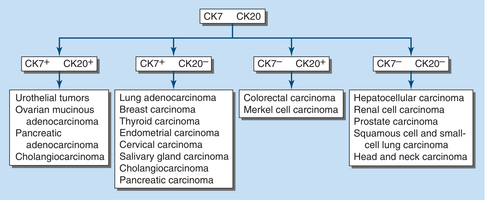
3. CK7/CK20 pattern in metastatic adenocarcinoma
The CK7/CK20 pattern is a triage tool, not a final diagnosis. It helps focus the next set of site-specific stains.
| CK7 / CK20 profile | Common diagnostic possibilities | Suggested confirmatory markers |
|---|---|---|
| CK7+ / CK20− | Lung adenocarcinoma, breast carcinoma, thyroid carcinoma, endometrial/serous ovarian carcinoma, pancreatobiliary carcinoma, cholangiocarcinoma, salivary carcinoma, many upper GI carcinomas | TTF-1, napsin A for lung; GATA3, ER, TRPS1 for breast; PAX8, WT1, ER for Müllerian origin; thyroglobulin, TTF-1 for thyroid |
| CK7− / CK20+ | Colorectal adenocarcinoma, Merkel cell carcinoma; occasional gastric/intestinal tumors | CDX2 and SATB2 for colorectal origin; correlate with nuclear beta-catenin. Merkel cell carcinoma usually has dot-like CK20 and is often TTF-1 negative. |
| CK7+ / CK20+ | Urothelial carcinoma, gastric adenocarcinoma, pancreaticobiliary carcinoma, cholangiocarcinoma, mucinous ovarian carcinoma, some pulmonary mucinous adenocarcinomas | GATA3, p63/p40, uroplakin II for urothelial; CDX2/SATB2 for intestinal phenotype; PAX8 in selected ovarian mucinous tumors |
| CK7− / CK20− | Hepatocellular carcinoma, renal cell carcinoma, prostate carcinoma, adrenal cortical neoplasm, squamous carcinoma, some neuroendocrine tumors | Arginase-1, HepPar-1 for hepatocellular; PAX8, CAIX for renal; NKX3.1, PSA/PSAP for prostate; SF1, inhibin for adrenal cortical origin |
High-yield examples
-
Colorectal adenocarcinoma: typically CK7− / CK20+ / CDX2+, often SATB2+.
Goldman-Cecil Medicine, 2-volume set, “Colon Cancer Profile.” -
Primary lung adenocarcinoma: usually CK7+ / TTF-1+ / napsin A+ / CK20−. Mucinous lung adenocarcinoma may be TTF-1 negative and can show CK20 expression.
-
Breast carcinoma: usually CK7+ / CK20− with GATA3, ER, TRPS1, mammaglobin, or GCDFP-15 as supportive markers.
-
Müllerian carcinoma: generally CK7+, commonly PAX8+. High-grade serous carcinoma is usually WT1+.
-
Pancreatobiliary carcinoma/cholangiocarcinoma: usually CK7+ / CK20−, though CK20 can be variable. Use morphology and markers such as S100P, SMAD4, MUC1, and site-specific clinical correlation.
-
Urothelial carcinoma: variable CK7/CK20 profile, often CK7 positive; use GATA3, p63/p40, uroplakin II/III, and thrombomodulin where appropriate.
-
Hepatocellular carcinoma: often CK7−/CK20− in tumor cells. Confirm using arginase-1, HepPar-1, glypican-3, and canalicular polyclonal CEA/CD10 pattern.
The CUP algorithm emphasizes that CK7/CK20 should direct a second-line organ-specific panel, rather than be used to assign an origin alone. Harrison's Principles of Internal Medicine, 22nd ed., p. 776.
4. CK profiles in common diagnostic differentials
| Differential diagnosis | Helpful CK pattern | Add-on markers |
|---|---|---|
| Lung adenocarcinoma vs metastatic colorectal carcinoma | Lung: CK7+/CK20−; colorectal: CK7−/CK20+ | TTF-1, napsin A versus CDX2, SATB2 |
| Mesothelioma vs adenocarcinoma | Mesothelioma often CK5/6+ and broad CK+ | Calretinin, WT1, D2-40 versus claudin-4, MOC31/BerEP4, CEA |
| Squamous carcinoma vs adenocarcinoma | Squamous: CK5/6+, high-MW CK+, p40/p63+ | TTF-1/napsin A for lung adenocarcinoma; site-specific markers |
| Merkel cell carcinoma vs small-cell lung carcinoma | Merkel: CK20+, characteristic paranuclear dot pattern; lung small-cell: often CK20− | Merkel cell polyomavirus, INSM1, synaptophysin, chromogranin; TTF-1 favors pulmonary small-cell carcinoma |
| Primary ovarian mucinous tumor vs colorectal metastasis | Primary ovarian mucinous tumor often CK7 diffuse with variable CK20; colorectal tumor often CK7−/CK20 diffuse | PAX8 may support ovarian origin; SATB2 and CDX2 favor lower GI origin |
| Prostate carcinoma vs urothelial carcinoma | Prostate usually CK7−/CK20−; urothelial tumors frequently CK7+ | NKX3.1, PSA, PSAP versus GATA3, p63/p40, uroplakin II |
5. Interpretation principles and pitfalls
-
Use internal controls. Native epithelium should stain appropriately. A negative CK result without an internal positive control is not interpretable.
-
Record distribution, intensity, and pattern. Diffuse strong staining is more meaningful than rare weak cells. CK20’s paranuclear dot pattern is diagnostically useful for Merkel cell carcinoma.
-
Do not diagnose a primary site from CK7/CK20 alone. Gastric, pancreatic, ovarian mucinous, urothelial, and mucinous pulmonary tumors show substantial overlap.
-
Expect altered profiles in poorly differentiated tumors. Loss of expected CKs, aberrant expression, and intratumoral heterogeneity occur, particularly in high-grade or metastatic lesions.
-
Beware entrapped benign epithelium. Small CK-positive glands in a largely CK-negative neoplasm may represent entrapped structures, not tumor staining.
-
Match the panel to the morphology. For example:
- gland-forming lesion: CK7/CK20 plus organ-specific markers;
- epithelioid pleural lesion: mesothelial versus epithelial carcinoma panel;
- spindle/epithelioid malignancy: include CK, SOX10/S100, CD45, desmin/myogenin, ERG/CD31, and selected molecularly informed markers.
A recent review of CK7/CK20 use found that these stains remain useful as a first-line orientation tool but have limited standalone value because profiles vary by histologic subtype and overlap across sites. The authors recommend integrating them with morphology and more specific markers, as summarized in this recent CK7/CK20 review.
From standard textbook For MD Pathology exams 10 marks question
Reading File
Finding Sources
Finding Sources
Searching PubMed
cytokeratin immunohistochemistry diagnostic pathology
Searching the Web
cytokeratin immunohistochemistry diagnostic pathology CK7 CK20 review
Immunostaining profiles of cytokeratins in diagnostic pathology
Introduction
Cytokeratins (CKs) are intermediate filament proteins expressed by epithelial cells. Their immunohistochemical demonstration is mainly used to:
- Establish epithelial differentiation in an undifferentiated neoplasm.
- Classify carcinomas by differentiation.
- Help identify the primary site of a metastatic carcinoma.
- Detect micrometastasis, especially in lymph nodes and bone marrow.
- Distinguish carcinoma from lymphoma, melanoma, sarcoma, and mesothelioma.
Cytokeratin positivity favors carcinoma, but is not diagnostic of carcinoma by itself, since mesothelioma and certain sarcomas may express keratins.
Bailey and Love's Short Practice of Surgery, 28th ed., p. 211.
Bailey and Love's Short Practice of Surgery, 28th ed., p. 211.
Classification
Cytokeratins are classified into:
| Group | Cytokeratins | Usual association |
|---|---|---|
| Type I: acidic, low molecular weight | CK9-20 | Simple and non-keratinizing epithelial differentiation |
| Type II: basic/neutral, high molecular weight | CK1-8 | Squamous, basal, and keratinizing epithelial differentiation |
CKs are normally expressed in pairs, for example:
- CK5/CK14: basal and squamous epithelium
- CK8/CK18: simple epithelium
- CK1/CK10: keratinizing epidermis
A. Broad-spectrum cytokeratin antibodies
| Antibody | Reactivity / use | Important application |
|---|---|---|
| AE1/AE3 | Broad-spectrum cocktail reacting with many low- and high-molecular-weight CKs | Most commonly used initial marker to establish epithelial differentiation |
| MNF116 | Broad-spectrum CK cocktail | Detects many epithelial tumors |
| OSCAR | Broad-spectrum epithelial marker | Helpful in poorly differentiated carcinoma |
| CAM5.2 | Mainly low-molecular-weight CKs, especially CK8 and CK18 | Useful in adenocarcinoma and neuroendocrine carcinoma |
| 34βE12 | High-molecular-weight CKs: CK1, 5, 10, 14 | Squamous/basal and urothelial differentiation; prostate basal cells |
| CK5/6 | Basal and squamous epithelial CKs | Squamous carcinoma and mesothelioma panel |
In an undifferentiated malignant neoplasm, a useful initial lineage panel is:
- Pancytokeratin/AE1-AE3: carcinoma
- CD45: lymphoma
- S100/SOX10: melanoma
- Desmin/SMA/myogenin: myogenic tumors
- CD31/ERG: vascular tumors
B. Cytokeratin profile of important tumors
1. Squamous differentiation
| Markers | Diagnostic implication |
|---|---|
| CK5/6+, CK14+, CK17+, 34βE12+ | Supports squamous or basal differentiation |
| p40 and p63 positive | Strong support for squamous differentiation |
| CK7 usually negative or variable | Depends on site and tumor type |
Squamous cell carcinomas usually express CK5/6 and p63/p40.
Yamada's Textbook of Gastroenterology, 7th ed., section “Squamous cell carcinoma.”
Yamada's Textbook of Gastroenterology, 7th ed., section “Squamous cell carcinoma.”
Uses
- Lung squamous carcinoma versus lung adenocarcinoma
- Squamous carcinoma versus mesothelioma
- Identification of basal phenotype in urothelial carcinoma
- Detection of basal cells in prostatic glands
2. CK7 and CK20 profile
The CK7/CK20 combination is particularly useful in the evaluation of metastatic adenocarcinoma and carcinoma of unknown primary (CUP).
| CK7 | CK20 | Common primary tumors | Confirmatory markers |
|---|---|---|---|
| + | − | Lung adenocarcinoma, breast carcinoma, thyroid carcinoma, endometrial carcinoma, ovarian serous carcinoma, pancreatobiliary carcinoma, cholangiocarcinoma | TTF-1/napsin A, GATA3, ER, PAX8, WT1, thyroglobulin |
| − | + | Colorectal adenocarcinoma, Merkel cell carcinoma | CDX2, SATB2; Merkel cell polyomavirus |
| + | + | Urothelial carcinoma, gastric carcinoma, pancreaticobiliary carcinoma, mucinous ovarian tumor, some mucinous lung adenocarcinomas | GATA3, uroplakin II, CDX2, SATB2, PAX8 |
| − | − | Hepatocellular carcinoma, renal cell carcinoma, prostate carcinoma, adrenal cortical carcinoma, many squamous carcinomas | Arginase-1/HepPar-1, PAX8/CAIX, NKX3.1/PSA, SF1/inhibin |
Important CK7/CK20 patterns
| Tumor | Typical profile |
|---|---|
| Colorectal adenocarcinoma | CK7−, CK20+, CDX2+, SATB2+ |
| Lung adenocarcinoma | CK7+, CK20−, TTF-1+, napsin A+ |
| Breast carcinoma | CK7+, CK20−, GATA3+, ER variable |
| Ovarian serous carcinoma | CK7+, CK20−, PAX8+, WT1+ |
| Pancreatic adenocarcinoma | CK7+, CK20− or focal+, usually MUC1+ |
| Cholangiocarcinoma | CK7+, CK19+, CK20 variable |
| Urothelial carcinoma | CK7+, CK20 variable, GATA3+, p63/p40+, uroplakin II+ |
| Hepatocellular carcinoma | Usually CK7−, CK20−; arginase-1+ and HepPar-1+ |
| Prostatic adenocarcinoma | Usually CK7−, CK20−; NKX3.1+, PSA/PSAP+ |
| Merkel cell carcinoma | CK20+, typically paranuclear dot-like staining; CK7−; TTF-1− |
High-yield point: The profile CK7−/CK20+/CDX2+/SATB2+ strongly supports a colorectal primary, especially in liver metastasis.
Goldman-Cecil Medicine, 2-volume set, “Colon Cancer Profile.”
Goldman-Cecil Medicine, 2-volume set, “Colon Cancer Profile.”
C. Other useful individual cytokeratins
| Cytokeratin | Principal expression / diagnostic value |
|---|---|
| CK1 and CK10 | Keratinizing stratified squamous epithelium |
| CK5 and CK14 | Basal cells, squamous epithelium, myoepithelial cells |
| CK6 and CK16 | Hyperproliferative squamous epithelium; squamous tumors |
| CK7 | Glandular/simple epithelium: lung, breast, pancreatobiliary tract, Müllerian tract |
| CK8 and CK18 | Simple epithelial cells, hepatocytes, renal tubular epithelium, endocrine/neuroendocrine tumors |
| CK19 | Biliary epithelium, pancreatic ducts, papillary thyroid carcinoma, many carcinomas; marker of ductular differentiation |
| CK20 | Gastrointestinal epithelium, urothelium, Merkel cells; colorectal and Merkel cell carcinoma |
| CK13 | Non-keratinizing squamous epithelium, including esophagus and cervix |
| CK17 | Basal/myoepithelial and squamous differentiation; often expressed in aggressive basal-type carcinomas |
D. Diagnostic applications
1. Carcinoma versus non-epithelial malignancy
Diffuse AE1/AE3 or pancytokeratin staining supports carcinoma. However, correlation with morphology and other lineage markers is mandatory.
2. Metastatic carcinoma of unknown primary
A practical approach:
- Confirm epithelial nature with AE1/AE3 or CAM5.2.
- Use CK7 and CK20.
- Add organ-specific markers based on the CK profile and morphology.
For example:
- CK7+/CK20−: TTF-1, napsin A, GATA3, ER, PAX8, WT1.
- CK7−/CK20+: CDX2 and SATB2.
- CK7+/CK20+: GATA3, uroplakin II, PAX8, CDX2/SATB2.
- CK7−/CK20−: arginase-1, HepPar-1, PAX8, PSA/NKX3.1, SF1.
In CUP, CK7/CK20 staining gives an overall direction, but no stain is completely sensitive or specific. IHC should be interpreted with tumor morphology, imaging, and clinical findings.
Harrison's Principles of Internal Medicine, 22nd ed., p. 776.
Harrison's Principles of Internal Medicine, 22nd ed., p. 776.
3. Mesothelioma versus adenocarcinoma
Mesothelioma may express pancytokeratin and CK5/6. Therefore, CK positivity cannot separate mesothelioma from carcinoma.
- Mesothelioma-favoring markers: CK5/6, calretinin, WT1, D2-40
- Adenocarcinoma-favoring markers: claudin-4, MOC31, BerEP4, CEA
4. Merkel cell carcinoma versus small-cell carcinoma of lung
- Merkel cell carcinoma: CK20 positive with paranuclear dot-like pattern; usually CK7− and TTF-1−.
- Small-cell carcinoma of lung: commonly TTF-1+; CK20 usually negative.
E. Limitations and pitfalls
-
No CK pattern is absolutely site-specific. CK7 and CK20 profiles overlap, particularly in gastric, pancreaticobiliary, mucinous ovarian, and urothelial carcinomas.
-
Poorly differentiated tumors may lose expected CK expression. Hence, a negative result does not exclude carcinoma.
-
Some non-epithelial tumors are keratin-positive, notably:
- Mesothelioma
- Synovial sarcoma
- Epithelioid sarcoma
- Desmoplastic small round cell tumor
- Chordoma
- Some melanomas and sarcomatoid tumors, usually focal
-
Entrapped benign epithelium may be mistaken for keratin-positive tumor cells.
-
Interpretation must include:
- Staining intensity
- Distribution: focal or diffuse
- Pattern: cytoplasmic, membranous, paranuclear dot-like
- Appropriate internal and external controls
Conclusion
Cytokeratin immunostaining is the primary tool for demonstrating epithelial differentiation. Pancytokeratin confirms epithelial phenotype, while individual CKs, especially CK7 and CK20, assist in narrowing the likely site of origin in metastatic carcinoma. Their greatest value lies in a morphology-guided, panel-based approach with site-specific markers, rather than isolated interpretation of a single cytokeratin stain.
This is a shared conversation. Sign in to Orris to start your own chat.