
Vancomycin tablet
I'll look up vancomycin tablets in the medical library right away.
Searching sources...
Reading File
Finding Sources
Searching the Web
vancomycin oral tablet clinical use indications pharmacology
Finding Sources
Finding Sources
Reading File
Reading File
Vancomycin tablet
Here is a focused overview of Vancomycin Oral (Tablet/Capsule) based on authoritative pharmacology textbooks and FDA labeling:
Vancomycin Oral Tablet / Capsule
Drug Class
Glycopeptide antibiotic - produced by Streptomyces orientalis (also called Amycolatopsis orientalis)
Key Principle: Why Oral?
Vancomycin is poorly absorbed from the GI tract - this is actually the therapeutic rationale for oral use. The drug stays in the gut lumen at high concentrations, acting locally on intestinal pathogens.
- IV vancomycin is NOT effective for intestinal infections (does not reach the gut lumen in sufficient concentrations)
- Oral vancomycin is NOT effective for systemic infections (not absorbed)
These two routes are essentially non-interchangeable.
FDA-Approved Oral Indications
| Indication | Notes |
|---|---|
| Clostridioides difficile (C. diff) colitis / pseudomembranous colitis | Primary use |
| Staphylococcal enterocolitis (including MRSA strains) | Including methicillin-resistant strains |
Mechanism of Action
Vancomycin inhibits cell wall peptidoglycan synthesis at an early stage - it binds to the D-Ala-D-Ala terminus of peptidoglycan precursors, blocking cross-linking. This is bactericidal against susceptible gram-positive organisms.
Dosing (Oral)
| Indication | Adult Dose |
|---|---|
| C. difficile (initial episode) | 125 mg orally 4 times daily x 10 days |
| C. difficile (severe/fulminant) | 500 mg orally 4 times daily |
| Staphylococcal enterocolitis | 500 mg - 2 g/day in divided doses x 7-10 days |
For fulminant C. diff with ileus or toxic megacolon: oral vancomycin 500 mg QID + IV metronidazole may be combined.
C. difficile Treatment Hierarchy (Current Guidelines)
- Fidaxomicin - preferred first-line (lower relapse rates vs. oral vancomycin)
- Oral Vancomycin - alternate first-line; preferred when cost is a concern
- Metronidazole - no longer recommended as monotherapy (inferior cure rates)
For recurrent C. diff: oral vancomycin in prolonged tapered/pulsed regimens is used.
Pharmacokinetics
| Parameter | Detail |
|---|---|
| Absorption | Minimal / negligible from intact GI tract |
| Distribution | Stays in gut lumen (local action) |
| Systemic exposure | May increase if gut mucosa is inflamed |
| Excretion | Feces (unabsorbed drug) |
Note: In patients with severe inflammatory bowel disease or intestinal inflammation, some systemic absorption may occur - monitoring may be warranted in such cases.
Adverse Effects (Oral Route)
Because oral vancomycin is not significantly absorbed, systemic toxicities (nephrotoxicity, ototoxicity - "Red Man Syndrome") seen with IV vancomycin are generally not a concern with oral use.
| Effect | Notes |
|---|---|
| GI upset, nausea | Most common |
| Dysbiosis / microbiome disruption | Significant - oral vancomycin is highly disruptive to the gut microbiome, which increases risk of C. diff recurrence paradoxically with long-term use |
| Superinfection | Prolonged use can lead to overgrowth of resistant organisms |
Resistance
- VRE (Vancomycin-Resistant Enterococci): Major concern; mediated by vanA and vanB gene transposons that alter the D-Ala-D-Ala target to D-Ala-D-Lac
- VISA / VRSA (S. aureus): Emerging resistance; isolated in patients with prolonged vancomycin therapy
- Resistance does not emerge rapidly from C. diff itself
Contraindications & Cautions
- Known hypersensitivity to vancomycin
- Not indicated for systemic infections when given orally
- Use with caution in inflammatory bowel disease (increased absorption)
- Avoid prophylactic use without clear evidence (routine prophylaxis during antibiotic therapy is discouraged due to microbiome disruption)
Available Formulations
| Form | Notes |
|---|---|
| Capsules (Vancocin) | 125 mg, 250 mg |
| Oral solution | Prepared from IV powder (compounded) |
| IV powder used orally | The injectable preparation can be dissolved and given orally - used in patients with feeding tubes or difficulty swallowing capsules |
Sources: Katzung's Basic & Clinical Pharmacology 16e | Jawetz Melnick & Adelberg's Medical Microbiology 28e | Goodman & Gilman's Pharmacological Basis of Therapeutics | Goldman-Cecil Medicine | Current Surgical Therapy 14e | FDA Vancocin Label
Arthroscopic fixation of ac joint subluxation
Arthroscopic fixation of AC joint subluxation involves stabilizing the acromioclavicular and coracoclavicular ligaments using arthroscopic or arthroscopically-assisted techniques, typically with suture button devices or anchors. It is used for Rockwood Grade III-V injuries.
Searching sources...
Reading File
Searching the Web
arthroscopic fixation AC joint subluxation technique suture button coracoclavicular
Finding Sources
Finding Sources
Campbell's Operative Orthopaedics (2026 edition) has a dedicated section. Let me read it in full.
Reading File
Finding Sources
Reading File
Reading File
Finding Sources
Reading File
Reading File
Reading File
Reading File
Reading File
Loading Image
Loading Image
Here is a comprehensive overview of arthroscopic fixation of AC joint subluxation/dislocation, drawn from Campbell's Operative Orthopaedics (15th Ed, 2026):
Arthroscopic Fixation of AC Joint Subluxation / Dislocation
Anatomy & Mechanism of Injury
The AC joint is stabilized by two ligament systems:
- Acromioclavicular (AC) ligaments - provide horizontal (anteroposterior) stability; the superior and posterior capsule are most important
- Coracoclavicular (CC) ligaments - the conoid (posteromedial) and trapezoid (anterolateral) provide vertical stability
The typical injury mechanism is a direct fall onto the dome of the shoulder, driving the acromion downward while the clavicle is held against the first rib. This progressively ruptures the AC ligament, then the CC ligaments, and may also tear the deltoid and trapezius muscle attachments.
Rockwood Classification
| Type | Ligaments Injured | Displacement | Treatment |
|---|---|---|---|
| I | AC ligament sprain only | None | Conservative |
| II | AC ruptured, CC intact | <clavicle width superiorly | Conservative |
| III | AC + CC both ruptured | 1–2 cm superior | Controversial (usually conservative initially) |
| IV | AC + CC + posterior clavicle displacement | Posteriorly into trapezius | Surgery |
| V | AC + CC + deltotrapezial fascia | 2–3× normal CC distance | Surgery |
| VI | Subcoracoid or subacromial clavicle | Inferior | Surgery |
Surgical indication: Types III (selected), IV, V, VI - with the goal of restoring both vertical AND horizontal stability.
Three Requirements for Any AC Repair/Reconstruction
As stated in Campbell's:
- The AC joint must be exposed and debrided
- The CC and AC ligaments must be repaired or reconstructed
- Stable reduction of the AC joint must be achieved
Surgical Options: Three Categories of Reconstruction
| Category | Examples |
|---|---|
| 1. Non-biologic fixation | Suture button devices (TightRope, EndoButton), suture loops, synthetic grafts |
| 2. Biologic CC reconstruction | Semitendinosus/gracilis allograft or autograft through coracoid and clavicle tunnels |
| 3. Ligament/tendon transfer | Weaver-Dunn (coracoacromial ligament transfer) - now largely out of favor |
Arthroscopic / Arthroscopically-Assisted Techniques
A. Suture Button (TightRope / EndoButton) Fixation
The most widely used modern technique - can be performed fully arthroscopically or arthroscopy-assisted.
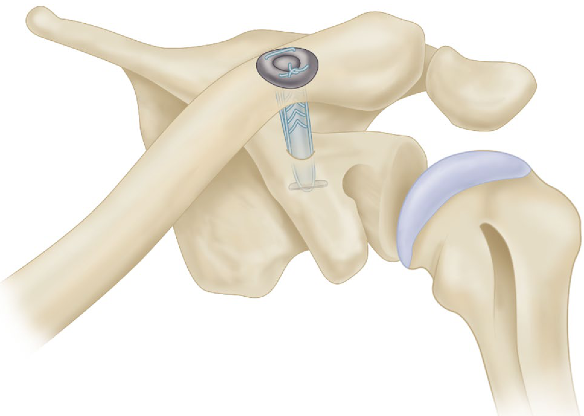
Key steps:
- Patient in beach-chair position; arthroscopic portals established (posterior, anterolateral, anteromedial)
- Arthroscopic exposure and identification of the coracoid base (subcoracoid space)
- A guide pin is drilled from the superior clavicle (approximately 2.5 cm medial to the lateral end) down through the base of the coracoid
- Tunnel is dilated over the guide pin
- A suture tape or FiberWire is passed through the tunnel carrying a small titanium button (e.g., EndoButton, TightRope)
- The subcoracoid button flips and seats under the coracoid base; the supraclavicular button is tightened and seated on the superior clavicle
- The AC joint is reduced and the construct is tensioned
- Optional: biologic graft augmentation (gracilis or semitendinosus allograft) is added to recreate conoid and trapezoid bundles for long-term stability
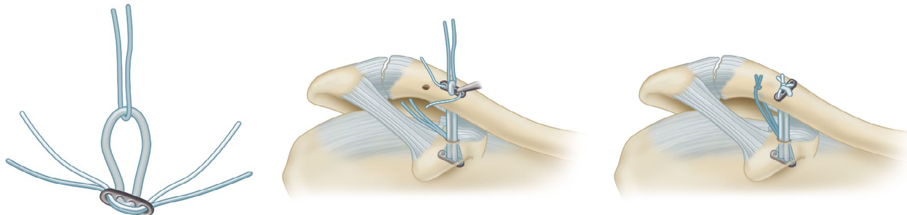
Variants:
- Single-bundle - one device (1 tunnel in coracoid, 1 in clavicle)
- Double-bundle (EndoButton) - two separate corocoid-to-clavicle constructs to recreate conoid and trapezoid separately
- Triple-bundle - adds AC ligament reconstruction for horizontal stability
B. Arthroscopic Coracoacromial Ligament Transfer (Modified Weaver-Dunn)
- The coracoacromial ligament is detached from the acromion (arthroscopically) and transferred to the resected distal clavicle
- Historically popular but now largely replaced by anatomic CC reconstruction
- Meta-analyses show inferior functional and radiographic outcomes vs. CC ligament reconstruction
C. Anatomic Double-Bundle CC Reconstruction (Open/Arthroscopic-Assisted)
- Uses the coracoacromial ligament + conjoined tendon, or semitendinosus/gracilis allograft
- Tunnels are placed to recreate the conoid (4 cm from lateral clavicle) and trapezoid (2.5 cm from lateral clavicle) footprints anatomically
- Graft tails are used to recreate the anterosuperior and posterosuperior AC capsular ligaments
Why Avoid Bone Tunnels?
Campbell's now recommends against clavicular bone tunnels due to a rare but catastrophic risk of clavicular fracture at the tunnel site. Modern suspensory button fixation and cerclage loops are preferred because they avoid tunnels and can provide both provisional and permanent fixation, augmented with biologic grafts.
Complications of Arthroscopic AC Fixation
| Complication | Notes |
|---|---|
| Loss of reduction / residual displacement | Seen in ~40% on post-op radiographs in some series |
| Implant failure / button migration | Suture breakage or button pull-through |
| Clavicular fracture | Risk with bone tunnels (now largely avoided) |
| AC joint re-dislocation | Especially if horizontal stability not restored |
| Hardware irritation | Requiring implant removal (less common vs. hook plate) |
| Infection, neurovascular injury | General surgical risks |
Hook Plate vs. Suture Button
| Feature | Hook Plate | Suture Button |
|---|---|---|
| Approach | Open | Arthroscopic/mini-open |
| Second surgery | Required (implant removal) | Not required |
| Clinical outcomes | Similar functional scores | Equivalent or slightly better |
| Complication profile | Subacromial impingement, rotator cuff injury | Button migration, fracture |
Recent comparative studies show equivalent short-term functional outcomes between hook plate and suture button fixation, but suture button avoids the need for a planned second surgery for hardware removal.
Postoperative Care (General Principles)
- Sling immobilization for 4-6 weeks
- Pendulum exercises at 2 weeks
- Active ROM at 6 weeks
- Return to sport at 4-6 months (biologic healing of graft required)
Sources: Campbell's Operative Orthopaedics 15th Ed 2026, Chapters 65 & 66 | Hook Plate vs. Suture Button comparative study (2024) | Arthroscopic AC fixation with suture tape, PMC 2018
Clavicle fracture classification
Clavicle fractures are most commonly classified by the Allman system (Groups I-III by location) and the Craig/Neer system for lateral-third fractures. Group I (middle third) accounts for ~80% of cases.
Searching sources...
Finding Sources
Finding Sources
Finding Sources
Finding Sources
Excellent - Rockwood and Green's Fractures in Adults (2025) and Miller's Review of Orthopaedics have dedicated sections. Let me read both simultaneously.
Reading File
Reading File
Reading File
Finding Sources
Finding Sources
Reading File
Finding Sources
Reading File
Loading Image
Loading Image
Excellent - I now have everything needed for a comprehensive answer with the key classification diagrams. Here is the full overview:
Clavicle Fracture Classification
Epidemiology
- Most common fracture of the shoulder girdle
- 80% middle third, 15% distal third, 5% proximal (medial) third
- Most common fracture in children; 90% of obstetric fractures involve the clavicle
Classification Systems
There are four major classification systems in clinical use:
1. Allman Classification (1967) - Anatomical / Location-Based
The original and most widely used clinical system - divides the clavicle into thirds.
| Group | Location | Frequency | Notes |
|---|---|---|---|
| Group I | Middle third (diaphysis) | ~80% | Most common; between CC ligaments medially and AC joint laterally |
| Group II | Distal (lateral) third | ~15% | Lateral to CC ligaments; highest nonunion risk |
| Group III | Proximal (medial) third | ~5% | Near sternoclavicular joint; often needs CT to define |
2. Craig / Neer Classification - Distal Third (Group II) Subclassification
This is the most clinically important subclassification because Type II has a high nonunion rate (~25-30%) due to loss of proximal fragment support.
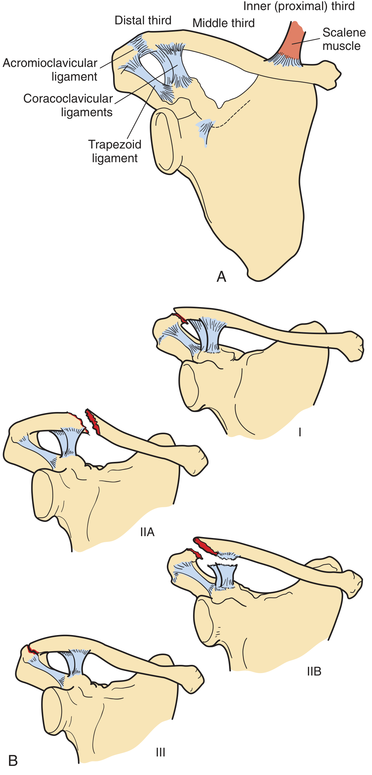
| Type | Description | Ligament Status | Clinical Significance |
|---|---|---|---|
| Type I | Minimal displacement, fracture between the CC ligaments | Both CC ligaments intact | Stable; treat conservatively |
| Type IIA | Proximal shaft displaced superiorly; fracture medial to both CC ligaments | Both conoid and trapezoid attached to distal fragment | Unstable; often needs surgery |
| Type IIB | Fracture between conoid and trapezoid; conoid ruptured | Trapezoid intact, conoid torn | Unstable; high nonunion risk |
| Type III | Fracture of articular surface (into AC joint) | No ligament disruption | Intra-articular; may lead to OA |
| Type IV | Ligament intact, periosteal sleeve fracture (in children) | Ligaments attached to periosteum | Pediatric; usually heals well |
| Type V | Comminuted; CC ligaments attached to inferior comminuted fragment | Ligaments on free fragment | Highly unstable |
3. Robinson Classification (1998) - Prognostic / Edinburgh System
Based on a prospective study of >1,000 patients - preferred by Rockwood & Green because it predicts outcomes and guides treatment. Uses a different numbering convention (confusingly, medial = Type 1, middle = Type 2, lateral = Type 3).
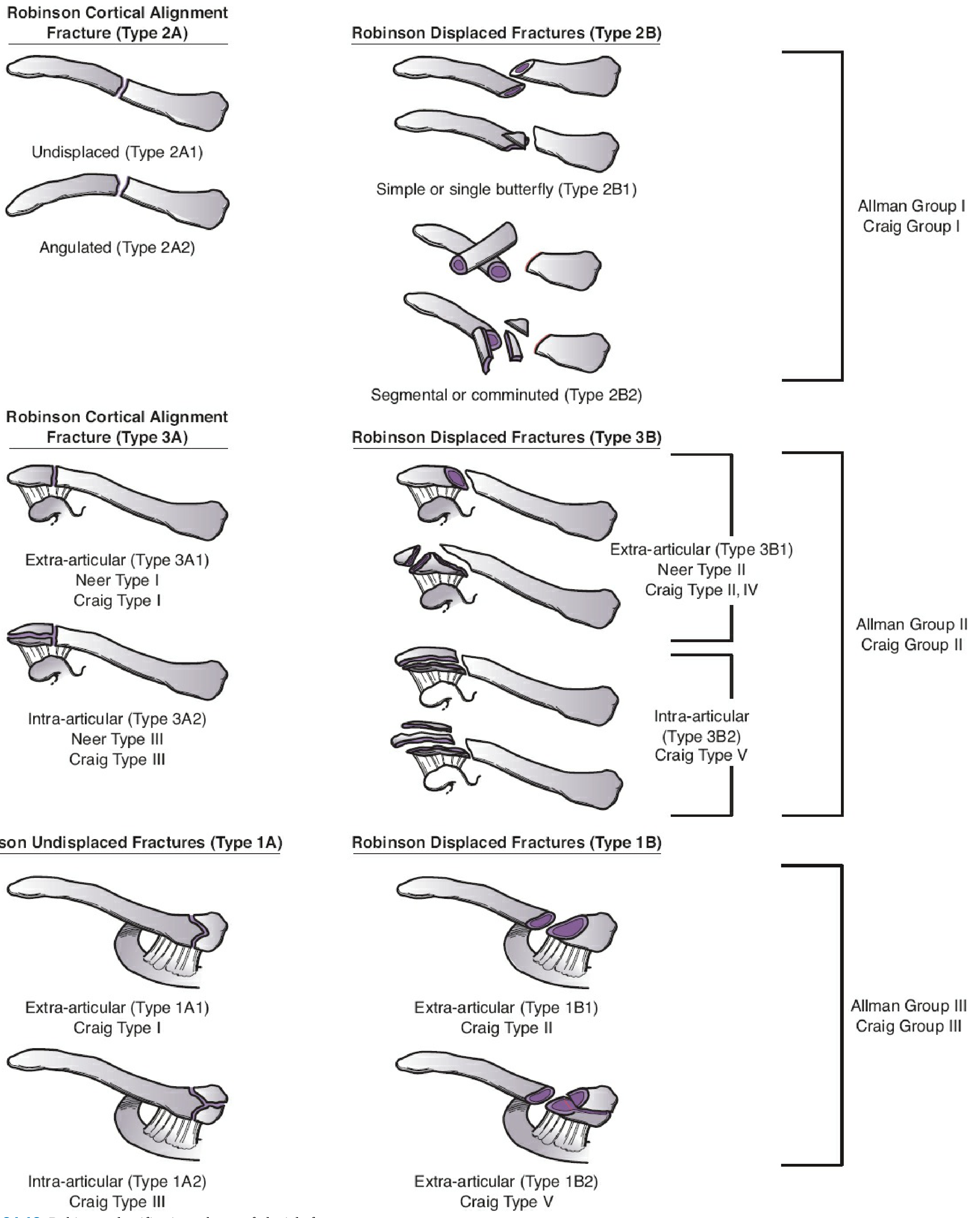
| Robinson Type | Location | Subtype A (Cortical Alignment = Undisplaced) | Subtype B (Displaced) |
|---|---|---|---|
| Type 1 | Medial (proximal) third | 1A1 = extra-articular; 1A2 = intra-articular | 1B1 = extra-articular displaced; 1B2 = intra-articular displaced |
| Type 2 | Middle third (diaphysis) | 2A1 = undisplaced; 2A2 = angulated | 2B1 = simple/single butterfly; 2B2 = segmental/comminuted |
| Type 3 | Distal (lateral) third | 3A1 = extra-articular (= Neer I, Craig I); 3A2 = intra-articular (= Neer III, Craig III) | 3B1 = extra-articular displaced (= Neer II, Craig II/IV); 3B2 = intra-articular displaced (= Craig V) |
Key prognostic points from Robinson:
- 2B2 (comminuted middle-third) = highest risk of nonunion and malunion
- 3B1 (displaced distal = Neer Type II) = highest nonunion risk in lateral fractures (~25%)
- Displacement >2 cm and comminution are the strongest predictors of poor outcome
4. OTA/AO Classification (Updated 2018)
The most detailed, research-oriented system. Clavicle = segment 15.
| AO Segment | Location | Types |
|---|---|---|
| 15.1 | Proximal (medial) metaphysis | A = extra-articular / epiphyseal; B = partial articular; C = complete articular |
| 15.2 | Diaphysis (shaft) | A = simple (15.2-B1); B = wedge (15.2-B2); C = complex/comminuted (15.2-B3) |
| 15.3 | Distal (lateral) metaphysis | A = extra-articular; B = partial articular; C = complete articular |
Note: AO metaphyseal segments are shorter than one-third of the bone length (uses the "AO rule of squares" - the segment defined by a square whose side = widest dimension of the metaphysis).
Cross-Reference Summary Table
| Region | Allman | Craig | Robinson | AO |
|---|---|---|---|---|
| Middle third | Group I | Group I | Type 2 | 15.2 |
| Distal third | Group II | Group II (Types I-V) | Type 3 | 15.3 |
| Proximal third | Group III | Group III (Types I-III) | Type 1 | 15.1 |
Key Clinical Points by Location
Middle Third (Group I / Robinson Type 2):
- Usually treated non-operatively (sling or figure-of-eight splint)
- Operative indications: open fracture, neurovascular injury, displacement >2 cm, shortening >1.5-2 cm, floating shoulder, polytrauma
- Nonunion rate: 1-3% (higher with displacement/comminution)
- Union expected: 6-8 weeks
Distal Third (Group II / Neer-Craig):
- Type I and III - conservative (ligaments intact or intra-articular without instability)
- Type II - often operative due to ~25% nonunion risk (proximal fragment displaced upward by sternocleidomastoid; distal fragment held down by weight of arm)
- Methods: hook plate, CC screw, suture button, open reduction + ligament repair
Proximal Third (Group III):
- Rare; may mimic sternoclavicular dislocation clinically
- Plain radiographs unreliable - CT is mandatory to define fracture type and posterior displacement
- Posterior displacement can threaten mediastinal structures (great vessels, trachea, esophagus) - requires urgent evaluation
Sources: Rockwood and Green's Fractures in Adults 10th Ed 2025 | Pfenninger & Fowler's Procedures for Primary Care 3rd Ed | Miller's Review of Orthopaedics 9th Ed | Campbell's Operative Orthopaedics 15th Ed 2026
Bone healing types
Bone healing occurs via two main types: indirect (secondary) healing through callus formation, and direct (primary) healing without callus when fragments are rigidly compressed. A third type - distraction osteogenesis - is sometimes considered separately.
Searching sources...
Finding Sources
Finding Sources
Reading File
Reading File
Reading File
Finding Sources
Reading File
Reading File
Loading Image
Loading Image
Here is the full, illustrated breakdown of bone healing types from authoritative orthopaedic and histology textbooks:
Bone Healing Types
Bone heals in two fundamental ways: direct (primary) and indirect (secondary). The type that occurs depends on the mechanical environment - primarily the degree of stability and compression at the fracture site.
TYPE 1: Indirect (Secondary) Bone Healing - with Callus
The most common type of fracture healing in clinical practice.
Occurs when there is relative stability with some movement (strain) at the fracture site. The body bridges the gap biologically through a sequence of tissue differentiation.
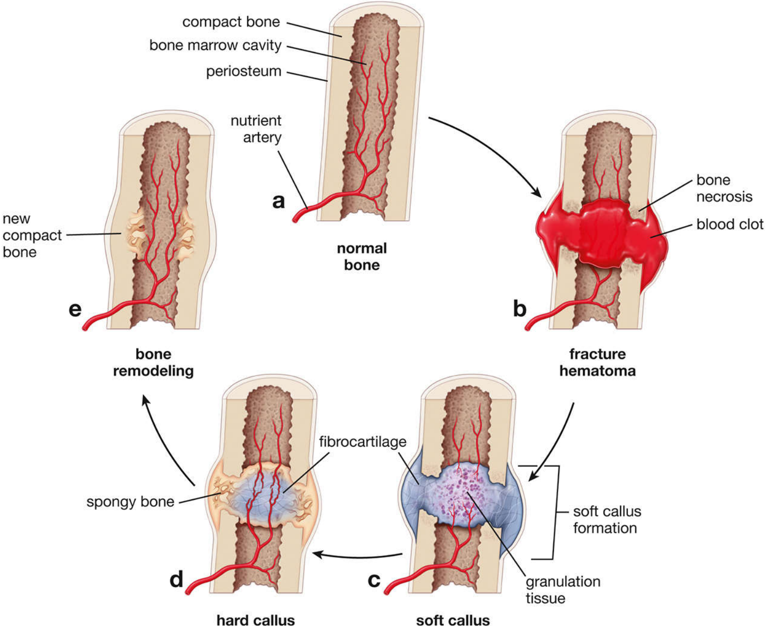
Phases of Indirect Bone Healing
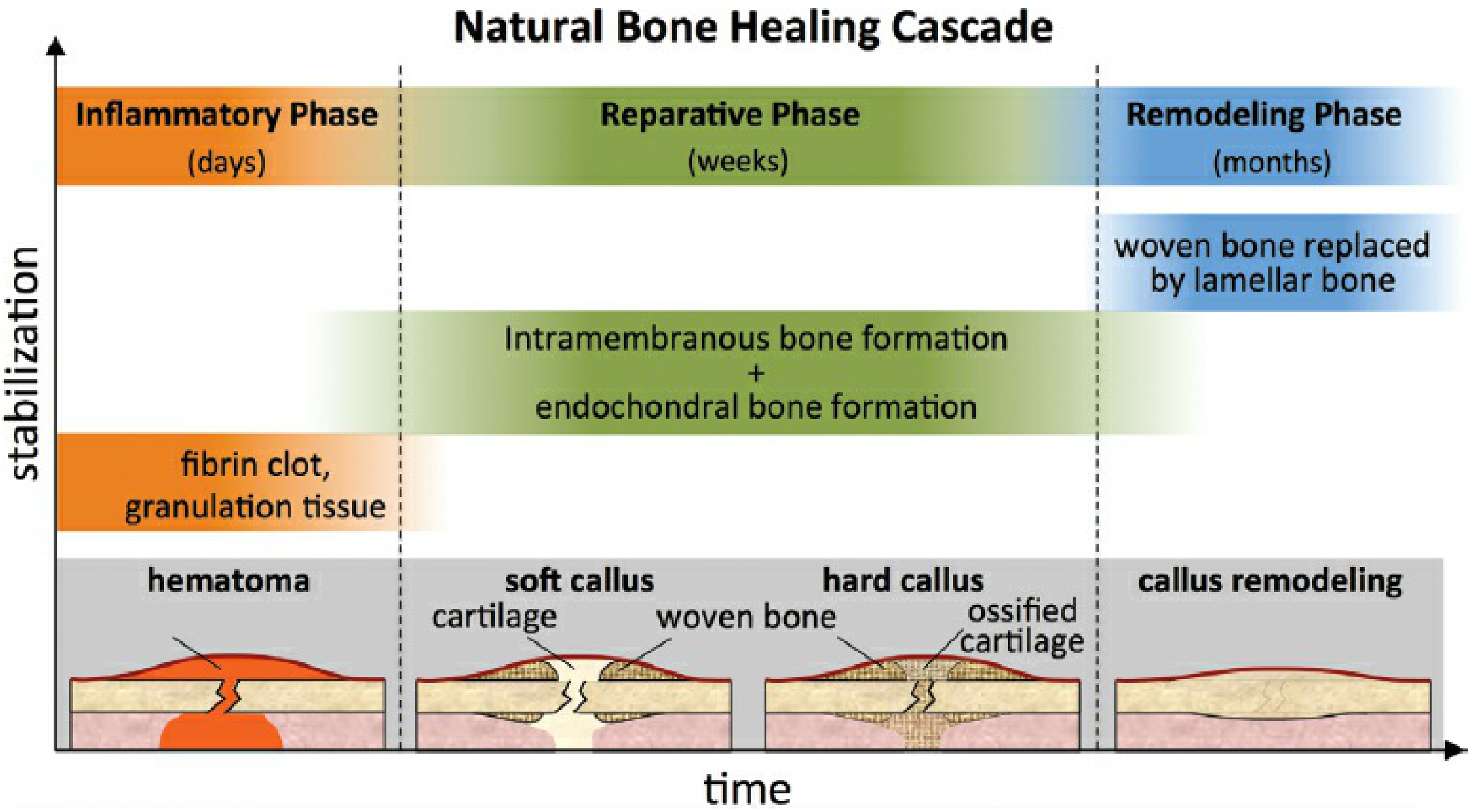
Phase 1 - Inflammatory Phase (Days)
- Fracture causes haematoma - blood fills the fracture gap and forms a fibrin clot
- Bone necrosis occurs at the cut ends of fragments
- Platelets degranulate; cytokines released: TNF-α, IL-1, IL-6, IL-11, IL-18
- Neutrophils infiltrate → followed by macrophages
- Fibroblasts and capillaries proliferate → granulation tissue replaces haematoma (contains collagen type III and II)
- Mesenchymal stem cells arrive from periosteum, endosteum, and bone marrow
- Tissue at this stage tolerates up to 100% strain (maximum deformability)
Phase 2 - Reparative Phase (Weeks)
Two simultaneous ossification processes occur:
a) Soft Callus formation (endochondral ossification)
- Periosteal cells differentiate into chondroblasts → lay down fibrocartilage matrix
- Fibrocartilage soft callus forms at the fracture site - binds and stabilizes the ends
- Soft callus is flexible; tolerates 10-12% strain; tensile strength 4-19 N/mm²
- The extramedullary callus contributes far more to stability than intramedullary callus (bending stiffness increases with the 4th power of distance from the neutral axis)
b) Hard Callus formation (intramembranous + endochondral)
- Osteoprogenitor cells from periosteum differentiate into osteoblasts → deposit woven bone on the outer surface (intramembranous)
- Simultaneously, the fibrocartilage calcifies and undergoes endochondral ossification → replaced by woven bone
- Hard callus: tolerates only 2% strain; tensile strength ~130 N/mm²
- Hard callus formation marks the end of the healing process and the beginning of the remodeling phase
Phase 3 - Remodeling Phase (Months)
- Woven bone is remodeled into lamellar (mature) bone by osteoclast/osteoblast units
- Callus volume decreases (visible on X-ray as callus index falls)
- Medullary canal is restored; cortical architecture is re-established
- Bone remodels along lines of mechanical stress (Wolff's law)
- Can continue for months to years until bone returns to original shape
Perren's Strain Theory (Key Concept)
The strain at the fracture site determines what tissue forms:
| Strain at Fracture Site | Tissue Formed |
|---|---|
| >100% | No tissue (gap too unstable - non-union) |
| 10-100% | Fibrous tissue |
| 2-10% | Fibrocartilage (soft callus) |
| <2% | Bone (hard callus / direct healing) |
"A little movement is good; too much movement is bad." - Bailey & Love
TYPE 2: Direct (Primary) Bone Healing - without Callus
Occurs only when there is absolute stability with cortical apposition and NO movement (or gap) between fragments. This is what compression plating (e.g., dynamic compression plate - DCP) aims to achieve.
Two subtypes of primary healing:
a) Contact Healing
- Fracture surfaces are in direct contact with < 0.01 mm gap
- Osteoclastic cutting cones tunnel directly across the fracture line
- Following osteoblasts lay down lamellar bone within the tunnel (osteons cross the fracture line)
- Bone is remodeled directly without an intermediate callus stage
- Requires compression forces and rigid fixation
b) Gap Healing
- Small gaps exist (0.01-1 mm) between fragments, too large for contact healing
- Gap is first filled with woven bone by direct osteoblastic deposition (intramembranous-like process)
- The woven bone is subsequently remodeled into lamellar bone by Haversian remodeling
- Occurs in areas where cutting cones have not yet bridged the gap
How Primary Healing Is Achieved Clinically
- Absolute compression at the fracture interface (e.g., lag screw, compression plate)
- No callus appears on post-op X-ray - this is normal and expected
- Rigid internal fixation that fails to achieve bone-to-bone contact (leaving a gap without compression) does not support primary healing
Comparison Summary
| Feature | Indirect (Secondary) | Direct (Primary) |
|---|---|---|
| Also called | Callus healing, natural healing | Contact healing |
| Mechanical environment | Relative stability (some movement) | Absolute stability (no movement) |
| Callus | Yes - visible on X-ray | No callus |
| Tissue sequence | Haematoma → fibrous → fibrocartilage → woven bone → lamellar bone | Direct osteonal remodeling across fracture |
| Ossification type | Endochondral + intramembranous | Intramembranous (direct) |
| Speed | Faster overall | Slower, but immediate stability |
| Clinical example | Cast immobilization, IM nail, external fixator | Compression plate (DCP), lag screw |
| Failure risk | Non-union if too much movement | Implant failure if fixation fails |
Failure of Healing: Terminology
| Term | Definition |
|---|---|
| Delayed union | Slow to heal; not yet healed in expected time frame |
| Non-union | Fracture has not healed and shows no potential to heal without further intervention; fracture failing to progress over 3 months; formally diagnosed at 6 months |
| Atrophic non-union | Biological problem - poor blood supply, lack of stimulus; no callus |
| Hypertrophic non-union | Mechanical problem - too much movement; abundant callus but no bridging ("elephant foot") |
| Consolidation | Follows union; bone has returned to normal strength; cortical pattern restored on X-ray |
| Remodelling | Bone reshaping based on mechanical forces; more complete in children |
Sources: Bailey and Love's Short Practice of Surgery 28th Ed | Rockwood and Green's Fractures in Adults 10th Ed 2025 | Histology: A Text and Atlas (Pawlina) | Cummings Otolaryngology Head and Neck Surgery
This is a shared conversation. Sign in to Orris to start your own chat.