
What are the differential diagnosis of bosellated enteric selwelling with white mucinous content intra abdominal
bosselated intra-abdominal swelling white mucinous content differential diagnosis
Differential Diagnosis: Bosselated Intra-Abdominal Swelling with White Mucinous Content
1. Mucocele of the Appendix (Appendiceal Mucinous Neoplasm) - TOP DIAGNOSIS
| Type | Features |
|---|---|
| Simple retention cyst | Non-neoplastic, <2 cm, incidental |
| Low-grade appendiceal mucinous neoplasm (LAMN) | Most common "mucocele"; slow-growing, can reach 8 cm+; bosselated wall with mucin |
| High-grade appendiceal mucinous neoplasm (HAMN) | Aggressive; high risk of rupture and peritoneal spread |
| Mucinous adenocarcinoma | Symptomatic; white mucin in peritoneum; high malignant potential |
- Wall calcification is characteristic on CT/plain X-ray
- If ruptured: leads to Pseudomyxoma Peritonei (see below)
- Treatment: open appendectomy (laparoscopic not recommended due to risk of spillage)
- Maingot's Abdominal Operations, p. 2863-2876
- Sabiston Textbook of Surgery, p. 1195-1197
2. Pseudomyxoma Peritonei (PMP)
- Gelatinous/mucinous ascites ("jelly belly")
- Cystic epithelial implants on peritoneal surfaces
- Bosselated/lobulated masses throughout the abdomen and pelvis
- Adhesions and omental involvement
- Most commonly arises from a ruptured LAMN of the appendix
- Acellular mucin
- Low-grade mucinous carcinoma peritonei (disseminated peritoneal adenomucinosis - DPAM)
- High-grade mucinous carcinoma peritonei (peritoneal mucinous carcinomatosis - PMCA)
- High-grade with signet ring cells (worst prognosis)
- Bailey and Love's Short Practice of Surgery 28th Ed., Table 76.4
- Berek & Novak's Gynecology, p. 2312
- Robbins, Cotran & Kumar Pathologic Basis of Disease, p. 3271
3. Ovarian Mucinous Cystadenoma / Cystadenocarcinoma
- Benign mucinous tumors of the ovary: classically lobulated (bosselated), smooth surface, multilocular, with mucoid/gelatinous material in each locule
- Can grow to enormous size (10-30 cm), filling the entire abdomen
- Bilateral in up to 10% of cases
- Malignant transformation in 5-10% (mucinous cystadenocarcinoma)
- Histologically may be indistinguishable from metastatic GI carcinoma - immunohistochemistry needed
- Associated with pseudomyxoma peritonei if primary GI origin is present
- Berek & Novak's Gynecology, p. 454-455
4. Pancreatic Mucinous Cystic Neoplasm (MCN) / IPMN
- MCN: Almost exclusively in women; unilocular or oligolocular cystic mass in the body/tail of the pancreas; lobulated surface; thick mucin inside
- Intraductal Papillary Mucinous Neoplasm (IPMN): Arises in the pancreatic ducts; forms macrocystic dilated mucin-filled ducts; can present as bosselated upper abdominal mass
- Both carry malignant potential and require resection
- Sabiston Textbook of Surgery (IPMN section)
5. Mucinous Colorectal Carcinoma
- Colorectal adenocarcinomas with mucinous histology (>50% extracellular mucin by definition)
- Can form large bosselated intra-abdominal masses
- Often present with peritoneal seeding and white gelatinous deposits on the peritoneum
- Associated with KRAS mutations and microsatellite instability
6. Intra-Abdominal Multicystic Lymphangioma (Chylous/Mucinous variant)
- Rare benign congenital anomaly of lymphatic channels
- Bosselated, multilocular cystic masses, typically in the mesentery or retroperitoneum
- Content is usually chylous (milky-white), but can mimic mucin
- Positive for D2-40, CD31 on immunohistochemistry
7. Mesenteric / Omental Cysts
- Derived from mesothelial or enteric tissue
- Can be multilocular and bosselated
- Content is usually clear serous or chylous fluid, but mucinous variant (enteric duplication cysts) contains mucin-secreting epithelium
- Enteric duplication cysts are lined by GI-type epithelium that actively secretes mucin
8. Retroperitoneal Mucinous Tumors (Primary Retroperitoneal Mucinous Tumors)
- Rare; arise from ectopic Mullerian tissue or enteric remnants
- Presents as large retroperitoneal or intra-abdominal cystic mass with mucinous content
- Usually in women
Summary Table
| Condition | Location | Surface | Content | Malignant risk |
|---|---|---|---|---|
| Appendiceal mucocele (LAMN/HAMN) | RLQ, appendix | Bosselated, calcified | White/gelatinous mucin | Low-High |
| Pseudomyxoma peritonei | Diffuse peritoneum | Multiple lobulated implants | Gelatinous mucin | Moderate-High |
| Ovarian mucinous cystadenoma | Pelvis/abdomen | Lobulated, multilocular | Mucoid in locules | Low (5-10%) |
| Ovarian mucinous cystadenocarcinoma | Pelvis/abdomen | Irregular, bosselated | Mucin + solid areas | High |
| Pancreatic MCN/IPMN | Upper abdomen | Lobulated cysts | Thick mucin | Moderate-High |
| Mucinous colorectal carcinoma | Colon + peritoneum | Irregular mass | Extracellular mucin pools | High |
| Multicystic lymphangioma | Mesentery | Lobulated cysts | Chylous/serous | Very low |
| Enteric duplication cyst | Mesentery/bowel | Smooth or lobulated | Mucin (GI-type) | Low |
| Retroperitoneal mucinous tumor | Retroperitoneum | Cystic, lobulated | Mucinous | Variable |
Clinical Approach
- CT abdomen/pelvis with IV contrast: First-line imaging; look for calcified appendiceal wall, "scalloping" of liver and spleen (classic PMP finding), organ of origin
- Serum markers: CEA, CA-125, CA 19-9 (elevated in mucinous tumors)
- Colonoscopy: Mandatory to exclude colorectal primary
- Tumor markers + IHC on biopsy: CK7/CK20, CDX2, PAX8 to distinguish ovarian vs. GI origin
- Laparoscopy/laparotomy: Definitive diagnosis and treatment at specialized center for PMP/CRS-HIPEC
Mesenteric cyst Marks answer
Mesenteric Cyst - Complete Marks Answer
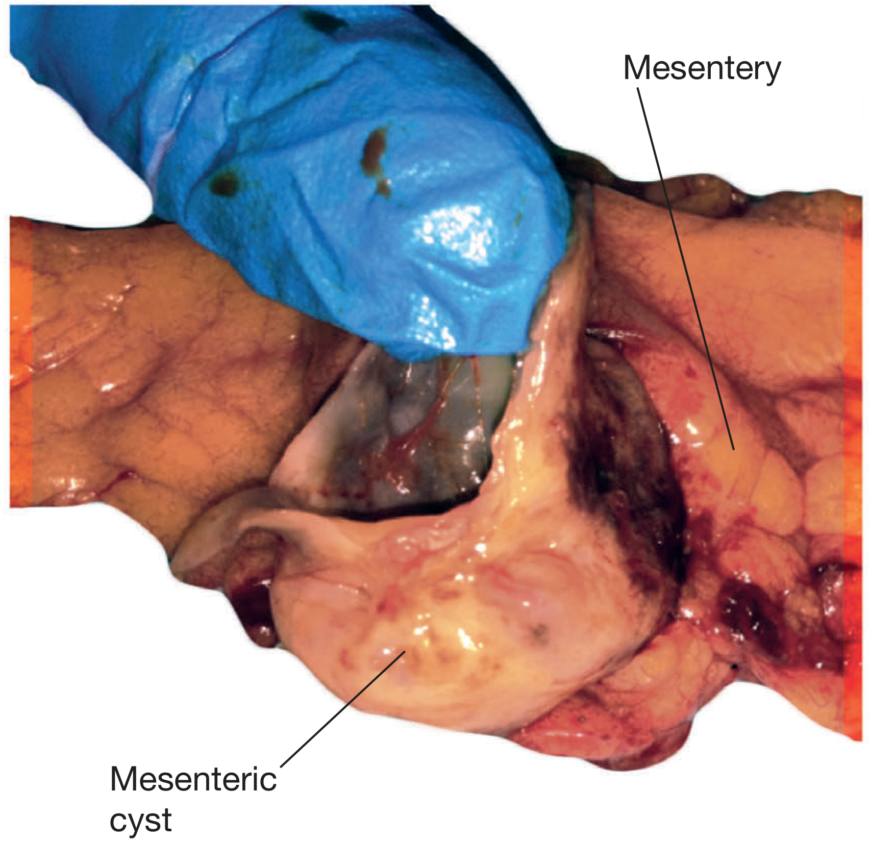
Definition
Incidence / Epidemiology
- Incidence: 1 in 27,000 to 1 in 250,000 hospital admissions
- Twice as common in women as in men
- Mean age: 45 years in adults
- ~1/3 of all cases occur in children < 15 years (mean age 5 years)
Classification (Bailey & Love)
| Type | Origin | Content |
|---|---|---|
| Chylolymphatic | Congenitally misplaced lymphatic tissue (most common) | Clear lymph or milky chyle |
| Enterogenous | Mesenteric border diverticulum / intestinal duplication | Mucinous - colourless or yellowish-brown |
| Traumatic | Post-traumatic disruption of lymphatics | Serous/haemorrhagic |
| Hydatid | Echinococcal infection | Hydatid fluid |
Most occur in small bowel mesentery (60%), rest in colonic mesentery (40%).
Pathology
- Chylolymphatic cyst: Thin wall of connective tissue lined by flat endothelium; filled with clear lymph or chyle. Usually unilocular, blood supply independent of bowel - so enucleation is possible without bowel resection.
- Enterogenous cyst (enteric duplication cyst): Thicker wall lined by mucous membrane (sometimes ciliated); content is mucinous. Shares blood supply with adjacent bowel - so bowel resection is always required.
- Usually lined by a single layer of columnar epithelial cells
- Most are unilocular; some are multiloculated
- Malignant in only 3% of cases (mostly sarcomatous)
Clinical Features
Symptoms
- Up to 45% found incidentally
- Most common symptom: abdominal pain (58%)
- Chronic: non-specific pain, nausea, vomiting, anorexia, altered bowel habits, abdominal distension
- Acute: pain from rupture, haemorrhage, torsion/volvulus of bowel, or infection
Signs
- Abdominal mass in 60% of patients
- Most common presentation: painless fluctuant swelling near the umbilicus
- Classic sign: Tillaux's sign - mass is mobile only in the lateral direction (perpendicular to root of mesentery), NOT superoinferiorly
- Distinguishes it from omental cysts (freely mobile in all directions)
- Mass is transilluminant (chylolymphatic type)
- Mass can be large (average ~13 cm, fluid ~2000 mL)
Investigations
| Investigation | Findings |
|---|---|
| Ultrasound | Thin-walled, anechoic/hypoechoic; internal septations; compresses adjacent bowel. Echogenic if chylous, infected, or haemorrhagic |
| CT abdomen | Fluid-filled homogenous cystic mass in mesentery; no solid components; water-to-fat attenuation; large mesenteric lymphangioma shows multiple cysts |
| MRI | High signal on T2W; enhancement of wall/septa post-contrast |
- CT/USS differentiates from solid tumours; solid components = higher malignancy risk
- Large mesenteric lymphangiomas differentiated from ascites by presence of septa, compression of bowel loops, and no fluid in dependent peritoneal recesses
Complications
- Rupture
- Haemorrhage into cyst
- Torsion of adjacent bowel loop / volvulus
- Infection / abscess
- Intestinal obstruction
- Malignant change (3%)
Treatment
- Small, incidental, asymptomatic cyst at laparotomy: no resection required
- Definitive treatment: complete surgical excision (enucleation)
- Can be open or laparoscopic
- Marsupialization and aspiration: high recurrence - generally discouraged
- Chylolymphatic cyst: enucleation alone - independent blood supply, bowel preservation possible
- Enterogenous (enteric duplication) cyst: excision always requires resection of adjacent bowel and primary anastomosis - shared mesenteric blood supply
- Malignant cysts: wide resection with clear margins + adjacent viscera if needed
Differential Diagnosis of Mesenteric Cyst
- Omental cyst (freely mobile in all directions)
- Ovarian cyst (in females - bimanual exam, USS)
- Retroperitoneal cyst / lymphangioma
- Hydronephrosis
- Pancreatic pseudocyst
- Choledochal cyst
- Enteric duplication cyst (part of the spectrum)
- Ascites (no septa, fluid in dependent recesses)
Key Exam Points
- Tillaux's sign = lateral mobility only (mesenteric attachment limits superoinferior movement)
- Most common type = chylolymphatic (lymphangioma)
- Mucinous content = enterogenous / enteric duplication cyst
- Enterogenous cyst requires bowel resection; chylolymphatic does not
- Malignancy in 3% (sarcoma)
- Treatment = complete excision; marsupialization leads to recurrence
Sources: Bailey and Love's Short Practice of Surgery 28th Ed., p. 4878-4925; Schwartz's Principles of Surgery 11th Ed., p. 1234-1249; Sleisenger & Fordtran's GI & Liver Disease, p. 3425-3427; Grainger & Allison's Diagnostic Radiology, p. 3908-3913
What are the sarcoma tumors of GIT other than GIST
Sarcomas of the GIT (Other Than GIST)
Classification of Non-GIST GIT Sarcomas
1. Leiomyosarcoma (LMS) - Most Important Non-GIST GIT Sarcoma
- Rectum (most common in the large intestine)
- Small intestine
- Stomach
- Colon
- Rare in GIT; when it occurs in the large intestine, rectum is the most common site
- Usually low grade in the rectum - difficult to distinguish from benign leiomyoma histologically
- Definitive diagnosis usually made after resection
- Symptoms: GI bleeding or obstruction
- Lesions >5 cm have high risk of malignancy and require radical resection
- Recurrence is common even after complete resection; prognosis is generally poor
- Imatinib-resistant (no c-KIT mutation; CD117 negative - key distinction from GIST)
- Schwartz's Principles of Surgery 11th Ed., p. 2164-2166 and 2234
- Sleisenger & Fordtran's GI & Liver Disease
2. Kaposi Sarcoma (KS)
- AIDS-related KS - most common GI form
- Classical (elderly Mediterranean men)
- Endemic (African)
- Iatrogenic (post-transplant, immunosuppression)
- Occurs in 40% of patients with AIDS-related KS
- Usually asymptomatic despite GI involvement
- Lesions appear as violaceous/reddish-purple submucosal nodules on endoscopy
- Complications (uncommon): GI bleeding, bowel perforation, obstruction
- Skin involvement almost always present concurrently
- Symptom to Diagnosis 4th Ed., p. 9266-9278
3. Angiosarcoma
- Extremely rare in GIT
- Most commonly involves small bowel and colon
- Presents with GI bleeding (often occult, leading to iron-deficiency anaemia), abdominal pain, mass
- Highly aggressive; poor prognosis
- Associated with prior radiation, vinyl chloride, thorotrast exposure, chronic lymphedema
4. Liposarcoma
- Very rare as a primary GIT tumor; more commonly a retroperitoneal tumor invading into the bowel
- Subtypes: well-differentiated, myxoid, pleomorphic (worst prognosis), dedifferentiated
- Presents as a large, bosselated intra-abdominal mass
- CT shows fat-density lesion with soft-tissue components
- Schwartz's Principles of Surgery 11th Ed.
5. Clear Cell Sarcoma of the GIT (Gastrointestinal Clear Cell Sarcoma / Malignant Gastrointestinal Neuroectodermal Tumor - GNET)
- Rare; distinct from GIST and melanoma
- Most common in small intestine (ileum) and stomach
- Presents in young adults
- Highly aggressive; poor prognosis
- Histology: nests of clear or pale cells; may show melanocytic differentiation
- Henry's Clinical Diagnosis and Management by Laboratory Methods
6. Inflammatory Myofibroblastic Tumor (IMT)
- Most common in lung; GIT involvement (mesentery, omentum, bowel wall) described
- Associated with ALK gene rearrangement (~50% of cases)
- Presents as a mass ± fever, anaemia, thrombocytosis (paraneoplastic)
- Low malignant potential but can recur locally; rare distant metastasis
7. Fibrosarcoma / Undifferentiated Pleomorphic Sarcoma (UPS / Malignant Fibrous Histiocytoma)
- Rare in GIT
- High-grade spindle cell sarcomas with complex karyotype
- Arise in mesentery or bowel wall
- Diagnosis by exclusion (after ruling out GIST, LMS, liposarcoma)
- Treatment: wide resection ± adjuvant chemotherapy (doxorubicin + ifosfamide)
8. Rhabdomyosarcoma
- Extremely rare in GIT in adults; more common in children (biliary tract, retroperitoneum)
- Embryonal and alveolar subtypes
- Highly aggressive
- Treatment: multimodal (surgery + chemotherapy + radiation)
Summary Table
| Sarcoma | Origin | Common GIT Site | Key Marker | Distinguishing Feature |
|---|---|---|---|---|
| Leiomyosarcoma | Smooth muscle | Rectum > small bowel | SMA+, Desmin+, CD117- | Most common non-GIST GIT sarcoma |
| Kaposi sarcoma | Vascular endothelium | Diffuse (40% in AIDS) | HHV-8, CD31+ | Violaceous lesions; AIDS-related |
| Angiosarcoma | Vascular endothelium | Small bowel, colon | CD31+, CD34+, ERG+ | GI bleeding; very aggressive |
| Liposarcoma | Adipocytes | Retroperitoneum/mesentery | MDM2+, CDK4+ | Fat-density on CT |
| Clear cell sarcoma of GIT | Neural crest | Ileum, stomach | EWSR1-CREB1, S100+, ALK- | Young adults; distinct from melanoma |
| IMT | Myofibroblasts | Mesentery, omentum | ALK+, SMA+ | Paraneoplastic fever, anemia |
| Fibrosarcoma/UPS | Fibroblasts | Mesentery/bowel | Diagnosis of exclusion | Complex karyotype |
| Rhabdomyosarcoma | Skeletal muscle | Biliary (pediatric) | MyoD1+, Myogenin+ | Rare; mainly children |
General Principles for ALL GIT Sarcomas (Non-GIST)
- Lymphatic spread is NOT the primary route - lymphadenectomy is not routinely performed
- Primary treatment = surgical resection with 2-4 cm clear margins
- Segmental bowel resection en bloc with mesentery for small/large intestinal sarcomas
- Imatinib does NOT work (these lack c-KIT/PDGFRA mutations)
- Chemotherapy: doxorubicin + ifosfamide (first-line for most high-grade sarcomas)
- No regional lymph node metastases on CT = typical of sarcoma (vs. adenocarcinoma which shows nodal spread)
Sources: Schwartz's Principles of Surgery 11th Ed., p. 2224-2236; Sleisenger & Fordtran's GI & Liver Disease; Yamada's Textbook of Gastroenterology 7th Ed.; Henry's Clinical Diagnosis by Laboratory Methods
Give me in detail about total mesorectal excision procedure.what is it,etymology, history,indications, contraindications,procedure
Total Mesorectal Excision (TME) - Complete Answer
1. Etymology
2. Definition
- The fascia propria (investing fascia of the mesorectum / mesorectal fascia, MRF) - innermost
- The presacral (parietal) fascia - outermost
3. Historical Background
| Year | Milestone |
|---|---|
| 1908 | Ernest Miles described abdominoperineal resection (APR) with wide perineal excision and removal of "highly dangerous tissues" (what we now call the mesorectum) |
| 1970s | Prof. Richard J. "Bill" Heald at Basingstoke Hospital, UK, began developing the concept of sharp mesorectal dissection in an anatomically defined plane |
| 1982 | Heald published his landmark paper describing TME with dramatically reduced local recurrence rates (~3-5% vs. the then-prevailing 30-40%) |
| 1993 | Heald published long-term results in The Lancet confirming survival and recurrence benefits; technique gained international attention |
| Late 1990s | Introduction of neoadjuvant chemoradiotherapy (CRT) combined with TME - further reducing local recurrence |
| 2000s | High-resolution thin-slice MRI of the pelvis (Brown, Blomquist et al.) enabled pre-operative visualization of the mesorectal fascia, enabling surgical planning |
| 2006-2010 | Laparoscopic TME validated in multiple RCTs (COLOR II, COREAN, ACOSOG Z6051) |
| 2010s | Robotic TME and Transanal TME (taTME) introduced for difficult pelvises |
Prof. Heald described the holy plane as "an embryologic redefinition of Miles' highly dangerous tissues" - recognizing that Miles was, intuitively, operating in the correct oncologic territory but without the anatomical precision of TME.
4. Relevant Anatomy
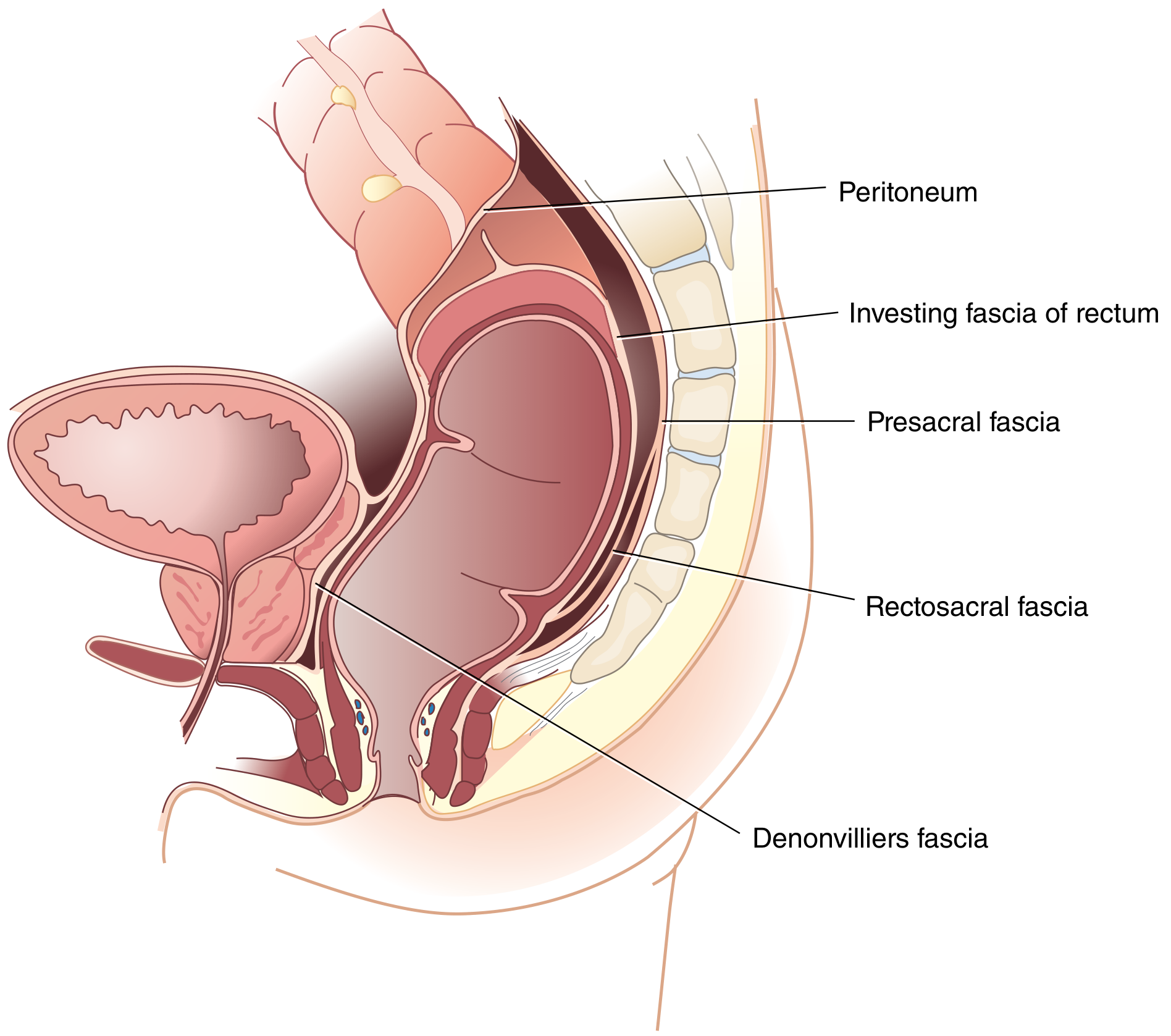
Key Fascial Layers
| Structure | Location | Significance |
|---|---|---|
| Fascia propria (mesorectal fascia, MRF) | Invests mesorectum circumferentially | Inner limit of TME dissection; contains lymphatics and vessels |
| Presacral fascia (Waldeyer's fascia superiorly) | Covers anterior sacrum and coccyx | Outer limit of TME dissection; contains presacral venous plexus deep to it |
| Rectosacral (Waldeyer's) fascia | Condensation at S4 connecting MRF to presacral fascia | Must be divided during posterior dissection to access deep pelvis |
| Denonvilliers' fascia | Between rectum and prostate/seminal vesicles (males); between rectum and vagina (females) | Critical anterior plane; taken with specimen in anterior cancers |
| Lateral stalks / ligaments | Connect rectum to pelvic sidewall | Contain middle rectal vessels in 25% of patients; pelvic autonomic nerves lie lateral to these |
The "Holy Plane"
- Embryologically defined (interface between midline mesodermal tube and paired genitourinary structures)
- Continuous circumferentially around the mesorectum
- Clearly visible under direct vision as a bloodless white areolar space
Structures at Risk
- Posteriorly: Presacral venous plexus (catastrophic haemorrhage if entered)
- Posterolaterally: Hypogastric nerves (at sacral promontory) - bladder/sexual function
- Laterally: Pelvic plexus / inferior hypogastric plexus - erectile and ejaculatory function
- Anteriorly (males): Seminal vesicles, prostate, urethra
- Anteriorly (females): Vaginal wall, cervix
- Throughout: Ureters
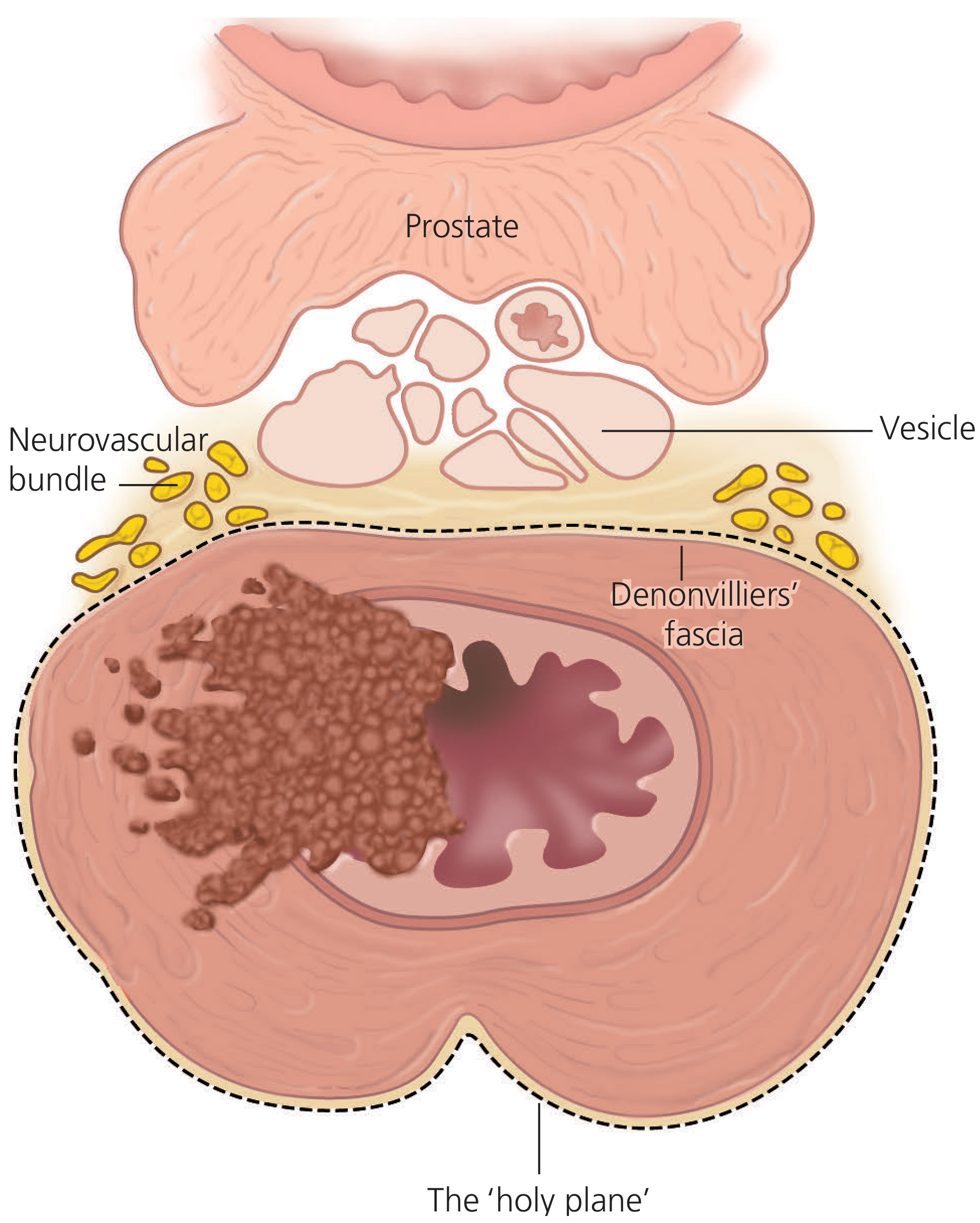
5. Oncological Rationale
- Rectal cancers spread via intramural, lymphatic, and perirectal mesorectal routes
- Mesorectal lymph nodes involved in up to 50% of rectal cancers penetrating the muscularis propria
- Tumor deposits in mesorectum rarely extend beyond 4-5 cm distal to the lower tumor edge
- Leaving mesorectal tissue behind creates a positive circumferential resection margin (CRM) - the single most important predictor of local recurrence
- A positive CRM (tumor ≤1 mm from cut mesorectal surface) is associated with local recurrence rates of 20-30%
- TME reduces local recurrence from 30-40% to 3-8%
6. Types of TME
| Type | Application | Mesorectal Extent |
|---|---|---|
| Complete TME | Mid and lower rectal cancers (≤10 cm from anal verge) | Entire mesorectum removed to pelvic floor |
| Tumor-Specific TME (TSME) | Upper rectal cancers (>10 cm from anal verge) | Mesorectum divided 5 cm distal to lower tumor edge (perpendicular - no coning) |
7. Indications
Primary Indications
- Carcinoma of the rectum - mid and lower third (primary indication)
- Upper rectal carcinoma - as TSME
- Rectal carcinoid / neuroendocrine tumor - large or invasive
- Locally invasive benign rectal tumors - large villous adenomas not amenable to local excision
Can be Used as Part of
- Low anterior resection (LAR) - TME + colorectal/coloanal anastomosis
- Abdominoperineal resection (APR) - TME + perineal dissection (very low cancers invading levator/external sphincter)
- Hartmann's procedure - TME + end colostomy (frail/elderly patients)
Neoadjuvant Therapy Prior to TME
- Threatened or involved CRM (<1-2 mm from MRF)
- T3/T4 tumors
- Node-positive disease
- Extramural vascular invasion (EMVI)
8. Contraindications
Absolute Contraindications
- Very distal rectal cancer invading the external anal sphincter and/or levator ani with inability to obtain negative CRM - requires APR, not sphincter-preserving TME
- Patient unfit for major surgery / high ASA grade (consider Hartmann's or defunctioning stoma only)
Relative Contraindications
- Metastatic disease - consider palliative intent (still may proceed for local control)
- Previous pelvic radiation with fibrosis obliterating planes
- Inflammatory or adhesive obliteration of the mesorectal plane
- Circumferential T4 tumor with fixation to all pelvic sidewalls
- Poor sphincter function pre-operatively (TME + anastomosis risks LARS; APR preferred)
9. Pre-operative Workup
| Investigation | Purpose |
|---|---|
| High-resolution pelvic MRI (T2W) | Gold standard; visualizes MRF (black line), CRM threat, tumor stage, EMVI, nodal disease |
| CT chest, abdomen, pelvis | Staging for distant metastases; nodes |
| Rigid sigmoidoscopy | Exact tumor height from anal verge; relationship to sphincter |
| Digital rectal examination (DRE) | Fixity, location, sphincter tone |
| Colonoscopy | Exclude synchronous lesions |
| CEA, CA19-9 | Baseline tumor markers |
| Bowel preparation | Mechanical + antibiotic prep |
| Stoma site marking | Enterostomal therapy nurse pre-op |
10. The TME Procedure - Step by Step
Patient Preparation
- Position: Modified Lloyd-Davies (lithotomy) position - hips minimally flexed and abducted; buttocks at table edge; Allen/Yellow Fin stirrups
- Access: Both abdominal and perineal operative fields available simultaneously
- Catheterisation: Foley catheter placed; draped over leg
- Ureteric stents: Consider bilaterally if previous pelvic surgery, hydronephrosis, dense adhesions, IBD, or post-radiation
- DVT prophylaxis: Sequential compression devices + subcutaneous heparin
- Antibiotics: Cefazolin + metronidazole pre-induction
- Incision: Low midline (umbilicus to pubis); may extend cephalad for splenic flexure mobilization
- Surgeon position: Typically stands on patient's left side; headlight facilitates deep pelvic visibility
Step 1 - Abdominal Exploration
- Assess liver and peritoneal surfaces for metastases
- Palpate rectum to assess primary mass
- Palpate colon for synchronous lesions
- DRE and rigid proctoscopy if needed to confirm distal tumor limits
Step 2 - Mobilization of the Left Colon
- Trendelenburg position; bowel packed into upper abdomen
- Incise white line of Toldt to mobilize sigmoid and left colon medially
- Identify and protect left ureter (crosses bifurcation of common iliac artery)
- Identify and protect right ureter
- Mobilize splenic flexure if extra colon length required for tension-free anastomosis
- Divide transverse colon from omentum along avascular plane
Step 3 - Vascular Ligation (High/Low Tie of IMA)
- Sigmoid held up; both sides of mesentery scored to sacral promontory
- Standard (low tie): Divide superior rectal (hemorrhoidal) artery just below the origin of left colic artery from IMA - preserves left colic artery
- High tie: Divide IMA close to aorta if enlarged nodes suspected at IMA origin, or if extra colon length is needed
- Doubly ligate vessels; suture ligate for hemostasis
- Divide bowel at sigmoid-descending colon junction using linear stapler
- Inferior mesenteric vein (IMV): Divide proximal to pancreas (ligament of Treitz) for added colon mobility if needed
Step 4 - Posterior Dissection (The Most Critical Step)
- Rectum retracted anteriorly
- Identify hypogastric (presacral) sympathetic nerves at pelvic brim - preserve posterolaterally
- Using electrocautery (long Bovie tip) or scissors, develop the avascular loose areolar plane (holy plane) posterior to the mesorectum
- Dissection proceeds between the fascia propria (anterior) and presacral fascia (posterior)
- Never enter the presacral fascia - catastrophic bleeding from presacral venous plexus
- Waldeyer's (rectosacral) fascia at S4 is a distinct condensation - must be sharply divided to gain access to deep retrorectal space
- Dissection carried inferiorly to the coccyx and pelvic floor (levator hiatus)
- Posterior mesorectum is thickest; sides are thinner; anteriorly it is very thin
Step 5 - Lateral Dissection
- Peritoneum incised on each side of the rectum
- Mesorectum separated from the pelvic sidewall by dividing thin areolar tissue
- Lateral stalks/ligaments divided flush with pelvic sidewall
- Inferior hypogastric plexus lies on pelvic sidewall just lateral to seminal vesicles (men) or cardinal ligaments (women) - must be preserved to protect urinary and sexual function
- Dissection carried anterolaterally to the pelvic floor
Step 6 - Anterior Dissection (Most Difficult)
- Peritoneum incised anteriorly at the rectovesical/rectouterine pouch (cul-de-sac)
- Males: Dissection through or anterior to Denonvilliers' fascia (two-layered membrane between rectum and seminal vesicles/prostate)
- For cancer: take both layers of Denonvilliers' fascia off seminal vesicles and upper prostate
- For benign: may dissect posterior to Denonvilliers' to preserve sexual nerve function
- Females: Careful dissection of posterior vaginal wall; avoid rectovaginal fistula especially with bulky anterior tumors
- Anterior mesorectum is very thin - risk of entering the bowel wall anteriorly if not careful
- Lipped St Mark's retractor elevates anterior structures while surgeon applies posterior traction on rectum
Step 7 - Point of Distal Transection
- Lower rectal cancers (≤10 cm): Complete TME - dissection carried to pelvic floor; rectum transected below the end of the mesorectum (distal to anorectal ring)
- Upper rectal cancers (>10 cm): TSME - mesorectum transected perpendicular (not coned) at 5-6 cm distal to lower tumor edge
- Distal bowel margin: Minimum 1 cm of normal rectal wall below tumor; 2 cm desirable
- Circumferential resection margin (CRM): Must be >1 mm on cut surface of specimen
- Rigid sigmoidoscopy used to verify distal limits of tumor after rectum is mobilized
Step 8 - Rectal Transection
- Double-stapling technique for most low anterior resections:
- First staple line: Linear stapler fires across rectum distal to tumor
- Anvil of circular stapler placed in proximal colon
- Second staple line: Circular stapler (29-33 mm) inserted transanally; trocar exits 2-3 mm from first staple line (posteriorly in women to avoid vagina); stapler fired creating end-to-end colorectal anastomosis
- Two intact tissue "donuts" (rings) from proximal and distal bowel confirmed after stapler removal
Step 9 - Anastomosis and Reservoir Construction
- Low colorectal anastomosis (LAR) or coloanal anastomosis (CAA)
- J-pouch reservoir or side-to-end anastomosis preferred for very low anastomoses - reduces urgency, frequency, and "anterior resection syndrome" (LARS)
- Anastomosis integrity test: Fill pelvis with saline; clamp bowel proximal to anastomosis; insufflate air via rigid sigmoidoscope - no bubbles = intact anastomosis
Step 10 - Defunctioning Stoma
- Loop ileostomy constructed if:
- Air-leak test positive
- Technically challenging anastomosis
- Post-neoadjuvant CRT
- Immunocompromised/malnourished patient
- Deep pelvic anastomosis with poor blood supply
- Patient preference
11. Variations / Approaches to TME
| Approach | Details |
|---|---|
| Open TME | Original Heald technique; best visualization; gold standard |
| Laparoscopic TME | Validated in COLOR II, COREAN, ACOSOG Z6051; less blood loss, earlier recovery; technically difficult in obese males with narrow pelvis |
| Robotic TME | Better visualization deep in pelvis; wristed instruments; reduced conversion rate; longer operative time; higher cost |
| Transanal TME (taTME) | Bottom-up approach via transanal platform; purse-string suture placed below tumor; dissection proceeds cephalad in "holy plane"; combined with synchronous laparoscopic top-down mobilization; useful in obese males with narrow pelvis; risk of urethral injury and multifocal recurrences noted; RCTs ongoing (COLOR III, TaLaR) |
12. Pathological Quality Assessment of TME Specimen
| Grade | Appearance | Significance |
|---|---|---|
| Grade 3 (Complete) | Intact mesorectal envelope; smooth surface; no defects >5 mm; no coning | Lowest local recurrence risk |
| Grade 2 (Near-complete) | Moderate mesorectal bulk; defects not reaching muscularis propria | Intermediate risk |
| Grade 1 (Incomplete) | Little mesorectum; defects down to muscularis propria; coning | Highest local recurrence risk |
- CRM positive (≤1 mm): ~22% local recurrence
- CRM negative (>1 mm): ~3-6% local recurrence
13. Outcomes of TME
| Outcome Measure | Pre-TME Era | Post-TME |
|---|---|---|
| Local recurrence | 30-40% | 3-8% |
| 5-year survival | ~40-50% | ~70-80% (early cancers) |
| Sexual dysfunction (males) | 50-80% | 10-20% (nerve-preserving TME) |
| Bladder dysfunction | 30-50% | 5-15% (nerve-preserving TME) |
| Anastomotic leak | N/A | 5-15% |
14. Complications
Intraoperative
- Presacral haemorrhage (if presacral fascia breached) - potentially life-threatening
- Ureteric injury
- Urethral injury (especially in taTME)
- Bladder injury
- Splenic injury (during splenic flexure mobilization)
- Bowel perforation with tumor cell spillage (increases local recurrence)
Early Post-operative
- Anastomotic leak (5-15%) - most serious complication; requires defunctioning/refashioning
- Pelvic sepsis / abscess
- Ileus
- DVT / PE
- Wound infection
Late / Functional
- Low Anterior Resection Syndrome (LARS): Urgency, frequency, clustering, soiling, inability to defer defecation (affects up to 80% of low anastomoses)
- Sexual dysfunction: Retrograde ejaculation, erectile dysfunction (hypogastric nerve/pelvic plexus injury)
- Urinary dysfunction: Retention, incontinence (pelvic plexus injury)
- Anastomotic stricture
- Local recurrence (3-8% in TME-specific centres)
Key Exam Points Summary
- Pioneered by: Prof. R.J. Heald, Basingstoke UK, 1970s-1982
- Dissection plane: Between fascia propria (MRF) and presacral fascia = "holy plane"
- Golden rule: Sharp dissection only - blunt dissection tears mesorectum, risks positive CRM and presacral bleeding
- TSME vs TME: Upper rectum (>10 cm) - 5 cm mesorectal clearance + perpendicular cut; Mid/lower rectum - complete TME to pelvic floor
- Distal margin: 1 cm bowel wall minimum; 2 cm ideal
- CRM: >1 mm = negative; ≤1 mm = positive - single most important predictor of local recurrence
- Nerves to preserve: Hypogastric nerves (posterolateral), inferior hypogastric plexus (lateral stalks)
- Local recurrence reduced from 30-40% → 3-8% by TME
Sources: Fischer's Mastery of Surgery 8th Ed.; Maingot's Abdominal Operations; Sabiston Textbook of Surgery; Bailey & Love's Short Practice of Surgery 28th Ed.; Mulholland & Greenfield's Surgery 7th Ed.
Similarly give me in detail about GI staplers
GI surgical staplers history types TA GIA EEA PPH applications surgery
https://www.lookmedchina.com/resources/history-of-surgical-s…
GI Surgical Staplers - Complete Detailed Answer
1. Definition
- Close a visceral lumen (transection / resection)
- Anastomose (join) two hollow viscera
- Ligate vessels and tissue pedicles
2. Etymology
- Stapler - from Middle Dutch stapel (post/pillar); a device that places metal clips (staples) in a regular row
- GIA - Gastro-Intestinal Anastomosis
- TA - Thoracic-Abdominal
- EEA - End-to-End Anastomosis
- PPH - Procedure for Prolapse and Hemorrhoids
- ENDO GIA - Endoscopic / Laparoscopic Gastro-Intestinal Anastomosis
3. History
| Year | Event |
|---|---|
| 1908 | Hümer Hültl (Hungarian professor/surgeon) invented the first surgical stapling device, designed by Victor Fischer (Hungarian instrument designer). The "Fischer-Hültl stapler" was the world's first surgical stapler - but was bulky, heavy, and expensive |
| 1920 | Aladár Petz (Hültl's student) refined the design into the lighter "Petz clamp" - a spring-loaded device placing two rows of staples |
| 1934 | Friedrich of Ulm designed a predecessor to the modern linear stapler |
| 1950s | Soviet Russia - Scientists at the Scientific Research Institute for Surgical Apparatus (NIIKHIAI) developed a range of mechanical suturing devices (UKL, UKZH, PKS, KTs); these heavily influenced Western surgeons |
| 1958 | Mark Ravitch (American) and Felicien Steichen brought Soviet staplers to the United States after attending a surgical conference in the USSR; they recognized the potential and began adapting the technology |
| 1960s | United States Surgical Corporation (USSC) developed the first widely used commercial surgical staplers for American surgeons; initially reusable devices |
| 1967 | TA stapler (Thoracic-Abdominal linear non-cutting stapler) introduced by USSC - first modern commercial surgical stapler |
| 1968 | GIA stapler (Gastrointestinal Anastomosis linear cutting stapler) introduced - combined stapling and cutting in one action |
| 1970s | Johnson & Johnson Ethicon entered the surgical stapler market, creating competition and improving designs |
| 1978 | EEA stapler (End-to-End Anastomosis circular stapler) introduced - landmark invention enabling deep pelvic colorectal anastomoses without manual suturing; described as "a milestone in modern surgery" |
| 1980s-1990s | Disposable single-use cartridges introduced; interchangeable reloads developed by Steichen, Ravitch, and Hirsch; proliferation of surgical stapler types |
| 1991 onward | ENDO GIA laparoscopic linear cutter introduced for minimally invasive surgery through trocars |
| 2000s | Powered/motorized staplers; articulating heads; robotic staplers; PPH stapler for hemorrhoids introduced |
| 2007 | USSC acquired by Tyco Healthcare → renamed Covidien (now Medtronic) |
| Present | Medtronic (Covidien) and Ethicon (J&J) are the two dominant manufacturers; fully powered intelligent staplers with tissue feedback available |
4. Staple Material and "B-shape" Formation
- Titanium alloy (most common) - inert, non-ferromagnetic (MRI-safe), lightweight
- Stainless steel - older devices
- Absorbable (PGA/PLA) - newer bioresorbable staples for specific applications
- This creates haemostasis without complete tissue ischaemia
- The gap inside the "B" allows tissue perfusion and ingrowth
- A staple that is too short (over-compressed) - tissue ischaemia, tearing, leak
- A staple that is too tall (under-compressed) - inadequate haemostasis, bleeding
5. Classification of GI Staplers
GI STAPLERS
│
├── LINEAR (NON-CUTTING) → TA Stapler
├── LINEAR CUTTING → GIA Stapler / ENDO GIA
├── CIRCULAR → EEA Stapler / PPH Stapler
└── SPECIALTY → Curved / Contour / Skin / Ligating Clip
6. Types of GI Staplers - In Detail
A. TA STAPLER (Thoracic-Abdominal Linear Non-Cutting Stapler)
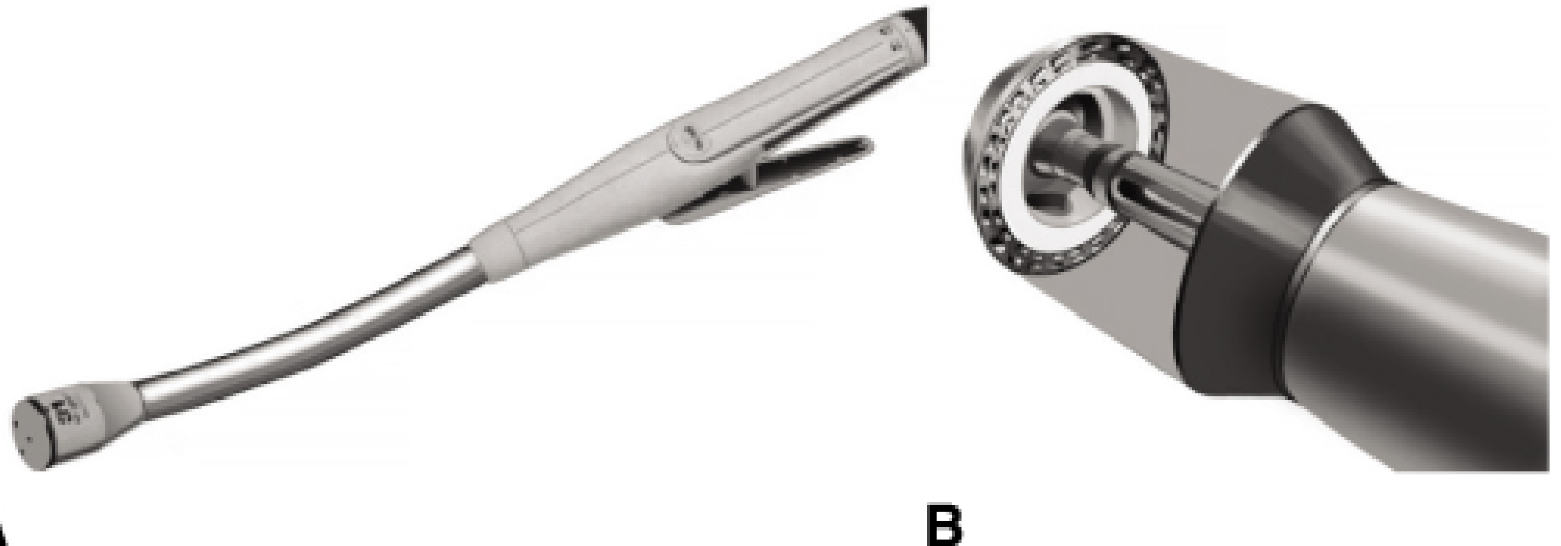
- Places 2 or 3 staggered rows of staples in a straight line
- Does NOT have a blade - tissue is NOT cut automatically
- After firing, the surgeon manually cuts the tissue with scissors or a knife along/adjacent to the staple line
- Fork-shaped device with upper and lower jaws
- Staple cartridge (disposable reload) snaps into jaws
- Safety pin to prevent accidental firing
- Firing handle/lever
| Model | Staple Line Length | Staple Heights | Port for Laparoscopic |
|---|---|---|---|
| TA-30 | 30 mm | 3.5 mm, 4.8 mm | 12 mm trocar |
| TA-55 | 55 mm | 3.5 mm, 4.8 mm | 12 mm trocar |
| TA-90 | 90 mm | 3.5 mm, 4.8 mm | External only |
- Closure of rectal stump (Hartmann's procedure)
- Closure of duodenal stump (Billroth II)
- Gastric closure
- Bronchial closure (thoracic surgery)
- Closure of bowel ends prior to anastomosis
- Appendix stump closure
- Bladder neck closure
- Simple mechanism; allows surgeon control over cutting line
- Longer staple lines available (up to 90 mm)
B. GIA STAPLER (Gastrointestinal Anastomosis / Linear Cutting Stapler)
- Places 4 staggered rows of staples (2 rows on each side)
- Contains an integral knife/blade that cuts between the two central rows simultaneously
- Result: two stapled edges, divided in the centre - creating a sealed, cut tissue on both sides
- The instrument both staples and cuts in one action
- Two opposing jaws (upper and lower)
- Cartridge half (containing staples) + Anvil half
- Central cutting blade running between the two central staple rows
- Locking mechanism and firing handle
- Side-to-side anastomosis (functional end-to-end) - e.g., ileotransverse anastomosis after right hemicolectomy
- Stomach division during gastrectomy and bariatric surgery (sleeve gastrectomy)
- Small bowel resection and anastomosis
- Esophageal division
- Lung resection (pulmonary)
- Creation of gastrojejunostomy pouch in bariatric surgery
- Hartmann's reversal anastomosis
- Two ends of bowel (cut with GIA) placed side-by-side, antiperistaltically aligned
- Small enterotomies made at anti-mesenteric corners
- GIA inserted into both lumens; fired to create the common channel
- The enterotomy openings at the apex are closed with a TA stapler or sutures
- Result is a wide-lumen, tension-free, well-vascularised anastomosis
C. ENDO GIA (Endoscopic / Laparoscopic Linear Cutting Stapler)
- Mounted on a long laparoscopic shaft
- Passes through a 10-12 mm trocar (some require 15 mm for thicker cartridges)
- Articulating head versions available - allow angulation for deep pelvic work
- Straight and curved reload options
- Straight-tip and curved-tip reload options
- Articulating heads (up to 45° articulation) - essential for low anterior resection in narrow pelvis
- Powered/motorized firing (e.g., Echelon Flex, Signia, iDrive) - provides even staple compression
- Tissue thickness indicators / real-time feedback systems
- Color-coded cartridges (see below)
- All laparoscopic and robotic bowel resections
- Laparoscopic gastrectomy, colectomy, anterior resection
- Rectal transection in TME (especially with articulating head in deep pelvis)
- Sleeve gastrectomy, Roux-en-Y gastric bypass
- Thoracoscopic lung resection (VATS)
- Laparoscopic appendectomy
D. EEA STAPLER (End-to-End Anastomosis / Circular Stapler)
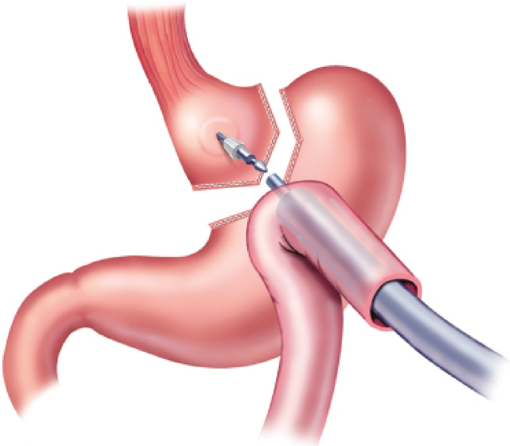
- Fires a circle of staples (2-3 concentric rows) in a doughnut pattern
- Contains a central circular knife that simultaneously excises a "donut" of tissue from inside the staple ring, creating the anastomotic lumen
- Two parts: Handpiece (contains staples + blade) + Anvil (detachable head)
- Handpiece/Body: Contains circular staple cartridge, central trocar/pin, and knife
- Anvil: Separate detachable mushroom-shaped head with a central pin-hole; sutured into one viscus via purse-string suture
- Central pin (trocar): Extends from handpiece; connects to anvil's central hole to align both rings of tissue
- Firing mechanism: Dial to close/approximate jaws → handle to fire
| Size | Diameter | Common Use |
|---|---|---|
| 21 mm | 21 mm | Esophageal anastomosis (narrow lumen) |
| 25 mm | 25 mm | Esophagojejunostomy; small lumen |
| 28 mm | 28 mm | Colorectal; ileoanal |
| 29 mm | 29 mm | Colorectal anastomosis |
| 31 mm | 31 mm | Low colorectal anastomosis |
| 33 mm | 33 mm | Standard colorectal anastomosis |
| 34 mm | 34 mm | PPH hemorrhoid procedure |
- Low colorectal anastomosis (LAR/TME)
- Colorectal anastomosis after left hemicolectomy
- Ileoanal pouch anastomosis (IPAA)
- Esophagojejunostomy (after total gastrectomy)
- Gastrojejunostomy (bypass surgery)
- Esophagogastrostomy (Ivor Lewis)
- Coloanal anastomosis
E. PPH STAPLER (Procedure for Prolapse and Hemorrhoids / Circular Hemorrhoid Stapler)
- A specialized circular stapler (33-36 mm diameter)
- Placed transanally above the dentate line
- Fires a complete circular ring of staples through the prolapsing rectal mucosa and hemorrhoidal tissue
- Simultaneously excises the donut of prolapsing mucosa/submucosa
- Lifts hemorrhoids back into the anal canal and reduces their blood supply
- Does NOT excise hemorrhoids directly - instead disrupts their blood supply and repositions them
- Less postoperative pain (staple line above dentate line - above pain-sensitive mucosa)
- Shorter hospital stay
- Faster return to normal activity
- Anastomotic leak / rectovaginal fistula (rare)
- Urgency
- Staple line bleeding
- Recurrence (higher than conventional)
F. CURVED / CONTOUR STAPLER (Specialty)
- Contour Curved Cutter Stapler (Ethicon/Covidien)
- Gently arched end-effector
- Places 3 rows of staples and transects between rows 2 and 3
- Narrower profile than standard linear stapler
- Designed specifically for deep pelvic rectal transection in LAR/TME - easier placement in narrow male pelvis
- Obviates need for manual post-staple scalpel division
7. Staple Heights and Color Coding
| Tissue Type | Closed Staple Height | ETHICON Color | COVIDIEN Color |
|---|---|---|---|
| Thin/vascular tissue (vessels, lung) | 1.0-1.5 mm | White | White |
| Thin bowel / esophagus | 1.5-2.0 mm | White/Blue | White |
| Standard bowel (small/large intestine) | 2.0-2.5 mm | Blue | Blue |
| Thick bowel / stomach | 3.5 mm | Green | Green |
| Very thick tissue / bronchus | 4.8 mm | Gold/Black | Purple/Black |
Note: Color coding is manufacturer-specific - surgeons must know the actual staple height, not rely on color alone as it varies between brands.
8. Stapling Techniques in GI Surgery
A. Purse-String Technique (for EEA)
- A concentric suture circumferentially placed around the lumen of the bowel end
- Tied down over the anvil shaft to secure the tissue evenly to the anvil
- Ensures all tissue is drawn uniformly into the circular stapler jaws
B. Single Stapling Technique
- Both ends of bowel secured with purse-string sutures around the EEA components
- Used when there is no pre-existing staple line
C. Double Stapling Technique (most common in LAR/TME)
- Rectum first transected with linear TA/ENDO GIA stapler → first staple line
- EEA anvil placed proximally; EEA pin advanced through the existing rectal staple line
- Only one purse-string (proximal); no purse-string on the distal rectum
- Simpler; faster; reliable for low pelvic anastomoses
D. Triple Stapling Technique
- Rarely used; involves additional staple lines in complex reconstructions
E. Functional End-to-End Anastomosis (side-to-side with GIA)
- Two cut ends placed side-by-side (antiperistaltic or isoperistaltic)
- GIA inserted into small enterotomies at anti-mesenteric corners
- Fired to create side-to-side common channel
- Enterotomy apex closed with TA or sutures
- Creates the widest, most blood-supply-rich anastomosis
- Preferred for right hemicolectomy, ileocolic anastomosis, small bowel anastomosis
9. Advantages of Surgical Staplers Over Hand-Sewn Anastomosis
| Advantage | Detail |
|---|---|
| Speed | 30 seconds vs. 20-30 minutes for hand-sewn |
| Reproducibility | Consistent staple formation regardless of tissue edema |
| Deep access | EEA can reach areas inaccessible by hand suturing (deep pelvis, posterior mediastinum) |
| Hemostasis | Simultaneous stapling seals vessels on both sides of cut |
| Lower infection risk | Minimal handling of bowel lumen |
| Everted anastomosis | Mucosal eversion avoided; serosal approximation achieved |
| Laparoscopic application | Allows minimally invasive surgery for complex resections |
10. Disadvantages and Complications of Staplers
| Complication | Mechanism | Prevention |
|---|---|---|
| Anastomotic leak (most feared) | Incomplete staple formation; wrong cartridge height; ischaemia; tension | Air-leak test; correct cartridge; tension-free; adequate blood supply |
| Haemorrhage from staple line | Torn tissue; wrong cartridge height (too high = inadequate haemostasis) | Oversew bleeding points; correct cartridge selection |
| Anastomotic stricture | Scar formation at staple line | Dilatation; correct sizing |
| Incomplete "donuts" | Purse-string too loose; tissue not fully approximated | Check donuts immediately; reinforce with sutures |
| Misfire | Mechanical failure | Always test fire; inspect cartridge before use; spare cartridge available |
| Injury to adjacent structures | Ureter/vagina/urethra caught in jaws | Inspect before closing; ureteric stents |
| Staple line dehiscence | Ischaemia; infection; distal obstruction | Defunctioning stoma for high-risk anastomoses |
| Cost | Each cartridge reload is expensive | Justified by reduced operative time and complications |
11. Specific Clinical Applications in GI Surgery
| Operation | Staplers Used | Technique |
|---|---|---|
| Right hemicolectomy | GIA / ENDO GIA | Functional end-to-end ileocolic anastomosis |
| Left hemicolectomy / sigmoid colectomy | ENDO GIA + EEA (29-33mm) | Linear division + circular EEA anastomosis |
| Low anterior resection (TME) | ENDO GIA (rectal transection) + EEA (anastomosis) | Double-stapling technique |
| APR / Hartmann's | TA or ENDO GIA | Rectal stump closure only |
| Total gastrectomy | GIA + EEA (25-28mm) | Roux-en-Y esophagojejunostomy |
| Subtotal gastrectomy (Billroth II) | TA (duodenal stump) + GIA (stomach) | Duodenal closure + gastrojejunostomy |
| Sleeve gastrectomy | ENDO GIA (multiple firings along greater curvature) | Gastric tube creation |
| Roux-en-Y Gastric Bypass | ENDO GIA + EEA | Gastrojejunostomy + jejunojejunostomy |
| Ivor Lewis esophagectomy | ENDO GIA + EEA (25mm) | Intrathoracic esophagogastrostomy |
| Whipple's procedure | GIA / TA + EEA | Multiple anastomoses |
| IPAA (ileal pouch-anal anastomosis) | GIA (J-pouch) + EEA (25mm) | J-pouch creation + pouch-anal anastomosis |
| Appendectomy | ENDO GIA or TA (30mm) | Appendix base stapling |
| Small bowel resection | GIA or ENDO GIA | Side-to-side anastomosis |
| Hemorrhoidectomy (PPH) | PPH stapler (33-36mm) | Circumferential mucosal excision above dentate line |
12. Powered Staplers (Modern Developments)
- Motorized firing mechanism (battery/electric powered)
- Automatic adjustment of firing speed based on tissue resistance
- Real-time tissue compression feedback - prevents premature firing in thick tissue
- Consistent, even staple formation across the entire cartridge length
- Reduces staple line failure rates
- Particularly useful in bariatric and thoracic surgery with thick, edematous tissue
13. Key Exam Points Summary
- 1908: Hültl (Hungary) - first surgical stapler
- 1967: TA stapler introduced (USSC)
- 1968: GIA stapler introduced (USSC)
- 1978: EEA circular stapler introduced - milestone in colorectal surgery
- TA: Staples only (no knife); 2-3 rows; for closing stumps
- GIA: Staples + cuts simultaneously; 4 rows; for anastomosis and division
- EEA: Circular; for end-to-end or end-to-side anastomosis transanally; sizes 21-33mm
- Staple shape: "B" - allows haemostasis without complete ischaemia
- Double stapling technique: TA/ENDO GIA for rectal transection + EEA transanally for anastomosis
- Donut check: Mandatory after EEA firing - incomplete donuts = anastomotic defect
- Air-leak test: After low anastomosis - saline fill + per-anal insufflation
- Staple height too low = ischaemia/tear/leak; too high = bleeding
- Colour coding is manufacturer-specific - know actual staple height
Sources: Fischer's Mastery of Surgery 8th Ed., p. 3991-4100; Maingot's Abdominal Operations; History of Surgical Staplers; MDPI Applied Sciences 2024 review of mechanical stapling devices
Similarly for colonoscopy
colonoscopy history invention Hirschowitz Overholt development fiber optic 1969
Colonoscopy - Complete Detailed Answer
1. Definition
2. Etymology
- Colon - Greek kolon (large intestine)
- -scopy - Greek skopein (to look/examine)
- Literally: "looking into the colon"
3. History
| Year | Event |
|---|---|
| 1853 | Antonin Desormeaux (France) - invented the first endoscope (cystoscope); used open-tube with mirror/candle light |
| 1895 | Howard Kelly introduced the rigid proctosigmoidoscope for rectal examination |
| 1930s-50s | Rigid sigmoidoscopy widely used; reach limited to 25-30 cm |
| 1954 | Harold Hopkins (UK physicist) developed the glass rod-lens system for better image transmission |
| 1956 | Basil Hirschowitz (University of Michigan) developed the first flexible fibre-optic bundle for light transmission - the revolution that made flexible endoscopy possible |
| Feb 1957 | Hirschowitz passed the first flexible fibre-optic endoscope down his own throat - proof of concept for flexible endoscopy |
| 1960s | Flexible fibre-optic upper GI endoscopes developed and refined |
| 1963 | Niwa and Yamagata (Japan) demonstrated the first flexible colonoscope |
| 1966-1969 | Bergein Overholt (USA) and Matsunaga (Japan) independently developed practical flexible colonoscopes; Overholt performed over 1,600 diagnostic colonoscopies from June 1969 to June 1972 using a specially designed flexible fibre-optic instrument |
| 1969 | Charge-Coupled Device (CCD) chip invented → basis for future video colonoscopy |
| 1970s | Polypectomy techniques developed; colonoscopy established as gold standard for colon examination |
| 1983 | Video colonoscopy (electronic/CCD-based) introduced - replaced fibre-optic; superior image quality; teachable via monitor |
| 1990s | Chromoendoscopy, injection-assisted polypectomy (EMR) introduced |
| 2000s | Narrow Band Imaging (NBI), high-definition (HD) colonoscopy, capsule colonoscopy introduced |
| 2010s | Virtual colonoscopy (CT colonography), AI-assisted polyp detection, water-exchange colonoscopy |
| Present | Robotic colonoscopy, single-use disposable colonoscopes, AI real-time detection systems |
4. The Colonoscope - Equipment and Parts
Types of Colonoscopes
| Type | Length | Diameter | Use |
|---|---|---|---|
| Standard colonoscope | 133-168 cm | 12-13 mm | Routine adult colonoscopy |
| Paediatric/slim colonoscope | 130-135 cm | 9-11 mm | Narrow strictures, paediatric, difficult sigmoid |
| Sigmoidoscope | 60-70 cm | 11-13 mm | Examination to sigmoid/descending colon only |
| Therapeutic colonoscope | 133-168 cm | 13-14 mm | Larger working channel (3.7 mm) for therapeutic procedures |
Parts of the Colonoscope
- Up/Down angulation dial (large)
- Left/Right angulation dial (small)
- Air/water button (insufflation and lens washing)
- Suction button
- Working channel port (biopsy/instrument entry)
- Light guide post
- Flexible shaft, 133-168 cm long
- Contains: fibre-optic bundles / video CCD chip, light-transmitting fibres, air/water channels, suction channel, working channel
- Outer layer: smooth, flexible braid
- Last 10-15 cm - actively deflectable in 4 directions (up/down/left/right) using control dials
- Maximum angulation: up 180°, down 180°, left/right 160°
- CCD chip (video) or fibre-optic lens (optical)
- Illumination fibres (light source)
- Air/water nozzle (for insufflation and lens cleaning)
- Working channel opening (2.8-3.7 mm diameter)
- Connects handle to light source and video processor
- Contains: light fibre cable, air/water tubes, suction tube, electrical connections
- Xenon or LED light source
- Digital video processor
- Monitor display
5. Indications for Colonoscopy
Diagnostic Indications
| Category | Specific Indications |
|---|---|
| Screening | Average-risk CRC screening from age 45 (ACS guidelines) or 50 (ACG); every 10 years if normal |
| Surveillance | Post-polypectomy surveillance; post-CRC resection surveillance; IBD surveillance for dysplasia; Lynch syndrome; familial adenomatous polyposis (FAP) |
| Lower GI bleeding | Haematochezia; melena with normal upper GI endoscopy; iron-deficiency anaemia; occult FOBT positive |
| Altered bowel habits | New-onset change in bowel habits in patients >40 years; unexplained chronic diarrhoea or constipation |
| Abdominal symptoms | Unexplained abdominal pain with high suspicion for colonic pathology |
| Abnormal imaging | Colonic lesion on CT/MRI/barium enema requiring biopsy |
| IBD | Diagnosis, grading of activity, surveillance, biopsy; assessment of extent |
| Colonic obstruction | Rule out mechanical obstruction vs. pseudo-obstruction (ACPO); decompression |
| Foreign body | Retrieval of ingested foreign body in colon |
| Family history | First-degree relative with CRC or adenoma - earlier/more frequent screening |
Therapeutic Indications
| Therapeutic Use | Detail |
|---|---|
| Polypectomy | Removal of polyps (sessile, pedunculated, flat) |
| EMR (Endoscopic Mucosal Resection) | Large flat/sessile polyps >10 mm |
| ESD (Endoscopic Submucosal Dissection) | En bloc resection of large flat lesions |
| Haemostasis | Bleeding diverticula, angiodysplasia, post-polypectomy bleeding, radiation proctitis |
| Decompression | ACPO (Acute Colonic Pseudo-Obstruction) |
| Stricture dilation | Balloon dilation of benign strictures (anastomotic, Crohn's) |
| Stent placement | Colonic stents for malignant obstruction (bridge to surgery or palliation) |
| Tattooing | India ink marking of lesions for surgical localization |
| Volvulus reduction | Sigmoid volvulus detorsion |
6. Contraindications
Absolute Contraindications
- Suspected or confirmed bowel perforation (free air on imaging)
- Fulminant colitis with toxic megacolon (risk of perforation)
- Recent (<6 weeks) colonic anastomosis (risk of dehiscence from air pressure)
- Severe haemodynamic instability (patient unfit for procedure)
- Patient refusal (no informed consent)
Relative Contraindications
- Acute diverticulitis - risk of perforation; CT preferred
- Recent acute MI (<3 months)
- Severe coagulopathy (INR >2.5; platelets <50,000) - particularly for polypectomy
- Large abdominal aortic aneurysm (manipulation risk)
- Pregnancy (first and third trimesters; second trimester relatively safe)
- Inadequate bowel preparation - reduces diagnostic yield; reschedule
- Severe COPD / cardiac failure - sedation risk
7. Pre-procedure Preparation
A. Bowel Preparation (Most Critical Step)
| Preparation | Regimen | Notes |
|---|---|---|
| Polyethylene Glycol (PEG) - GoLYTELY, MoviPrep | 4 L (split-dose: 2 L evening before + 2 L morning of procedure) | Gold standard; safe in renal failure; no significant electrolyte shifts |
| Low-volume PEG (2 L) + ascorbic acid | 2 L total; better tolerated | Same efficacy; improved patient compliance |
| Sodium Picosulfate + Magnesium Citrate (PicoPrep) | Two sachets + 2L water | Better tolerated; avoid in renal impairment |
| Sodium Phosphate (NaP) solution | 2 doses × 45 mL | Effective but risk of acute phosphate nephropathy; avoid in renal failure, elderly, on ACE inhibitors |
| Sodium Phosphate tablets | 32 tablets | Same risks as NaP solution |
| Tap water enema | Before therapeutic colonoscopy for ACPO | Minimal preparation; not ideal for diagnostic |
- Split-dose preparation (half the evening before, half 4-6 hours before the procedure) gives superior bowel cleanliness
- Patients remain on clear liquid diet for 24 hours before
- NPO for 6-8 hours before procedure for solids; 2 hours for clear fluids
- Patient eats regular diet (low-fibre) until the day before
B. Medication Management
| Drug | Action Before Colonoscopy |
|---|---|
| Anticoagulants (warfarin) | Stop 5 days before for polypectomy; bridge with LMWH if high risk |
| NOACs (rivaroxaban, apixaban) | Omit on day before and day of procedure |
| Antiplatelet (clopidogrel) | Stop 5-7 days before for polypectomy |
| Aspirin | Continue; low bleeding risk |
| Metformin | Stop 48h before if contrast needed |
| Iron supplements | Stop 5-7 days before (turns stool dark, worsens prep) |
| NSAIDs | Continue |
| Insulin / oral hypoglycaemics | Reduce dose on prep day (reduced food intake) |
C. Informed Consent
- Risks: perforation, bleeding, missed lesions, sedation risks
- Alternatives: CT colonography, sigmoidoscopy, faecal occult testing
- Therapeutic aspects (polypectomy) if likely
D. Sedation Options
| Option | Drugs | Notes |
|---|---|---|
| Conscious sedation (most common) | Midazolam 1-5 mg IV + Pethidine (meperidine) 25-100 mg IV or Fentanyl 50-100 mcg | Patient responsive to commands; most common worldwide |
| Deep sedation / MAC | Propofol IV (anaesthetist-administered) | Deeper sedation; faster recovery; preferred for complex or prolonged procedures |
| General anaesthesia | Propofol + intubation | Only for paediatric or uncooperative patients |
| Unsedated | None | Possible in experienced hands with CO₂; minimal discomfort |
| Entonox | 50% N₂O / 50% O₂ (inhaled) | Used in some centres as alternative |
8. Step-by-Step Procedure
Set-Up and Positioning
- Colonoscope tested: air/water button, suction, tip deflection in all 4 directions, white balance
- Monitoring: pulse oximeter, blood pressure, ECG leads attached
- IV access secured
- Resuscitation equipment available
Step 1 - Digital Rectal Examination
- Performed before scope insertion
- Assesses: anal tone, masses, haemorrhoids, prostate
- Lubricates anus
Step 2 - Insertion and Rectal Examination (0-15 cm)
- Lubricate scope tip generously
- Insert gently with slight posterior angulation following curve of sacrum
- Insufflate air (or CO₂ - preferred) until rectal lumen visible
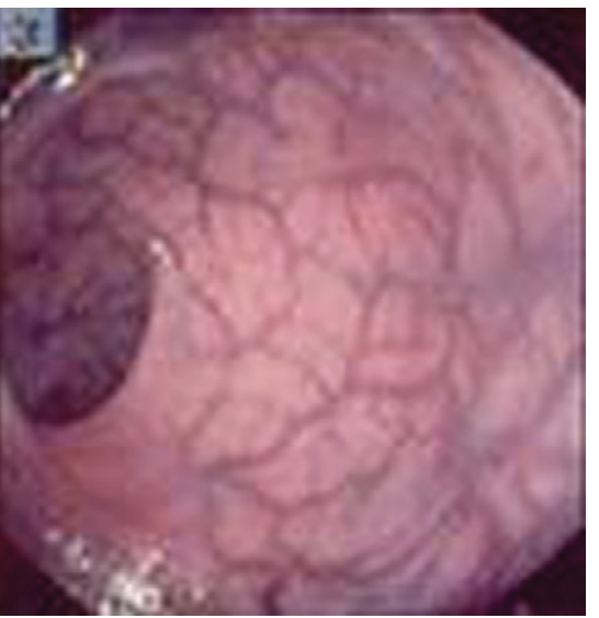
- Rectal landmarks:
- Three valves of Houston (prominent mucosal folds/shelves) - seen at 5, 9, and 14 cm
- Haemorrhoidal plexus - submucosal vascular network clearly visible on rectum mucosa
Step 3 - Rectosigmoid Junction (15-18 cm)
- Abrupt angulation; most painful area for patient
- Advance by torquing right + withdrawal to reduce loop formation
Step 4 - Sigmoid Colon (18-50 cm) - Most Difficult Segment
- Most common site of difficulty and most common site of perforation
- Sigmoid is mobile, redundant, may be fixed by diverticular disease or post-surgical adhesions
- Key manoeuvres:
- Right torque + withdrawal = most important manoeuvre; straightens sigmoid loops
- Abdominal pressure (applied by assistant): usually in left iliac fossa or left lower quadrant
- Repositioning patient: supine, right lateral
- CO₂ insufflation: gas absorbed more rapidly, reduces distension and discomfort
- Slide-by technique: used when lumen not well visualised around acute angles - scope advanced while mucosa slides past; mucosa should appear reddish (blanching = excess pressure - STOP and withdraw)
- Essential principle: when scope impeded → torque RIGHT + WITHDRAW (do not push!)
Step 5 - Descending Colon (50-80 cm)
- Relatively straight; circular (round) lumen - recognisable landmark
- Easy passage; minimal patient discomfort
Step 6 - Splenic Flexure (70-80 cm)
- Abrupt angulation; may require slight slide-by
- May require: left lateral to supine position change
- Landmark: "Blue shadow" of spleen occasionally visible through colon wall
Step 7 - Transverse Colon (80-130 cm)
- Recognised by characteristic triangular lumen (haustra create triangular shape)
- Contains prominent haustral folds
- Hangs down in mid-abdomen; may sag ("tent" effect on fluoroscopy)
- Advance by maintaining right torque
Step 8 - Hepatic Flexure (110-130 cm)
- Recognised by "liver shadow" - blue/green discolouration through bowel wall from adjacent liver
- Right turn required to enter ascending colon
Step 9 - Ascending Colon and Cecum (130-150 cm)
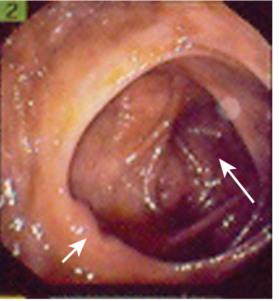
- Wide lumen, haustral folds
- Cecal landmarks (confirms cecal intubation):
- Ileocecal valve - prominent semi-lunar fold with central slit, usually in posteromedial wall
- Appendiceal orifice - small round/slit orifice at the apex of the cecum ("crow's foot" - convergence of three taeniae coli pointing toward appendix orifice)
- Transillumination - light from scope visible on right iliac fossa skin surface (not always reliable)
- Typical cecal appearance on fluoroscopy (if used)
Step 10 - Intubation of Terminal Ileum (Optional)
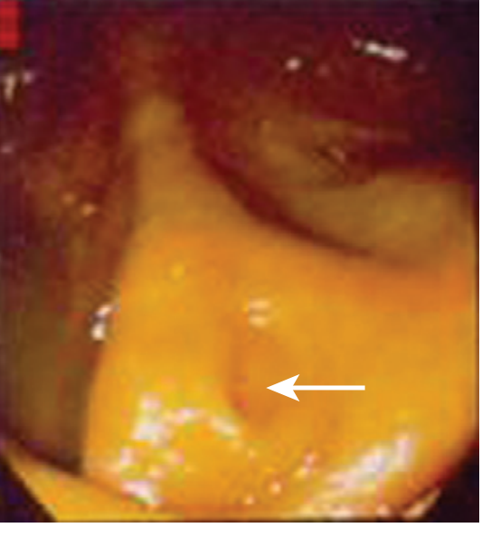
- Attempted when clinically indicated (Crohn's disease, malabsorption, ileal pathology)
- ICV intubated by deflecting scope tip into the valve opening
- Terminal ileum: villi visible, circular folds (plicae circulares), no haustral folds
- Normal terminal ileum mucosa appears granular/velvety from villi
Step 11 - Withdrawal Phase (Most Important for Diagnosis!)
- Withdrawal time should be minimum 6-8 minutes from cecum to anus (studies show lower polyp detection rate if <6 minutes)
- Scope withdrawn slowly with continuous 360° rotation to examine all mucosal folds
- Suction excess air/fluid
- Systematically inspect behind folds; use tip deflection to look behind haustral folds
- Biopsy/polypectomy performed during withdrawal
Step 12 - Retroflexion in Rectum
- At anorectal junction (5-10 cm from anus), scope tip deflected 180° back on itself (retroflexion/turnaround manoeuvre)
- Allows inspection of distal rectum and anal canal from inside
- Visualises internal haemorrhoids, distal rectal lesions that may be missed on forward view
- Control wheels both rotated counterclockwise to achieve retroflexion
- Shaft torqued to examine all around anorectal junction
9. Endoscopic Landmarks - Summary
| Structure | Distance from Anus | Characteristic Appearance |
|---|---|---|
| Anal canal | 0-4 cm | Squamous epithelium; dentate line |
| Rectum | 4-15 cm | Vascular plexus visible; valves of Houston |
| Rectosigmoid junction | 15-18 cm | Acute angulation |
| Sigmoid colon | 18-50 cm | Crescent-shaped lumen; tortuous |
| Descending colon | 50-80 cm | Circular lumen; straight |
| Splenic flexure | 70-80 cm | Acute turn; blue splenic shadow |
| Transverse colon | 80-130 cm | Triangular lumen (haustral folds create triangle) |
| Hepatic flexure | 110-130 cm | Acute turn; liver shadow (blue-green) |
| Ascending colon | 130-145 cm | Wide lumen; prominent haustrae |
| Cecum | 145-150 cm | ICV + appendiceal orifice + crow's foot |
| Terminal ileum | >150 cm | Villi; plicae circulares; no haustra |
10. Polypectomy
Types of Polyps
- Pedunculated: on a stalk; easier to remove
- Sessile: flat-based; more challenging
- Flat / depressed: lie at or below mucosal surface; harder to detect; higher malignancy risk
Polypectomy Methods
| Method | Use | Technique |
|---|---|---|
| Cold forceps biopsy | Diminutive polyps <5 mm | Forceps grasp and remove tissue; no electrocautery |
| Hot forceps biopsy | Small polyps 5-8 mm | Forceps + electrocautery (rarely used now) |
| Cold snare polypectomy | Polyps 4-10 mm | Loop snare placed around polyp; closed and cut without electrocautery; preferred for small polyps |
| Hot snare polypectomy | Pedunculated polyps; larger sessile | Snare + electrocautery; haemostasis achieved simultaneously |
| Endoscopic Mucosal Resection (EMR) | Flat/sessile polyps >10 mm | Submucosal injection (saline ± adrenaline ± dye) → raises polyp → snare resection; piece-meal or en bloc |
| Endoscopic Submucosal Dissection (ESD) | Large flat lesions; early mucosal cancer | Submucosal injection → circumferential mucosal incision → submucosal dissection with knife; en bloc resection regardless of size; technically demanding |
Pre-injection for EMR
- Saline alone - most basic
- Saline + methylene blue - delineates submucosal plane (turns blue)
- Saline + 1:100,000 adrenaline - vasoconstriction reduces bleeding
- Hyaluronic acid - maintains submucosal cushion longer
- "Lift sign": if polyp doesn't lift with injection → deep invasion → do NOT attempt EMR
11. Therapeutic Procedures During Colonoscopy
| Procedure | Indication | Technique |
|---|---|---|
| Haemostasis | Diverticular bleeding, angiodysplasia, post-polypectomy bleeding | Adrenaline injection, endoclips, bipolar coagulation, APC, heater probe |
| Argon Plasma Coagulation (APC) | Angiodysplasia, radiation proctitis, superficial bleeding | Non-contact electrocautery via argon gas ionisation |
| Endoclip | Perforation closure, bleeding vessel | Metal clips deployed through working channel |
| Tattooing | Mark lesion site for future surgery/surveillance | India ink injected submucosally; performed proximal or distal to lesion (not on tumour) to prevent peritoneal seeding |
| Colonic decompression | ACPO | Pass scope to hepatic flexure; suction air; may place decompression tube over guidewire |
| Volvulus reduction | Sigmoid volvulus | Scope advanced into twisted segment; air suctioned; rectal tube left in situ to prevent recurrence |
| Balloon dilation | Anastomotic stricture, Crohn's stricture | Balloon introduced through working channel and inflated under direct vision |
| Stent placement | Malignant obstruction | Self-expanding metallic stent (SEMS) deployed over guidewire |
| Foreign body retrieval | Ingested foreign body | Retrieval forceps, Roth net, snare |
12. Complications
Overall complication rate: 0.28% for diagnostic colonoscopy
A. Perforation (Most Serious)
- Rate: 0.05-0.3% (diagnostic); higher with therapeutic procedures
- Mechanisms:
- Mechanical tear - excessive force on antimesenteric border of sigmoid colon (most common site)
- Barotrauma - excessive air insufflation (most common site: cecum - widest diameter, highest wall tension)
- Electrosurgical injury - polypectomy burn
- Presentations: Abdominal pain, distension, shoulder-tip pain (referred diaphragmatic irritation), peritonitis
- Management:
- Small recognised perforation: endoclips + antibiotics + close observation (if no peritonitis)
- Large tear / peritonitis: urgent surgical repair (primary repair or resection)
- Delayed presentation: usually requires surgery
B. Haemorrhage
- Rate: 0.1-0.6% overall; 0.87% with polypectomy
- Immediate (during procedure) or delayed (up to 2 weeks post-polypectomy)
- Risk factors: polyp size, right colon location, anticoagulants, cardiovascular disease
- Management: endoscopic haemostasis (adrenaline injection, clips, coagulation); rarely angiography or surgery
C. Post-Polypectomy Electrocoagulation Syndrome (PPES)
- Also called: "post-polypectomy syndrome" or "transmural burn syndrome"
- Occurs after hot snare/electrocautery polypectomy
- Mechanism: Transmural thermal injury without frank perforation → localised peritoneal irritation
- Presentation: Fever, localized abdominal pain and tenderness 1-5 days post-procedure; no free air on CT
- Management: Conservative (IV fluids, antibiotics, bowel rest, monitoring)
- Distinguishable from perforation by absence of free air on CT
D. Cardiopulmonary Complications
- Most common overall complication category
- Hypoxia/hypotension from sedation
- Aspiration pneumonia (especially under deep/MAC sedation)
- Vagal reactions (bradycardia from air insufflation/distension)
- Prevention: careful patient selection, O₂ monitoring, titrated sedation
E. Complications of Bowel Preparation
- Hyponatraemia/hypernatraemia (especially sodium phosphate preparations)
- Acute phosphate nephropathy (sodium phosphate in patients with renal impairment)
- Electrolyte disturbances (hypokalaemia, hyperphosphataemia)
- Nausea, vomiting, abdominal discomfort
- Mallory-Weiss tear (from retching with large-volume prep)
F. Missed Lesions
- Adenoma miss rate: 6-12% overall; up to 26% for polyps <5 mm
- Causes: poor prep, withdrawal time <6 min, behind folds
- Prevention: adequate withdrawal time ≥6-8 min; HD colonoscopy; cap-assisted; water exchange technique
G. Other Complications
- Splenic injury (0.1%) - from over-traction of splenocolic ligament during sigmoid manoeuvres
- Mesenteric laceration
- Air embolism (rare)
- Bacteraemia (transient; usually benign; antibiotic prophylaxis not routinely required)
- Incarceration of hernia
- Drug reactions to sedation agents
13. Colonoscopy Quality Indicators
| Indicator | Target |
|---|---|
| Adenoma Detection Rate (ADR) | ≥25% (men), ≥20% (women) - key quality metric |
| Cecal intubation rate | ≥90% (≥95% in elective screening) |
| Withdrawal time | ≥6 minutes |
| Bowel prep adequacy | ≥85% of procedures with adequate prep |
| Perforation rate | <0.1% (diagnostic), <0.2% (therapeutic) |
| Polyp retrieval rate | ≥90% of detected polyps |
14. Screening Intervals (Post-Colonoscopy Surveillance)
| Finding | Next Colonoscopy |
|---|---|
| Normal / no polyps | 10 years |
| 1-2 small (<10 mm) tubular adenomas | 7-10 years |
| 3-4 adenomas OR any ≥10 mm | 3-5 years |
| 5-10 adenomas | 3 years |
| High-grade dysplasia, villous adenoma | 3 years |
| >10 adenomas | <3 years; consider polyposis workup |
| Serrated polyp <10 mm, no dysplasia | 5 years |
| Sessile serrated polyp ≥10 mm | 3 years |
| Colorectal cancer resected | 1 year post-resection |
| IBD (ulcerative colitis/Crohn's with colonic involvement) | Every 1-2 years from 8-10 years after diagnosis |
15. Special Colonoscopy Techniques
| Technique | Description | Advantage |
|---|---|---|
| Chromoendoscopy | Topical dye (indigo carmine, crystal violet) sprayed on mucosa | Enhances flat/depressed lesions; IBD dysplasia surveillance |
| Narrow Band Imaging (NBI) | Filtered light (415 nm blue, 540 nm green) | Enhances surface pattern and vascular architecture; no dye needed |
| Cap-assisted colonoscopy | Transparent cap fitted on scope tip | Flattens folds; reduces blind spots behind folds; may improve ADR |
| Water-exchange colonoscopy | Water (not air) used for insufflation during insertion; suctioned on withdrawal | Less pain; better prep removal; may improve ADR |
| CO₂ insufflation | Carbon dioxide instead of air | Absorbed 150× faster; significantly less post-procedure bloating and pain |
| Fluoroscopy-guided | Real-time X-ray guidance | Demonstrates scope loops; guides decompression tube placement |
| Magnetic endoscope imaging (MEI) | Real-time 3D scope position mapping | Helps reduce loop formation without fluoroscopy radiation |
| AI-assisted detection | Computer-aided detection (CADe) algorithms | Real-time polyp detection; reduces miss rate |
16. Key Exam Points Summary
- Most common site of difficulty on insertion = sigmoid colon
- Most common site of perforation = sigmoid colon (mechanical); cecum (barotrauma)
- Key manoeuvre when impeded = torque right + withdraw (never push blind)
- Cecal landmarks = ICV + appendiceal orifice + crow's foot
- Transverse colon = triangular lumen; descending colon = circular lumen
- Withdrawal time ≥6-8 minutes mandatory
- Retroflexion in rectum - examines distal rectum and haemorrhoids
- Perforation rate: 0.05-0.3% diagnostic; barotrauma in cecum; mechanical in sigmoid
- PPES - post-polypectomy syndrome: fever + abdominal pain + NO free air on CT
- EMR: submucosal injection lifts polyp → snare resection; "non-lift sign" = deep invasion → do NOT resect
- ADR ≥25% (men) / ≥20% (women) = key quality indicator
- CO₂ preferred over air: absorbed faster, less bloating
- Split-dose PEG = gold standard bowel prep
Sources: Pfenninger & Fowler's Procedures for Primary Care 3rd Ed., p. 4635-4788; Clinical GI Endoscopy Expert Consult 3rd Ed.; Sleisenger & Fordtran's GI & Liver Disease; Current Surgical Therapy 14th Ed.; Yamada's Textbook of Gastroenterology 7th Ed.; History of Colonoscopy - PMC
Similarly give me answer for minilaparoscopy
minilaparoscopy definition history indications procedure needlescopic surgery 3mm 2mm trocar
minilaparoscopy needlescopic surgery history indications advantages disadvantages procedure 2mm 3mm instruments
minilaparoscopy needlescopic surgery indications outcomes
minilaparoscopy needlescopic surgery
PMID: 23052524
PMID: 35986221
minilaparoscopy 3mm needlescopic surgery definition history advantages disadvantages complications applications review
PMID: 36314589
minilaparoscopy cholecystectomy appendectomy hernia repair outcomes pain cosmesis
minilaparoscopy versus standard laparoscopy pain recovery
https://pmc.ncbi.nlm.nih.gov/articles/PMC5206838/
PMID: 25895688
PMID: 27777499
Minilaparoscopy (Needlescopic Surgery) - Complete Detailed Answer
1. Definition
2. Etymology
- Mini - Latin minimus (least/smallest)
- Laparo - Greek lapara (flank/abdomen)
- -scopy - Greek skopein (to look/examine)
- Needlescopic - surgical instruments as thin as a needle
- Acuscopic - from Latin acus (needle) + Greek skopein (to look)
- Microlaparoscopy - microscale laparoscopy
3. Historical Background
| Year | Milestone |
|---|---|
| 1901 | George Kelling (Germany) performed the first "coelioscopy" in a dog using a cystoscope |
| 1910 | Jacobaeus (Sweden) performed first human laparoscopy |
| 1987 | Philippe Mouret (Lyon, France) performed the first laparoscopic cholecystectomy - the landmark that opened the minimally invasive era |
| Early 1990s | Standard laparoscopy using 10-12 mm and 5 mm trocars became established |
| 1992-1994 | Gotz, Pier, and Bacher pioneered the first true needlescopic/minilaparoscopic procedures using 2-3 mm instruments |
| 1995 | Cuschieri and colleagues described minilaparoscopy for cholecystectomy using 2 mm instruments |
| 1996-1998 | Wider adoption: reports of minilaparoscopic appendectomy, herniorrhaphy, fundoplication |
| Late 1990s - early 2000s | First-generation instruments limited by poor optics, weak instrument strength, fragility of scopes - early enthusiasm waned |
| 2000s | Parallel development of SILS (Single Incision Laparoscopic Surgery) and NOTES (Natural Orifice Transluminal Endoscopic Surgery) temporarily overshadowed minilaparoscopy |
| 2010s onward | Second-generation minilaparoscopic instruments with improved optics (HD, chip-on-tip technology), stronger instrument materials (titanium alloys), better insulation and energy delivery re-established minilaparoscopy as a serious approach |
| 2012-2022 | Carvalho, Dapri, Lobe and others extensively described minilaparoscopic cholecystectomy ("clipless technique"), hernia repair, appendectomy, bariatric surgery, gynaecological procedures |
| 2022 | SAGES published formal technology assessment of mini-laparoscopic instrumentation, confirming value particularly in paediatric patients and as adjuncts in adult surgery |
4. Terminology Clarification
| Term | Notes |
|---|---|
| Minilaparoscopy (MINI) | Most widely used; preferred term |
| Needlescopic surgery | Emphasises needle-like instrument diameter |
| Microlaparoscopy | Sometimes implies even smaller instruments (1-2 mm); also used in office-based procedures under local anaesthetic |
| Acuscopic surgery | Less common; used in European literature |
| Reduced-port laparoscopy | Broader term including MINI but also SILS |
The term "minilaparoscopy" specifically refers to the technique using instruments 3 mm or less in diameter - distinct from SILS (single incision) and NOTES (no abdominal incision).
5. Instruments and Equipment
A. Trocars
| Type | Size | Features |
|---|---|---|
| Standard mini-trocar | 2 mm, 3 mm | Smaller diameter; lower profile; minimal fascial defect |
| Trocar-less instruments | 2-3 mm | Inserted directly without trocar; reduced friction; preserves peritoneal gas |
| Low-friction trocars | 3 mm | Precision-engineered; enhance surgical precision during knotting and suturing |
B. Optics / Laparoscopes
| Generation | Features |
|---|---|
| First generation (1990s) | Fibre-optic rod-lens systems; poor image quality; fragile; short lifespan |
| Second generation (2010s) | Chip-on-tip (CCD at distal end) technology; HD image quality; comparable to 5 mm scopes |
| Current | 3 mm HD 0° and 30° telescopes; some with 2 mm diameter; 175-180° field of view |
C. Working Instruments (3 mm diameter)
| Instrument | Available in 3 mm |
|---|---|
| Dissectors (Maryland, hook) | Yes |
| Graspers (atraumatic, bowel) | Yes |
| Scissors | Yes |
| Needle holders | Yes |
| Clip appliers | Yes (limited clips size) |
| Bipolar forceps | Yes |
| Hook electrocautery | Yes |
| Irrigation/suction cannula | Yes |
| Fan retractors | Limited |
| Specimen retrieval bags | Yes |
D. Energy Devices
- Monopolar electrocautery through 3 mm instruments: produces less lateral thermal spread than through larger 5 mm instruments - a genuine advantage in tissue protection
- Bipolar forceps available in 3 mm
- Harmonic (ultrasonic) scalpel: not available in 3 mm (limitation)
- LigaSure and advanced energy devices: currently only available in ≥5 mm - a major limitation
E. Hybrid Technique
- 10-12 mm camera port (standard laparoscope for best optics)
- 3 mm working ports (minilaparoscopic instruments)
- Allows use of larger energy devices or clip appliers through the camera port if needed
6. Principles of Minilaparoscopy
Core Surgical Principles (Same as Standard Laparoscopy)
- Pneumoperitoneum - CO₂ insufflation to 12-15 mmHg
- Triangulation - maintained despite smaller instruments
- Direct vision at all times
- Tissue planes - identical anatomical dissection as standard laparoscopy
- Haemostasis - achieved via electrocautery, clips, sutures
Unique Principle - The "Clipless Technique" (Carvalho et al.)
- Structures (cystic duct, cystic artery) ligated with extracorporeal or intracorporeal knots instead of metal clips
- Advantages: eliminates clip-related complications (clip migration, bile duct injury); truly scarless specimen
- Requires proficiency in laparoscopic knot-tying
- Now widely adopted in minilaparoscopic cholecystectomy programmes
7. Indications
Well-Established / Most Evidence
| Procedure | Notes |
|---|---|
| Laparoscopic cholecystectomy | Most extensively studied; RCT evidence confirms less pain; same safety |
| Diagnostic laparoscopy | Minimal trauma; office-based under local anaesthesia (microlaparoscopy) |
| Appendectomy | Safe and feasible; excellent cosmesis |
| Inguinal hernia repair (TEP/TAPP) | RCT shows reduced early pain and operative time vs. standard |
| Paediatric surgery | Ideal patient population; small abdominal wall; instruments proportionate to anatomy |
Emerging / Reported
| Procedure | Notes |
|---|---|
| Laparoscopic Nissen fundoplication | Feasible; published series |
| Sleeve gastrectomy (needlescopic) | Reported; pushing technical limits |
| Colectomy / colorectal surgery | Hybrid MINI + NOTES colectomy described |
| Gynaecological procedures (ovarian cystectomy, salpingectomy, adnexal surgery) | Popular in gynaecology; smaller pelvic incisions |
| Cholecystectomy + CBD exploration | Reported |
| Hepatic cyst resection | Published (giant hepatic cyst by hybrid minilaparoscopy) |
| Splenectomy | Reported in paediatric cases |
| Varicocelectomy / orchidopexy | Paediatric urology applications |
| Pelvic floor procedures | Gynaecology |
Paediatric Applications (Primary Domain)
- Instruments are proportionately sized to the small abdominal wall
- Minimal fascial trauma matters more
- Cosmesis is highly valued by families
- Applications include: appendectomy, cholecystectomy, fundoplication, hernia repair, orchidopexy
8. Contraindications
Absolute
- Haemodynamic instability / shock (same as for standard laparoscopy)
- Uncorrected severe coagulopathy
- Conditions requiring larger instrument capabilities not achievable in MINI (e.g., complex hepatic resection)
Relative
- Severe obesity (BMI >50) - abdominal wall thickness limits trocar placement; instrument leverage compromised
- Dense adhesions from prior surgery - fine 3 mm instruments may lack the force needed for adhesiolysis
- Acute severe inflammation (e.g., gangrenous cholecystitis, perforated appendix) - fragile instruments may be insufficient for safe dissection
- Need for advanced energy devices (LigaSure, Harmonic) only available in ≥5 mm
- Large specimen retrieval required - limited by port size
- Surgeon inexperience with minilaparoscopy - steep additional learning curve beyond standard laparoscopy
- Intraoperative bleeding requiring rapid haemostasis with large sutures
9. Pre-operative Preparation
- Same bowel preparation protocols
- Same fasting (NPO) guidelines
- Same anaesthetic assessment
- Informed consent specifically covering: possibility of conversion to standard laparoscopy (port-size upgrade) or open surgery; slightly longer operative time possible
- No special pre-operative preparation unique to minilaparoscopy
- Patient counselling regarding cosmetic expectations (essentially invisible scars)
10. The Procedure - Step by Step (Minilaparoscopic Cholecystectomy as Model)
Setup
- General anaesthesia (same as standard laparoscopy)
- CO₂ pneumoperitoneum established: Veress needle or Hasson open technique
- Insufflation pressure: 12-15 mmHg (same as standard)
- Patient position: reverse Trendelenburg, left lateral tilt (same as standard)
Port Placement (Classic 4-port MINI Cholecystectomy)
- Umbilical port (10-12 mm) - for laparoscope (10 mm or 5 mm camera); or 3 mm if using mini camera
- Epigastric port (3 mm) - right-angle grasper/retractor for gallbladder fundus
- Right hypochondrium port (3 mm) - Hartmann's pouch grasper
- Right lateral port (3 mm) - main dissecting/working instrument
Hybrid approach (most common): 10-12 mm umbilical camera port + three 3 mm working ports
Pneumoperitoneum and Port Insertion
- 3 mm ports require only a 3 mm stab incision - no fascial closure needed (contrast with 10-12 mm ports requiring fascial closure)
- Trocars inserted under direct vision after establishment of pneumoperitoneum
- Low-friction precision trocars used to minimise gas leak and enhance instrument movement
Dissection - Critical View of Safety (CVS)
- Gallbladder fundus elevated cephalad with 3 mm grasper
- Hartmann's pouch retracted laterally
- Critical View of Safety (CVS) achieved:
- Hepatocystic triangle cleared of fat and fibrous tissue
- Two structures only (cystic duct + cystic artery) entering gallbladder identified
- Cystic artery and cystic duct controlled
Structure Control - The "Clipless Technique"
- Small titanium clips applied to cystic duct and artery via 3 mm clip applier
- Limitation: 3 mm clip appliers may be fragile; limited to smaller clips
- Cystic duct and artery ligated with extracorporeal knot (Roeder knot) or intracorporeal knot via 3 mm needle holder
- Structures then divided with scissors or hook electrocautery
- Avoids clip-related complications entirely
- Large clip applier introduced through the 10-12 mm umbilical port after temporarily removing the camera
Gallbladder Extraction
- 10-12 mm umbilical port allows specimen retrieval in most cases
- For very large gallbladders with stones: specimen bag used; stones may need crushing
Wound Closure
- 3 mm stab wounds: No fascial closure required; skin closed with Steri-strips or single absorbable subcuticular stitch or cyanoacrylate glue
- 10-12 mm umbilical port: Fascial closure with 0-Vicryl to prevent port-site hernia
- Result: essentially invisible scars at 3 mm sites
11. Advantages of Minilaparoscopy over Standard Laparoscopy
| Advantage | Evidence / Mechanism |
|---|---|
| Less postoperative pain | RCT (Planells Roig 2016): significantly less pain (P=0.026) and less analgesic consumption (P=0.006) in MINI cholecystectomy vs. standard |
| Superior cosmesis | Essentially invisible 3 mm scars vs. visible 5-12 mm scars; critical in adolescents, young women, cosmesis-conscious patients |
| No fascial closure needed at working ports | 3 mm ports leave a fascial defect too small to require closure → reduces risk of port-site hernia; saves operative time |
| Less tissue trauma | Smaller instrument shaft → less abdominal wall damage; reduced postoperative wall pain |
| Reduced lateral thermal spread | 3 mm monopolar instruments transmit electrocautery with less lateral spread than 5 mm → less inadvertent tissue injury |
| Better surgical field visualisation | Smaller instruments occupy less visual space; improved view of operative field particularly in narrow spaces |
| Less trocar-site bleeding | Smaller incision → smaller vessels encountered → less incision-related haemorrhage |
| Potential day-case / ambulatory surgery | Less pain facilitates same-day discharge |
| Paediatric benefits | Instruments proportionate to child's anatomy; less trauma to small abdominal wall |
| Enhanced precision | Low-friction precision trocars enhance surgeon precision during delicate tasks (knotting, suturing fine structures) |
| Faster recovery | Less incision pain; patient mobilises and returns to normal activities sooner |
12. Disadvantages and Limitations
| Limitation | Explanation |
|---|---|
| Poor first-generation optics | Early 3 mm scopes had inferior image quality - now largely resolved with chip-on-tip technology |
| Instrument fragility | 3 mm instruments are more prone to bending/breaking under force; limited lifespan |
| Limited energy device availability | LigaSure, Harmonic scalpel, advanced staplers only available in ≥5 mm - major operative limitation |
| Reduced instrument force/strength | 3 mm instruments have less mechanical advantage for tough dissection (dense adhesions, fibrotic tissue) |
| No clip applier for large structures | Standard 10 mm clips cannot fit through 3 mm port; smaller clips may be inadequate for wide cystic ducts |
| Learning curve | Additional learning curve beyond standard laparoscopy; requires specific training in minilaparoscopic techniques and knot-tying |
| Longer operative time | Particularly in early learning phase; clipless technique adds complexity |
| Limited specimen extraction | Only small specimens retrievable through 3 mm port; larger specimens require 10-12 mm port or extended incision |
| Higher cost (historically) | First-generation instruments were expensive and fragile; newer instruments more cost-effective |
| Not suitable for all anatomies | Obese patients, dense adhesions, severe acute inflammation may require conversion to standard instruments |
| Less haptic feedback | Thinner instruments transmit slightly less tactile feedback through the shaft |
13. Comparison: Minilaparoscopy vs. Other Minimally Invasive Approaches
| Feature | Open Surgery | Standard Laparoscopy | Minilaparoscopy (MINI) | SILS | NOTES |
|---|---|---|---|---|---|
| Incision size | 5-20 cm | 5-12 mm ports | ≤3 mm ports | 1 port (2-4 cm) | No incision |
| Scars | Large | Visible (3-4 scars) | Near-invisible | Single scar (umbilical) | None |
| Port-site hernia risk | High | Yes (>10 mm ports) | Minimal (no fascial closure at 3 mm) | Yes | No |
| Pain | High | Moderate | Lower than standard | Similar to standard | Lowest |
| Triangulation | Full | Full | Full | Limited (clashing) | Limited |
| Range of motion | Full | Moderate | Full | Severely limited | Limited |
| Learning curve | Low | Moderate | Moderate-high | High | Very high |
| Energy devices available | All | All | Limited (≤3 mm) | All (but clashing) | Limited |
| Evidence base | High | Very high | Moderate | Moderate | Low |
| Current status | Standard | Gold standard | Growing; niche applications | Declining | Experimental |
14. Clinical Evidence Summary
RCT - Minilaparoscopic vs. Standard Cholecystectomy
- 40 patients randomised: MINI (3 mm) vs. standard multiport cholecystectomy
- MINI = less postoperative pain (P=0.026), less analgesic consumption (P=0.006)
- Similar ambulatorization rate (85%), operative time, technical difficulty, haemorrhage
- Only 15% partial conversion rate in MINI group
Prospective Study - Minilaparoscopic Inguinal Hernia Repair (TEP)
- 58 patients: 22 MINI TEP vs. 36 standard TEP
- MINI = reduced early postoperative pain and reduced operative time
- Confirms feasibility and aesthetic benefits of MINI TEP
SAGES Technology Assessment 2022 (PMID 35986221):
- Exhaustive review of literature 1990-2021
- Best suited for paediatric populations - instruments proportionate to anatomy
- Valuable as adjuncts to adult procedures (e.g., working ports in otherwise standard laparoscopy)
- Smaller-diameter instruments beneficial additions to minimally invasive toolbox
Minilaparoscopic Clipless Cholecystectomy (Carvalho et al. 2022, PMID 36314589):
- 20+ years of experience
- "MINI has been proven to offer more than just better cosmesis"
- Advantages: better surgical field visualization; less lateral thermal spread; enhanced precision with low-friction trocars; less stress and higher efficiency
- Even with rising popularity of robotic surgery (8 mm instruments), minilaparoscopy remains an attractive, non-obsolete option
15. Complications
Same as standard laparoscopy plus MINI-specific risks:
| Complication | Rate / Notes |
|---|---|
| Conversion to standard laparoscopy | Most common "complication" - upgrading from 3 mm to 5 mm ports; NOT a failure; ~15% partial conversion in series |
| Conversion to open | Rare; same rate as standard laparoscopy for uncomplicated procedures |
| Instrument breakage | 3 mm instruments may fracture under excessive force; intra-abdominal instrument fragment |
| Bile duct injury (cholecystectomy) | Same rate as standard laparoscopy when CVS achieved |
| Port-site infection | Very low (smaller wounds); no fascial closure complications |
| Port-site hernia | Essentially zero at 3 mm ports (no fascial closure needed) |
| Pneumoperitoneum complications | Same as standard laparoscopy (CO₂ embolism, subcutaneous emphysema, gas pain) |
| Visceral/vascular injury | Theoretically less likely (smaller instruments) but same risks during dissection |
16. Position of Minilaparoscopy in the Spectrum of Minimally Invasive Surgery
LESS INVASIVE ◄────────────────────────────────────────► MORE INVASIVE
NOTES → SILS → Minilaparoscopy → Standard Laparoscopy → Robotic → Open
(no scar) (1 scar) (invisible scars) (3-4 visible scars) (8mm ports) (large scar)
- More feasible and safer than SILS or NOTES
- Better cosmesis than standard laparoscopy
- Maintains full triangulation and range of motion (unlike SILS)
- Achieves near-scarless surgery without the technical constraints of NOTES
- Complementary to robotic surgery (MINI instruments can be used alongside robotic platforms)
17. Key Exam Points Summary
- Definition: Minimally invasive surgery using instruments ≤3 mm (1-4 mm) in diameter
- Synonyms: Needlescopic surgery, microlaparoscopy, acuscopic surgery
- Pioneer: Gotz, Pier, Bacher (1992-94); Cuschieri popularised
- Best population: Paediatric patients (SAGES recommendation); also cosmetically conscious adults
- Key advantage over standard laparoscopy: Less pain; better cosmesis; no fascial closure needed at 3 mm ports; less lateral thermal spread; better surgical field visualisation
- Key limitation: No advanced energy devices (LigaSure, Harmonic) in 3 mm; instrument fragility; learning curve
- Clipless technique: Carvalho's innovation - knots replace clips in MINI cholecystectomy; avoids clip-related complications
- Hybrid approach: 10-12 mm camera port + 3 mm working ports = most practical compromise
- RCT evidence: Less postoperative pain (P<0.05) and less analgesic use vs. standard cholecystectomy (Planells Roig 2016)
- Conversion: Upgrading to 5 mm or 12 mm is NOT failure; safest option when anatomy is unclear
- No fascial closure needed at ≤3 mm ports - no port-site hernia risk
- CO₂ insufflation pressure: same as standard laparoscopy (12-15 mmHg)
- Chip-on-tip technology: resolved the early problem of poor optics
Sources: Krpata & Ponsky, Surgical Endoscopy 2013 [PMID 23052524]; Lobe et al. SAGES Assessment, Surgical Endoscopy 2022 [PMID 35986221]; Carvalho et al. Surgical Technology International 2022 [PMID 36314589]; Planells Roig et al. RCT, Cir Esp 2016 [PMID 25895688]; Malcher et al. JSLS 2016 [PMID 27777499]
HIV and opportunistic infection according to CD4 counts