
Lateral wall of nose
Reading File
The lateral wall of the nose bears the three turbinates (conchae) and their corresponding meatuses, and is formed by contributions from several bones. Let me pull the full textbook content on this topic.
Searching sources...
Finding Sources
Finding Sources
Reading File
Finding Sources
Reading File
Finding Sources
Loading Image
Loading Image
Finding Sources
Now let me get the blood supply and nerve supply content from Scott-Brown's ENT textbook:
Reading File
Finding Sources
Reading File
Finding Sources
Reading File
Loading Image
Loading Image
Reading File
I now have comprehensive information. Let me compose the detailed answer.
Lateral Wall of the Nose
The lateral wall is the most complex wall of the nasal cavity. Its complexity comes from the three projecting turbinates (conchae) and the multiple sinus ostia that open onto it.
Bony Constituents
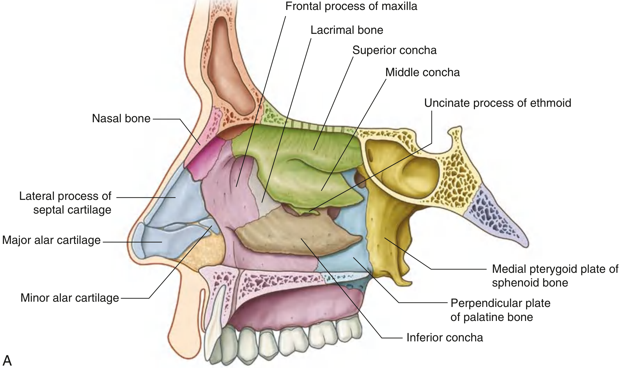
The lateral wall is formed by contributions from several bones (from anterior to posterior):
| Bone | Contribution |
|---|---|
| Nasal bone | Anterior support |
| Frontal process of maxilla | Anterosuperior portion |
| Lacrimal bone | Medial wall of orbit / anterosuperior |
| Ethmoidal labyrinth (ethmoid) | Superior & middle conchae, uncinate process |
| Inferior concha | An independent bone (not part of ethmoid) |
| Perpendicular plate of palatine bone | Posterior portion |
| Medial pterygoid plate of sphenoid | Posteroinferior |
In the external nose, the lateral wall is also supported by cartilage - specifically the lateral process of the septal cartilage, and the major and minor alar cartilages.
Turbinates (Conchae) and Meatuses
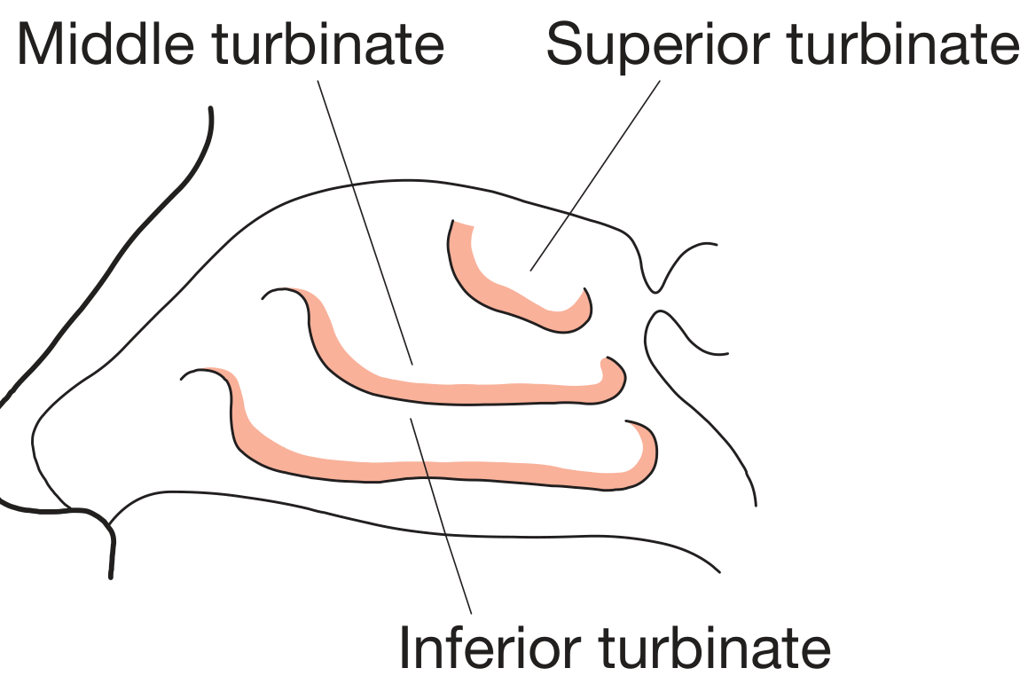
The surface of the lateral wall is interrupted by three nasal conchae - inferior, middle, and superior - which extend medially and divide the nasal cavity into four air channels:
The Four Channels
- Inferior meatus - between inferior concha and floor
- Middle meatus - between middle and inferior conchae
- Superior meatus - between superior and middle conchae
- Spheno-ethmoidal recess - above and behind the superior concha
The anterior end of each concha curves inferiorly to form a lip that overlies the end of the related meatus.
Key Features of the Middle Meatus (Ostiomeatal Complex)
The middle meatus is of greatest clinical importance as it receives drainage from the anterior group of paranasal sinuses:
- Ethmoidal bulla: A dome-shaped elevation on the lateral wall of the middle meatus, formed by the underlying middle ethmoidal air cells. It lies just inferior to the attachment of the middle concha.
- Semilunar hiatus: A curved gutter inferior to the ethmoidal bulla, formed by mucosa spanning the gap between the ethmoidal bulla above and the uncinate process below.
- Ethmoidal infundibulum: The anterior end of the semilunar hiatus forms this channel, into which the frontonasal duct drains the frontal sinus and anterior ethmoidal cells.
Openings on the Lateral Wall
This is a high-yield topic - most paranasal sinuses drain onto the lateral wall:
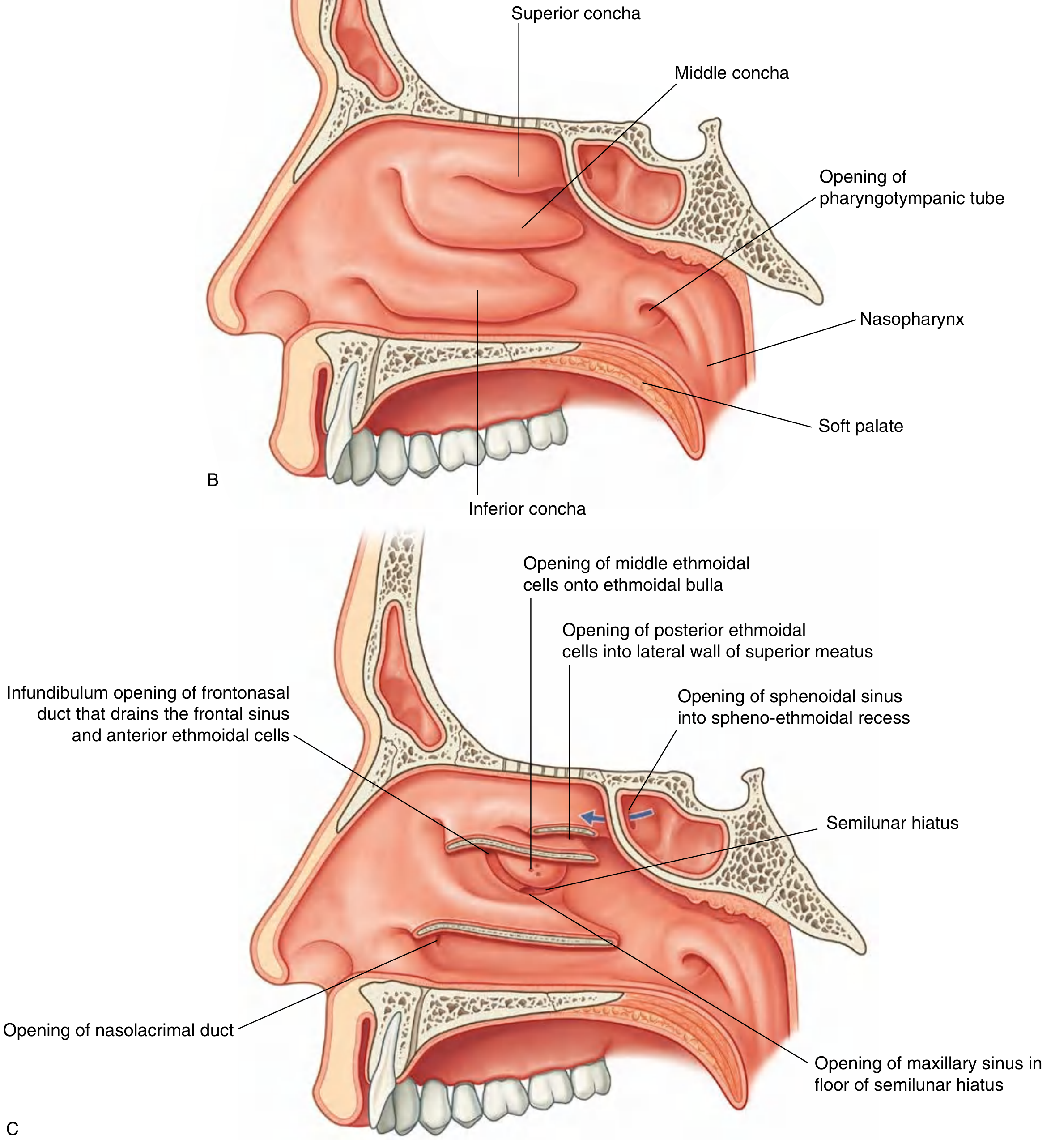
| Structure | Opens into |
|---|---|
| Nasolacrimal duct | Inferior meatus (under anterior lip of inferior concha) |
| Maxillary sinus | Semilunar hiatus in middle meatus (near roof of sinus) |
| Frontal sinus | Via frontonasal duct into ethmoidal infundibulum → anterior semilunar hiatus |
| Anterior ethmoidal cells | Frontonasal duct / ethmoidal infundibulum |
| Middle ethmoidal cells | Onto or just above the ethmoidal bulla |
| Posterior ethmoidal cells | Lateral wall of superior meatus |
| Sphenoidal sinus | Does NOT drain onto lateral wall - opens into the spheno-ethmoidal recess on the sloping posterior roof |
Mnemonic for anterior sinuses: Frontal, Maxillary, and Anterior ethmoidal all drain into the Middle meatus (FMAM = Foramen Middle Anterior/Maxillary).
Blood Supply
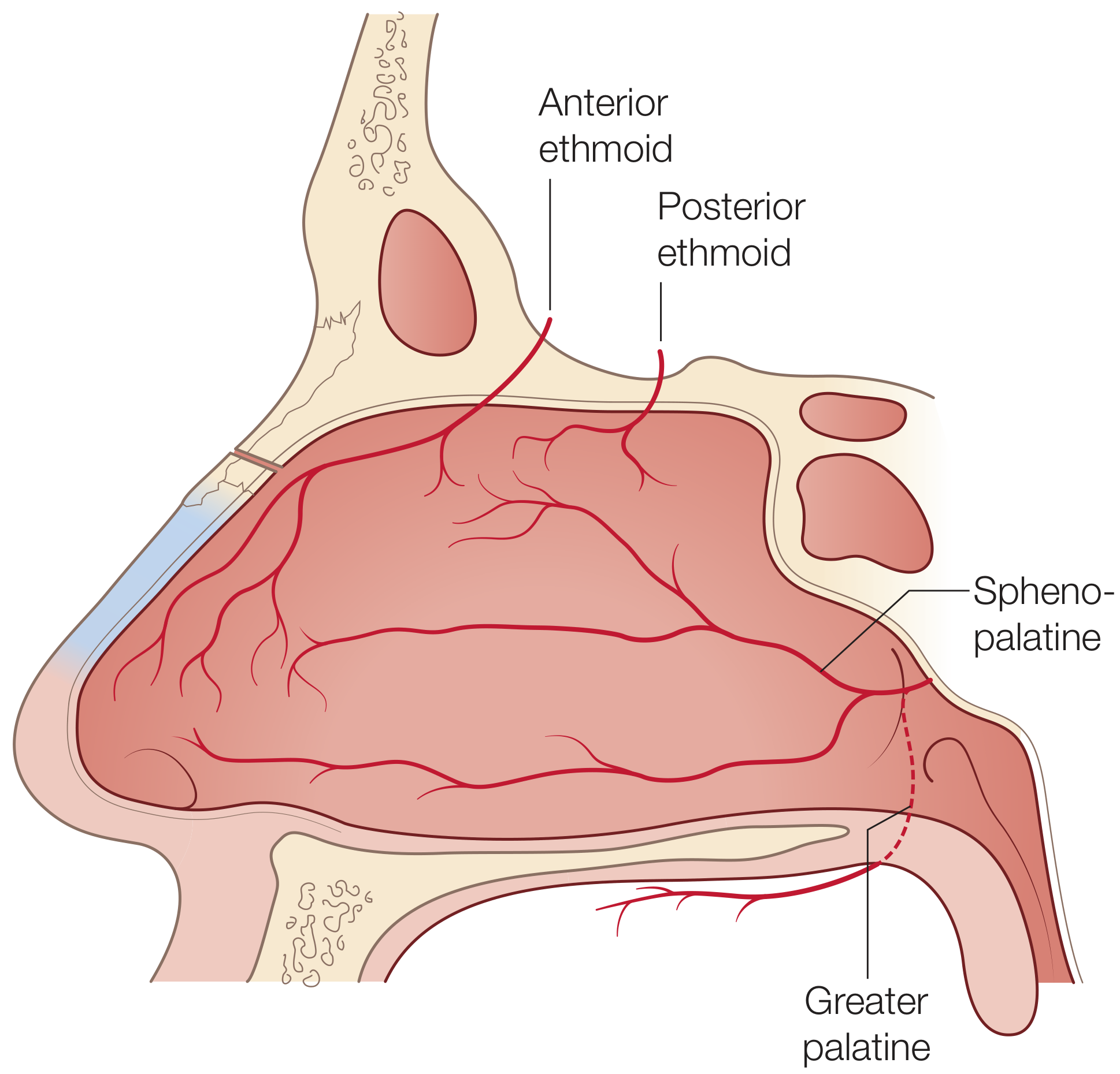
Both internal and external carotid arteries contribute:
External Carotid System
- Sphenopalatine artery (terminal branch of internal maxillary artery): The dominant supply. Enters through the sphenopalatine foramen, which lies just inferior to the horizontal attachment of the middle turbinate. Divides into lateral nasal branches supplying turbinates and most of the lateral wall.
- The crista ethmoidalis (a crest on the perpendicular plate of palatine bone) is the key surgical landmark to identify this vessel.
- Greater palatine artery: Supplies the inferior portion of the lateral wall adjacent to the palate.
- Facial artery: A small branch supplies a small anterior area.
Internal Carotid System
- Anterior ethmoidal artery (branch of ophthalmic artery): Supplies the superior and anterior lateral wall.
- Posterior ethmoidal artery (branch of ophthalmic artery): Supplies the superior posterior lateral wall.
Venous Drainage
Via the ophthalmic and facial veins and the pterygoid and pharyngeal plexuses. Intracranial drainage into the cavernous sinus via the ophthalmic vein is clinically important - it allows intracranial spread of nasal infection.
Nerve Supply
Innervation has autonomic and sensory components:
Sensory (Trigeminal)
- Lateral wall and turbinates: Posterolateral nasal nerves from V2 (maxillary division) arising from the sphenopalatine foramen, plus ethmoidal nerves from V1 (ophthalmic division).
Autonomic
- Parasympathetic (secretomotor): Pre-synaptic fibers travel via the vidian nerve (= greater superficial petrosal nerve [parasympathetic] + deep petrosal nerve [sympathetic]) and synapse in the sphenopalatine ganglion. Post-synaptic fibers innervate the nasal mucosa and regulate nasal secretions.
- Sympathetic (vasomotor): Post-synaptic fibers pass through the sphenopalatine ganglion and regulate vascular tone and turbinate congestion.
Clinical Correlates
- Nasolacrimal duct opening: Located in the inferior meatus - hence watery eyes (epiphora) can produce sniffing/nasal discharge; used when probing/irrigating the lacrimal system.
- Sphenopalatine artery ligation: The artery is targeted endoscopically (identified via the crista ethmoidalis) for refractory epistaxis. Multiple branches exist medial to the crista in ~97% of specimens, so all must be controlled.
- Ostiomeatal complex (OMC): The middle meatus region (uncinate process, ethmoidal infundibulum, ethmoidal bulla, semilunar hiatus) is the functional unit for anterior sinus drainage. OMC obstruction leads to recurrent sinusitis.
- Vidian nerve surgery: Used for vasomotor rhinitis - cutting the vidian nerve reduces parasympathetic drive to the nasal mucosa and decreases secretions.
Sources: Gray's Anatomy for Students, p.1217-1219; Scott-Brown's Otorhinolaryngology Head & Neck Surgery Vol.1, pp.1015-1017; Bailey and Love's Short Practice of Surgery 28th Ed., p.785
This is a shared conversation. Sign in to Orris to start your own chat.