
Lower back pain
"low back pain"[MeSH Terms] AND management
lumbar spine anatomy low back pain diagram
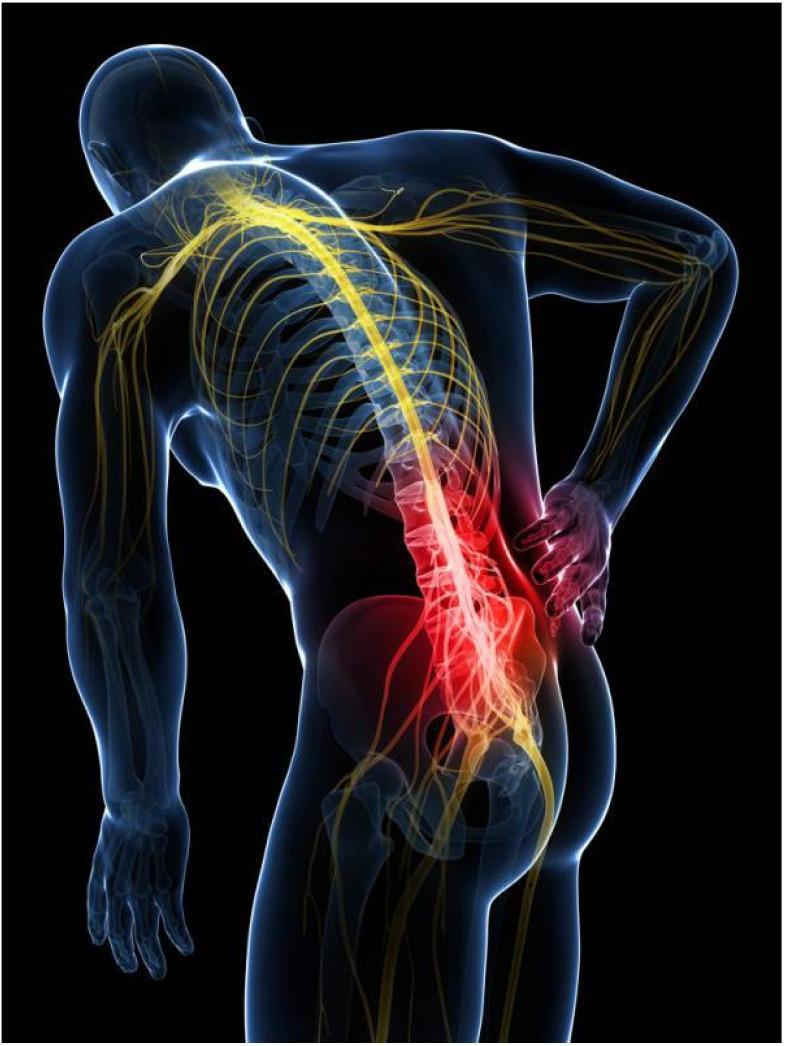
This medical illustration features a translucent anatomical diagram of a human male in a posterior oblique view, emphasizing the skeletal and nervous systems. The image illustrates axial midline low back pain, likely associated with lumbar discogenic disease. The vertebral column is shown from the thoracic region through the sacrum, with a complex network of yellow-coded nerve pathways branching from the spinal cord to the upper and lower extremities. A distinct red glow is concentrated over the lower lumbar spine (L3-L5) and the superior aspect of the pelvis, signifying the site of pain or inflammation. A hand is placed over this lumbar region in a classic clinical gesture for back pain. The diagram effectively demonstrates the relationship between the vertebral structures, neural distribution, and the clinical manifestation of somatic referred pain or discogenic pathology. It is designed for educational use to visualize spinal anatomy and common areas of musculoskeletal distress.
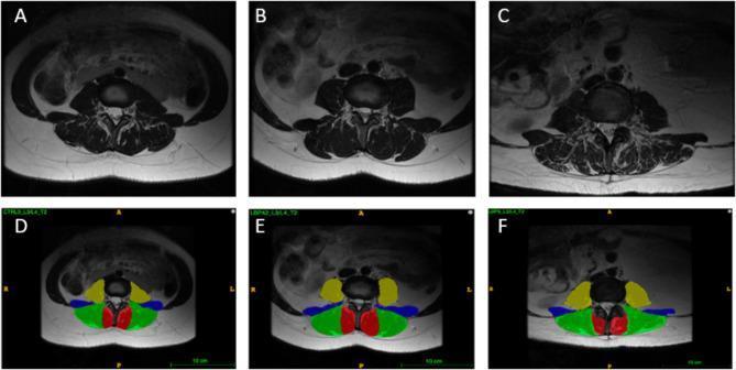
This set of six T2-weighted axial magnetic resonance images (MRI) demonstrates the anatomy of the lumbar spine at the L3/L4 vertebral level. The images compare three female subjects: a healthy control (A, D), an age-matched individual with low back pain (LBP) (B, E), and an older individual with LBP (C, F). Rows A-C show raw radiological data, while rows D-F provide color-coded anatomical segmentations of the paraspinal and core musculature. The segmentations identify the lumbar multifidus (red) adjacent to the spinous process, the erector spinae (green) located postero-laterally, the quadratus lumborum (blue) situated laterally, and the psoas major (yellow) positioned anterior-laterally to the vertebral body. These diagnostic images are designed to illustrate muscular morphology, including variations in muscle cross-sectional area and fatty infiltration associated with aging and chronic pain. High T2 signal intensity (bright areas) within the muscle compartments indicates intramuscular fat or fluid, aiding in the assessment of sarcopenia and tissue quality in clinical and research settings.
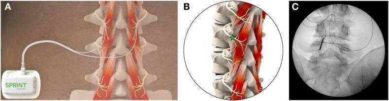
Educational panel illustrating percutaneous peripheral nerve stimulation (PNS) for chronic low back pain. Panel A is a clinical diagram showing a body-worn stimulator (SPRINT system) connected to two fine-wire leads targeting the paraspinal muscles of the lumbar spine. Panel B provides an anatomical illustration of the lumbar vertebrae and paraspinal musculature in an oblique view, highlighting the target location (green star) where the medial branches of the dorsal rami course over the lamina, medial and inferior to the facet joint, to innervate the multifidus muscles. Panel C is an anteroposterior (AP) fluoroscopic diagnostic image demonstrating the intraprocedural placement of a radiopaque introducer needle and lead. The lead is positioned adjacent to the vertebral lamina, confirming appropriate clinical targeting for medial branch stimulation. This content is designed for pain management specialists and radiologists to understand the technical and anatomical approach for treating axial chronic low back pain via neuromodulation.
Lower Back Pain (LBP)
Epidemiology
Classification
| Type | Duration |
|---|---|
| Acute | < 4-6 weeks |
| Subacute | 6 weeks - 3 months |
| Chronic | > 3 months |
Etiologies
1. Myofascial / Mechanical Pain
2. Discogenic Pain
3. Facet Joint Pain
4. Sacroiliac (SI) Joint Pain
5. Radiculopathy (Disc Herniation / Nerve Root Compression)
6. Lumbar Spinal Stenosis
7. Spondylolisthesis / Spondylolysis
8. Inflammatory (e.g., Ankylosing Spondylitis)
9. Referred / Visceral Pain
Red Flags (Require Urgent Evaluation)
| Red Flag | Possible Cause |
|---|---|
| Age >50 or <20 at first episode | Malignancy, fracture |
| History of cancer | Metastatic disease |
| Unexplained weight loss, night sweats | Malignancy, infection |
| Fever / IV drug use | Spinal epidural abscess, discitis |
| Bladder/bowel dysfunction + saddle anesthesia | Cauda equina syndrome (surgical emergency) |
| Bilateral leg weakness/numbness | Cord compression |
| Severe trauma | Fracture |
| Pain unrelieved by rest / nocturnal pain | Malignancy, infection |
| Corticosteroid use or osteoporosis risk | Vertebral fracture |
Diagnosis
- Plain X-rays: first-line for suspected fracture, malignancy, or spondylolisthesis
- MRI: preferred for suspected disc herniation, radiculopathy, infection, malignancy, or spinal stenosis - should not be routine for non-specific LBP
- CT myelography: if MRI contraindicated
- Note: MRI findings of disc degeneration are present in >50% of asymptomatic adults by age 40 and do not reliably correlate with symptoms
Management
Acute Non-specific LBP (< 6 weeks)
- Staying active (bed rest is harmful and delays recovery)
- Heat application
- Physical therapy / targeted exercises
- Patient education and reassurance
- NSAIDs (e.g., ibuprofen, naproxen) - first-line analgesic
- Muscle relaxants (non-benzodiazepine, e.g., cyclobenzaprine, tizanidine) - short-term for spasm
- Acetaminophen - no longer recommended as first-line (insufficient evidence for back pain)
- Opioids - only for severe debilitating pain, lowest dose, shortest duration; avoid with benzodiazepines (respiratory depression risk)
- Gabapentinoids (gabapentin, pregabalin) - insufficient evidence; not recommended
Chronic LBP (> 3 months)
- Exercise therapy (core strengthening, aerobics) - strong evidence
- Cognitive-behavioral therapy (CBT) - addresses catastrophizing, fear-avoidance
- Mindfulness-based stress reduction
- Pilates (recent meta-analysis, PMID 37632387 confirms effectiveness)
- Cognitive Functional Therapy - 2024 systematic review supports benefit in chronic LBP
- Massage, acupuncture, TENS
- NSAIDs (continued as needed)
- Duloxetine (SNRI) - approved for chronic musculoskeletal pain
- Tricyclic antidepressants (e.g., amitriptyline, low-dose) - some evidence
- Fluoroscopic epidural steroid injections (radiculopathy/stenosis)
- Facet joint injections or medial branch blocks
- Radiofrequency ablation of medial branch nerves (facet pain)
- SI joint injections
- Microdiscectomy - for disc herniation with persistent radiculopathy after 6-12 weeks of conservative therapy
- Laminectomy/decompression - for symptomatic spinal stenosis
- Spinal fusion - selected cases; evidence is modest for non-specific LBP
Psychological Co-morbidities
- Depression: 33-67%
- Anxiety: 10-30%
- Substance misuse: 13-40%
- Personality disorders: >50% in some studies
Sleep Posture
Pt was complaining of lower back pain since 6 months I have advised lumbosacral MRI and given treatment is Tab gabapentin GRS 300 BD Tab ultracet BD Cap Bcomplex Tab pantocid DSR OD
"gabapentin" AND "low back pain" AND efficacy
PMID: 37379868
Clinical Review: Chronic LBP Prescription (6 months duration)
Prescription Analysis
1. Tab Gabapentin GRS 300 mg BD (Gabapentin Gastric Retentive System, twice daily)
- Gabapentin is licensed for neuropathic pain - it works by binding alpha-2-delta subunits of voltage-gated calcium channels, reducing neuronal excitability.
- For pure non-specific chronic LBP without radiculopathy or neuropathic features, evidence is weak. The Maudsley Deprescribing Guidelines state: "Systematic reviews have found no benefit of gabapentinoids over placebo in low back pain, sciatica, spinal stenosis."
- A 2023 systematic review (PMID 37379868) of gabapentin/pregabalin for chronic LBP without radiculopathy found only 5 qualifying trials with 242 participants - evidence quality was very low to moderate, and the conclusion was that quality information to support routine use is lacking.
- However, if this patient has a neuropathic/radicular component (shooting leg pain, dermatomal distribution, burning/tingling) - gabapentin is a reasonable and indicated choice.
- Textbook of Family Medicine states: "Anticonvulsant medications such as gabapentin may be indicated for neuropathic pain but do not have evidence of effectiveness in more general musculoskeletal pain."
- Both gabapentin and tramadol/opioid combinations carry additive CNS depression risk
- The Maudsley guidelines explicitly warn: "Co-prescription [of gabapentinoids with opioids] should usually be avoided because of increased risk of respiratory depression, accidental overdose and death."
- The MHRA has issued warnings on this combination. You should counsel the patient about sedation, dizziness, and avoid driving/operating machinery.
2. Tab Ultracet BD (Tramadol 37.5 mg + Paracetamol/Acetaminophen 325 mg)
- Ultracet = Tramadol 37.5 mg + Acetaminophen 325 mg per tablet
- Tramadol is a weak opioid agonist + serotonin-norepinephrine reuptake inhibitor (SNRI), giving it a dual mechanism useful in pain with a neuropathic component
- The combination with acetaminophen provides synergistic analgesia
- Textbook of Family Medicine confirms: "Combination analgesic therapy with acetaminophen and tramadol was found to decrease pain significantly"
- Tramadol is a good choice for chronic LBP as it provides moderate analgesia with lower addiction potential than stronger opioids
- Tramadol + Gabapentin = additive sedation risk (as above)
- Tramadol lowers seizure threshold - of note if patient has any seizure history
- Tramadol is a serotonergic drug - avoid combination with SSRIs/SNRIs (serotonin syndrome risk)
- BD dosing of Ultracet is appropriate for chronic use; monitor for tolerance and dependence with long-term use
- Tramadol is not ideal as a long-term standalone strategy without a plan to introduce stronger disease-modifying therapies (physiotherapy, exercise)
3. Cap B-Complex (Vitamin B Complex)
- B vitamins (B1, B6, B12) support peripheral nerve health and are commonly used as adjuncts in back pain, especially when there is a neuropathic/radicular component
- B12 deficiency can itself cause neuropathic pain and should be excluded
- Low risk, reasonable add-on, especially with the neuropathic pain medication on board
- No major interactions
4. Tab Pantocid DSR OD (Pantoprazole 40 mg + Domperidone 30 mg SR, once daily)
- Tramadol can cause nausea/vomiting and GI upset; domperidone component helps with this
- Pantoprazole (PPI) provides gastric protection - particularly relevant if the patient is also taking NSAIDs or has GI risk factors
- Domperidone SR component is particularly helpful for tramadol-induced nausea, which is one of the most common reasons for discontinuation
- Good choice as a cover for this prescription
Summary Scorecard
| Drug | Indication | Evidence | Concern |
|---|---|---|---|
| Gabapentin GRS 300 BD | Neuropathic/radicular LBP | Weak for non-radicular; reasonable for neuropathic | Sedation + tramadol interaction |
| Ultracet BD | Moderate chronic LBP | Moderate - reasonable | Sedation + seizure threshold |
| B-Complex | Nerve support | Supportive | None |
| Pantocid DSR OD | GI protection, antiemetic | Appropriate | None |
Suggestions to Strengthen Management
-
Await MRI results before finalising pharmacotherapy - if there is disc herniation with nerve root compression (L4-L5, L5-S1), gabapentin is strongly justified and dose can be optimised.
-
Add an NSAID (e.g., Tab Etoricoxib 60 mg OD or Tab Aceclofenac 100 mg BD with food) if not contraindicated - NSAIDs remain first-line for chronic LBP with inflammatory/mechanical component and are more evidence-based than gabapentin for non-neuropathic pain.
-
Physiotherapy referral is mandatory - Exercise therapy (core strengthening, stretching) has the strongest long-term evidence for chronic LBP. Medication alone without physiotherapy rarely leads to lasting improvement.
-
Counsel on gabapentin + tramadol CNS interaction - warn patient about dizziness, sedation, avoid driving, no alcohol.
-
Set a 4-6 week review - Reassess pain scores, functional improvement, and side effects. If no meaningful improvement with gabapentin, consider tapering it, especially if MRI shows no radiculopathy.
-
Consider Duloxetine 30-60 mg OD as an alternative/add-on - it has FDA approval for chronic musculoskeletal pain and addresses any co-morbid depression/anxiety (which is highly prevalent in chronic LBP).
-
Screen for psychological co-morbidities - Depression and anxiety co-occur in 33-67% of chronic LBP patients and worsen outcomes. Brief PHQ-9 / GAD-7 screening is worthwhile.