
Vestibular apparatus for Physiology mbbs 1st year student
Reading File
Finding Sources
Finding Sources
Searching Images
vestibular apparatus semicircular canals utricle saccule diagram
Guyton and Hall has a dedicated section, perfect for physiology. Let me pull the full content from that block (lines 492-644 covers structure, function, and CNS connections).
Reading File
Searching Images
crista ampullaris hair cells cupula
Searching Images
macula utricle otolith organ hair cells
Reading File
Searching PubMed
vestibular system physiology
Reading File
I have enough content now. Let me compile the complete answer.
Vestibular Apparatus — Physiology (MBBS I)
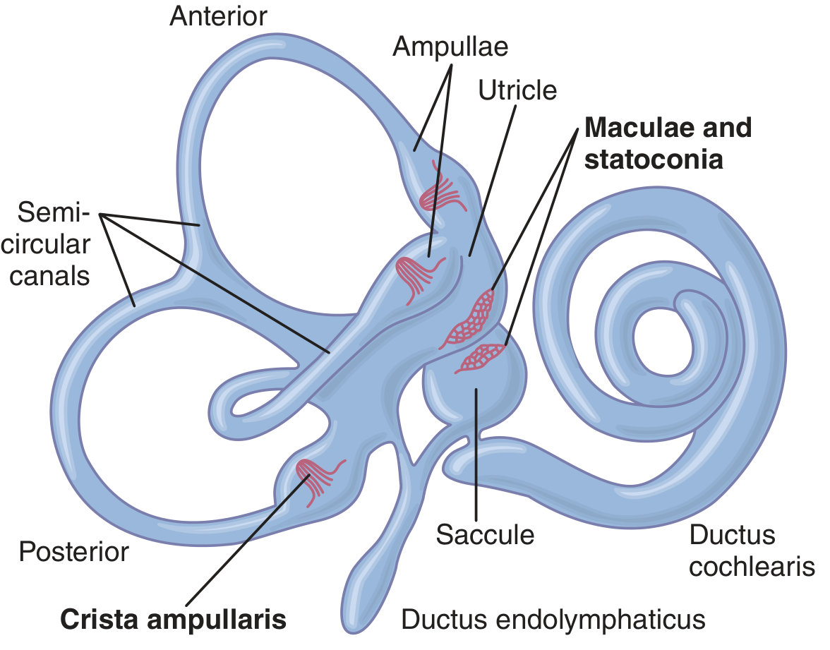
The vestibular apparatus is the sensory organ for equilibrium (balance). It lies in the petrous part of the temporal bone, inside the bony labyrinth, which houses the fluid-filled membranous labyrinth - the functionally active part.
Components
The membranous labyrinth consists of:
- Cochlea (ductus cochlearis) - concerned with hearing, not equilibrium
- Three semicircular ducts (anterior, posterior, lateral/horizontal)
- Utricle and saccule - two membranous sacs
The semicircular ducts, utricle, and saccule together form the equilibrium mechanism (Guyton and Hall Textbook of Medical Physiology, p. 694).
1. Maculae of the Utricle and Saccule - Static Equilibrium
Each utricle and saccule has a sensory patch called the macula (~2 mm diameter):
- Macula of the utricle lies in the horizontal plane -> senses head orientation when upright
- Macula of the saccule lies in the vertical plane -> senses orientation when lying down
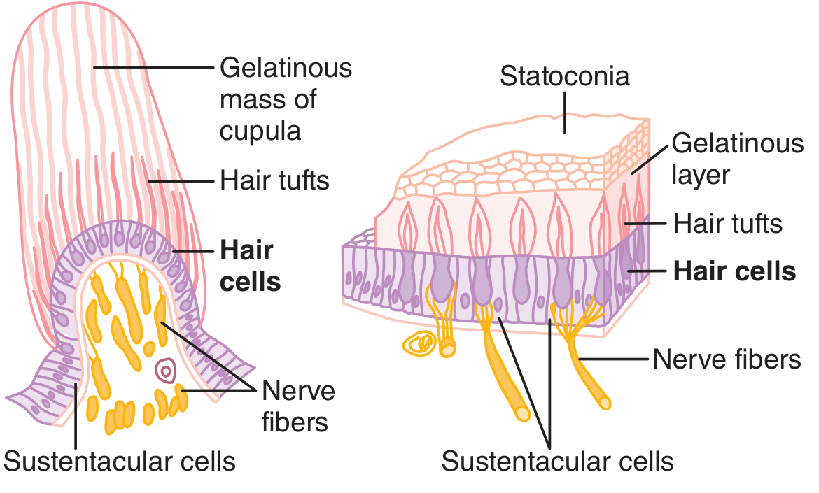
Each macula is covered by a gelatinous layer embedded with calcium carbonate crystals called statoconia (otoliths), which are denser than the surrounding endolymph. Gravity pulls the statoconia, bending the hair cell cilia beneath them.
Hair cell mechanism (kinocilium/stereocilia):
Each hair cell has ~100 stereocilia and one large kinocilium on one side. Fine filaments link each stereocilium to the next longer one.
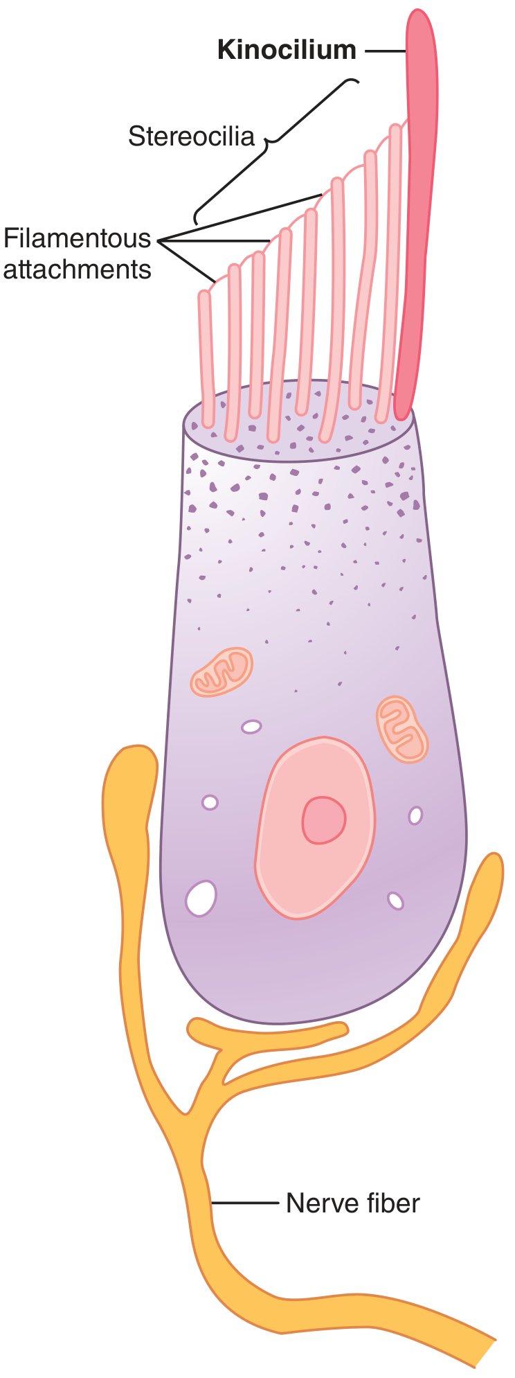
- Bending stereocilia toward the kinocilium -> opens cation channels -> depolarization -> increased nerve firing (baseline ~100 impulses/sec, rising to several hundred)
- Bending away from the kinocilium -> channels close -> hyperpolarization -> decreased/silenced firing
Since hair cells within a macula are oriented in many different directions, each head position produces a unique "pattern" of excitation across the vestibular nerve fibers - this pattern tells the brain the head's orientation in space (Guyton and Hall, p. 695).
Detection of linear acceleration: When the body accelerates forward, inertia causes the statoconia to lag backward, bending the cilia and signaling "falling backward" - prompting a compensatory forward lean. Note the maculae detect linear acceleration, not constant linear velocity.
2. Semicircular Ducts - Detection of Head Rotation (Dynamic Equilibrium)
The three ducts (anterior, posterior, lateral) are arranged at right angles, representing all three planes in space. Each has a dilated end, the ampulla, containing a crest called the crista ampullaris, topped by a gelatinous cupula into which hair cell cilia project.
When the head rotates (angular acceleration), the inertia of the endolymph causes it to lag behind, generating relative flow that bends the cupula - deflecting the hair cells and altering their firing rate (increased if bent toward the kinocilium orientation, decreased if away).
Key features of the semicircular canal response (predictive function):
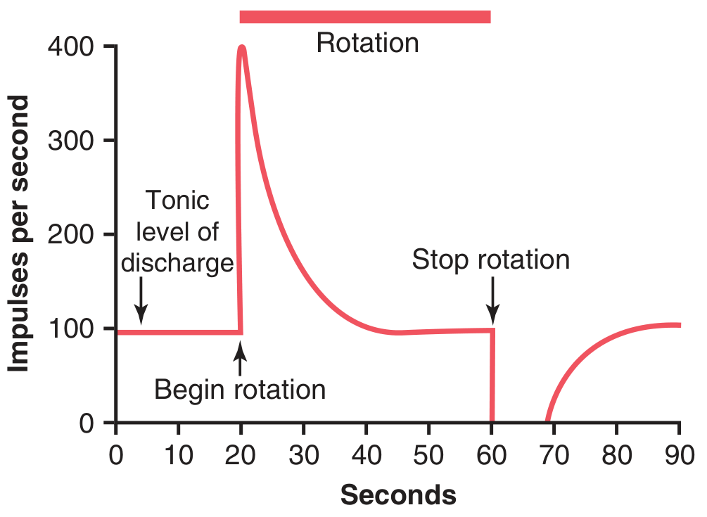
- Resting discharge: ~100 impulses/sec
- Onset of rotation: sharp increase in discharge
- With continued constant rotation, discharge decays back to baseline over seconds (adaptation) because the endolymph catches up with the rotating duct and the cupula springs back
- Stopping rotation causes the opposite effect - the endolymph continues moving, bending the cupula the other way, causing a transient decrease below baseline (this is why you feel like you are spinning the opposite way after stopping - post-rotatory nystagmus/vertigo)
This makes the semicircular ducts a predictive apparatus - they detect that the head is beginning to turn and allow the CNS to pre-emptively correct posture and eye position (vestibulo-ocular reflex) before disequilibrium actually occurs, unlike the maculae which detect equilibrium after it has already been disturbed.
3. Central Connections
Signals from the vestibular apparatus travel via the vestibular nerve to the vestibular nuclei in the brainstem, and are integrated with the cerebellum (especially the flocculonodular lobe) and reticular formation. These centers coordinate:
- Postural reflexes (antigravity muscle tone)
- Eye movements (vestibulo-ocular reflex, nystagmus)
- Conscious sense of orientation, integrated with visual and proprioceptive input
Quick summary table
| Structure | Stimulus detected | Mechanism |
|---|---|---|
| Utricle macula | Head tilt (static), linear acceleration (horizontal) | Statoconia shift with gravity/inertia, bend cilia |
| Saccule macula | Head tilt (static), linear acceleration (vertical) | Same, vertical plane |
| Semicircular ducts (3, at right angles) | Angular (rotational) acceleration | Endolymph inertia bends cupula in crista ampullaris |
Source: Guyton and Hall Textbook of Medical Physiology, Ch. 56 "Vestibular Sensations and Maintenance of Equilibrium," pp. 694-696.
This is core exam content - expect questions on hair cell polarization (kinocilium/stereocilia direction), the difference between static (maculae) vs dynamic (semicircular canal) equilibrium, and the adaptation/post-rotatory response of the crista ampullaris.
This is a shared conversation. Sign in to Orris to start your own chat.