
single shot epidural anaesthesia
epidural needle insertion technique loss of resistance Tuohy needle
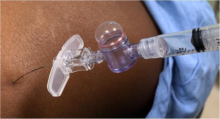
This procedural photograph illustrates the Loss of Resistance (LOR) technique for identifying the epidural space using an Epidrum device. A 25-gauge Tuohy needle is inserted through the skin of a patient's back. The needle hub is connected to an Epidrum, a transparent, air-filled diaphragm device designed to provide a visual indication of pressure changes. Positioned at the distal end of the assembly is a clear plastic syringe containing fluid, used to inflate the device's dome-shaped silicone diaphragm. In this specific clinical scenario, the Epidrum diaphragm remains inflated and spherical, signifying high resistance and a 'negative' result, indicating that the needle tip has not yet reached the epidural space. The image demonstrates the integration of objective visual monitoring tools in neuraxial anesthesia to enhance the accuracy of epidural needle placement, particularly when using smaller-gauge needles where tactile feedback is reduced.
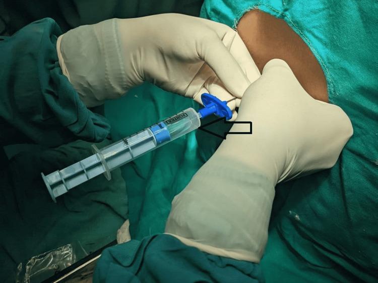
Clinical photograph demonstrating a medical procedure in an anesthesiology context. The image shows a practitioner's gloved hands performing an epidural or spinal intervention on a patient's back. A Tuohy needle is inserted into the vertebral interspace, connected via a blue hub to a 'Loss of Resistance' (LOR) syringe. The syringe, marked with clear graduations, contains transparent, colorless fluid, which is clinically identified as cerebrospinal fluid (CSF). This visual demonstrates an accidental dural puncture during an attempted epidural block, where the presence of CSF in the LOR syringe confirms the needle has entered the intrathecal space. The procedural field is maintained with sterile green drapes, and the practitioner is wearing sterile surgical gloves and gown. This image serves as an educational tool for identifying the 'Loss of Resistance' technique and the visual confirmation of dural breach in regional anesthesia.
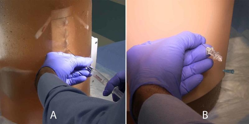
Two clinical photographs (A and B) demonstrate the use of a high-fidelity 3D-printed neuraxial block task trainer for medical simulation. Image A shows an operator in blue gloves performing a simulated epidural procedure. A 20G Tuohy needle is inserted into a translucent ballistic gelatin matrix that reveals the underlying 3D-printed lumbar vertebral anatomy (T10-L5). A syringe is attached to the needle hub, simulating the loss of resistance technique. Image B depicts a simulated spinal block on the same model, now covered with a beige silicone skin to mimic human tissue for landmark palpation. A 22G Whitacre spinal needle is inserted perpendicularly, and clear saline (simulating cerebrospinal fluid) is seen egressing from the clear needle hub, confirming successful dural puncture. This content illustrates anesthesiology training, specifically the procedural steps for neuraxial anesthesia, landmark-based needle insertion, and the verification of needle placement in the epidural and subarachnoid spaces.
Single-Shot Epidural Anaesthesia
Definition and Concept
- Pfenninger and Fowler's Procedures for Primary Care, 3rd ed. - "Single-shot epidural injections may be used for surgical procedures of short duration. If a longer-duration procedure is anticipated or if postoperative pain relief is desired, place an epidural catheter."
- Miller's Anesthesia, 10e - Single-shot epidural blocks are appropriate for many pediatric surgeries, especially when adjuvants such as clonidine are co-administered.
Indications
- Short-duration surgical procedures (anorectal, lower limb, perineal, inguinal, lower abdominal)
- Pediatric surgeries below the umbilicus (lumbar or caudal approach)
- Situations where catheter placement is technically difficult or undesirable
- Diagnostic epidural injections (e.g., foraminal epidural steroid injection for radiculopathy)
- Labor analgesia when only a single bolus is needed (e.g., instrumental delivery)
Anatomy of the Epidural Space
Equipment
- Tuohy needle: 16-18 gauge for adults; curved bevel (Huber tip) reduces risk of dural puncture and guides catheter passage (though in SSE no catheter is used)
- Loss-of-resistance (LOR) syringe (glass or low-friction plastic)
- 18-gauge skin puncture needle (introducer)
- Sterile drapes, antiseptic, local anaesthetic for skin infiltration
- Preservative-free drug solutions for epidural injection
Patient Positioning
- Lateral decubitus (fetal position): most common; flexion of spine widens interspinous spaces
- Sitting position: easier for landmark identification in obese patients; useful in conscious patients
- Neck and back maximally flexed; spine straight and not rotated
- An assistant stands in front to maintain patient position
Technique (Step-by-Step)
1. Preparation
- IV access established; IV fluid preload (500-1000 mL crystalloid to minimize hypotension)
- Monitoring: BP (every 2-3 min), ECG, pulse oximetry
- Emergency drugs and equipment (ephedrine, atropine, lipid emulsion) immediately available
- Sterile technique throughout; preservative-free drugs only
2. Skin Infiltration
- Identify the appropriate interspace (L2-3, L3-4, or L4-5 for lumbar epidural)
- Infiltrate skin and deeper tissues with 1% lidocaine along the planned needle trajectory
- Skin puncture with an 18G introducer needle to ease passage of the Tuohy needle
3. Needle Insertion
- The Tuohy needle is advanced via a midline approach (most common) or paramedian approach
- Midline: perpendicular to skin at L4-5 (~90°), angled slightly more cephalad at L2-3 (~70°)
- Paramedian: useful when midline approach is obstructed by calcified ligaments or scoliosis
- The needle passes through: skin → subcutaneous fat → supraspinous ligament → interspinous ligament → ligamentum flavum → epidural space
4. Identifying the Epidural Space
- Attach LOR syringe containing saline or air to needle hub
- While needle tip is in ligamentum flavum, injection meets firm resistance
- Advance needle slowly, millimetre by millimetre, with continuous or intermittent pressure on plunger
- On entering the epidural space, resistance suddenly disappears - the plunger "collapses"
- A drop of saline placed at the needle hub is sucked inward as the needle tip enters the epidural space, due to negative pressure
- More reliable for thoracic than lumbar insertion
- Less commonly used; risk of false-negative if needle becomes plugged
- Air: simpler, easier to detect accidental dural puncture; but intracranial air or patchy block possible
- Saline: meta-analysis shows no clear clinical superiority of either; clinician preference governs
- Barash's Clinical Anesthesia, 9e - "Clinical outcomes are similar when anesthesiologists use the technique of their choice."
5. Test Dose (MANDATORY before injection)
- Aspiration test first - no blood (intravascular) or CSF (intrathecal) should return
- Classic test dose: 3 mL of 1.5% lidocaine + 15 mcg epinephrine (1:200,000)
- 45 mg lidocaine → will produce rapid spinal block if intrathecal
- 15 mcg epinephrine → will cause ≥20% heart rate rise if intravascular
- Aspiration alone is insufficient - false-negatives occur even with experienced practitioners
- Morgan & Mikhail's Clinical Anesthesiology, 7e - "Rescue lipid emulsion (20% Intralipid 1.5 mL/kg) must be available whenever epidural blocks are performed in the event of local anesthetic systemic toxicity."
6. Main Injection (Single Shot)
- After confirming correct placement, inject the full dose slowly and incrementally
- Volume guidelines:
- Adults: 1-2 mL per segment to be blocked (e.g., T4 level from L4-5 requires ~12-24 mL)
- Children: approximately 0.1 mL/year of age per neuromere; usual range 0.5-1 mL/kg (up to 20 mL)
- Inject slowly, pausing to reassess if any systemic symptoms appear
Drugs Used
| Agent | Concentration | Onset | Duration | Notes |
|---|---|---|---|---|
| Bupivacaine | 0.5% (surgical), 0.25% (analgesic) | Slow (15-20 min) | Long (3-4 h) | Most widely used; avoid >0.75% (cardiac toxicity) |
| Ropivacaine | 0.5-0.75% | Moderate | Long | Less cardiotoxic; less motor block than bupivacaine |
| Lidocaine | 1.5-2% | Fast (10-15 min) | Moderate (90 min) | Adding epinephrine prolongs and intensifies block |
| Chloroprocaine | 3% | Fastest | Short (45-60 min) | Low systemic toxicity; good for short procedures |
- Epinephrine (1:200,000): prolongs block, reduces systemic absorption, acts as marker for intravascular injection
- Clonidine (1-2 mcg/kg in children): extends duration, reduces LA dose needed
- Morphine (30 mcg/kg in children, up to 2-4 mg adults): prolonged postoperative analgesia
- Hydromorphone (10 mcg/kg in children): alternative opioid adjuvant
- Sodium bicarbonate (1 mEq/10 mL): alkalinises solution → faster onset (not used with bupivacaine - precipitates above pH 6.8)
- Miller's Anesthesia, 10e - single-shot blocks are appropriate especially when adjuvants such as clonidine 1-2 mcg/kg and morphine 30 mcg/kg are co-administered.
Factors Affecting Level of Block
- Volume: primary determinant of spread in epidural (unlike spinal where baricity matters)
- Age: dose requirement decreases with age (reduced epidural space compliance)
- Height: minor correlation; less predictable than in spinal anaesthesia
- Position: patient positioning during/after injection affects spread
- Injection site level: lumbar, thoracic, or caudal
- Speed of injection: faster injection → slightly wider spread
- Morgan & Mikhail, 7e - "Factors affecting the level of epidural anesthesia may not be as predictable as with spinal anesthesia."
Comparison: Single-Shot vs. Catheter Technique
| Feature | Single-Shot Epidural | Continuous Catheter |
|---|---|---|
| Duration | Limited to drug's duration | Indefinite (infusion/PCEA) |
| Procedure length | Short operations | Long operations / ICU |
| Hypotension control | Abrupt onset | More titratable |
| Postoperative analgesia | Not suitable | Excellent |
| Technical complexity | Simpler | Higher (catheter migration) |
| Infection risk | Lower | Higher (prolonged catheter) |
| Risk of intravascular migration | N/A | Catheter can migrate at any time |
Contraindications
- Patient refusal
- Coagulopathy / anticoagulation (within specified time intervals)
- Infection at injection site
- True local anaesthetic allergy
- Raised intracranial pressure (relative)
- Severe spine deformity
- Haemodynamic instability
- Intraspinal tumours or lesions
- Prior spinal surgery at the level
- Tethered cord syndrome
- Sepsis (risk of epidural abscess)
Complications
| Complication | Mechanism | Management |
|---|---|---|
| Hypotension | Sympathetic blockade | IV fluids, ephedrine/phenylephrine |
| Accidental dural puncture (ADP) | Needle through dura | Convert to spinal if LOR confirmed; PDPH if unrecognised |
| Post-dural puncture headache (PDPH) | CSF leak | Conservative/epidural blood patch |
| Total spinal | Intrathecal injection of epidural dose | ABCDE resuscitation, intubation |
| Local anaesthetic systemic toxicity (LAST) | Intravascular injection | Lipid emulsion 20% (Intralipid 1.5 mL/kg IV) |
| Epidural haematoma | Venous plexus trauma | Urgent surgical decompression |
| Epidural abscess | Infection | Antibiotics + surgical drainage |
| Nerve injury | Direct trauma or ischaemia | Neuromonitoring, neurology referral |
| Inadequate/unilateral block | Misplacement, septum | Repositioning, supplementation |
Special Considerations
Paediatrics
- Caudal approach preferred in infants/young children for surgery below the umbilicus; lumbar in older children
- Distance from skin to epidural space: approximately 1 mm/kg (6 months-10 years)
- Ultrasound guidance strongly recommended in children - allows real-time visualisation of ligamentum flavum, dura, and local anaesthetic spread
- Miller's Anesthesia, 10e
Obstetrics
- Single-shot epidural opioids (e.g., morphine 2.5 mg) used for post-caesarean analgesia
- Single-shot neuraxial techniques for labour: lower opioid dose preferred; caution with repeated dosing re: neonatal effects
Thoracic Epidural (Single Shot)
- Requires higher expertise due to proximity of spinal cord
- Hanging drop technique preferred by some at this level
- Used for thoracotomy, oesophagectomy when catheter not feasible
Key Points Summary
- Single-shot epidural is suitable for short procedures where prolonged analgesia is not required
- The Tuohy needle is inserted midline at the appropriate lumbar interspace, with the epidural space identified by loss of resistance to saline or air
- A test dose (lidocaine + epinephrine) is mandatory before the main injection to exclude intrathecal or intravascular placement
- Main drug volume: 1-2 mL per segment in adults; 0.5-1 mL/kg in children
- Bupivacaine 0.5% and ropivacaine 0.75% are the most commonly used agents; adjuvants extend duration and improve quality of block
- Complications include hypotension, dural puncture, total spinal, LAST, haematoma, and infection - preventable with test dose and incremental injection
- Lipid emulsion 20% (Intralipid) must always be immediately available