
Cellulitis
cellulitis skin infection erythema swelling lower leg
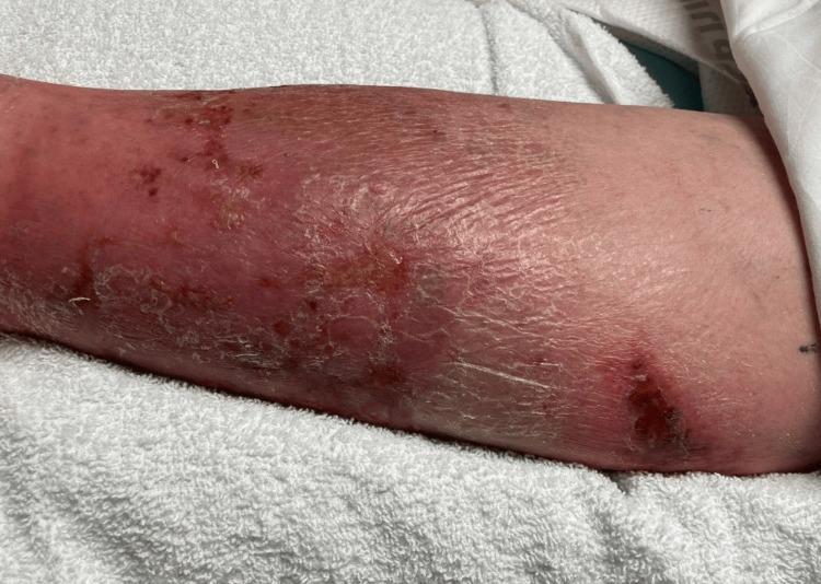
This clinical photograph displays the lower extremity of a patient, demonstrating classic physical examination findings of cellulitis. The skin exhibits diffuse erythema and swelling extending across the anterior and lateral aspects of the leg. The texture is notably compromised, showing signs of desquamation (peeling), xerosis (dryness), and lichenification, particularly in the mid-to-lower portion. Multiple discrete skin lesions are visible, including areas of darker, crusted scabbing and potential ulceration that may serve as a portal of entry for infection. Small petechiae or ecchymotic spots are scattered within the erythematous region. The presentation is highly characteristic of an acute bacterial skin and soft tissue infection (SSTI), emphasizing the visual transition from healthy proximal skin to distal inflamed, edematous, and scaling tissue. This image is an educational resource for identifying inflammatory dermatological conditions and assessing secondary skin changes associated with chronic or severe cellulitis.
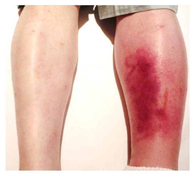
This clinical photograph shows a posterior view of a patient's lower legs, providing a side-by-side comparison between a healthy limb and a symptomatic one. The left lower leg (right side of the image) exhibits classic clinical signs of cellulitis, characterized by significant unilateral edema and diffuse erythema. The affected skin shows a spectrum of inflammatory discoloration, ranging from bright red to deep purplish hues, particularly over the calf region. The skin texture appears tense and glossy due to underlying swelling. A thin, vertical line of serous fluid exudate is visible on the posterior-lateral aspect of the left calf. In contrast, the right lower leg (left side of the image) appears normal with healthy skin tone and no evidence of swelling. This visual demonstrates the acute presentation of a soft tissue infection, useful for teaching the differential diagnosis of unilateral leg swelling and redness, which may include cellulitis, deep vein thrombosis (DVT), or localized vasculitis.
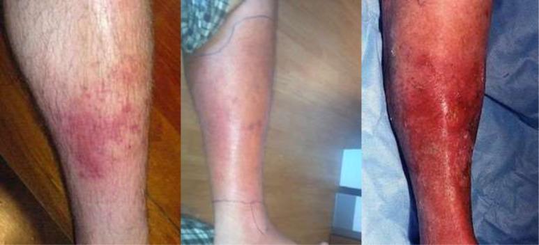
A sequence of three clinical photographs illustrating the progression of cellulitis on the lower leg, categorized by clinical severity. The 'Mild' stage (left) shows a localized, erythematous patch with poorly defined borders and scattered red papules. The 'Moderate' stage (center) demonstrates expanding confluent erythema and edema covering a larger surface area; notably, ink markings are visible on the skin to monitor the spread of infection. The 'Severe' stage (right) depicts intense, deep red to violaceous erythema involving the majority of the limb, accompanied by significant inflammatory swelling, a shiny skin texture suggestive of severe edema, and focal areas of potential skin breakdown or desquamation. This comparison highlights key diagnostic features of skin and soft tissue infections (SSTIs), including changes in color intensity, surface area involvement, and signs of tissue tension associated with worsening bacterial infection.
cellulitis skin AND infection management
Cellulitis
Definition & Pathophysiology
- Nonpurulent - most common form; typically caused by beta-hemolytic streptococci (Group A Streptococcus accounts for ~75% of cases)
- Purulent - drains freely; primarily caused by Staphylococcus aureus, including CA-MRSA
- Andrews' Diseases of the Skin, p. 14.17
- Rosen's Emergency Medicine, p. 2741
Etiology by Clinical Setting
| Setting | Common Pathogens |
|---|---|
| Typical nonpurulent cellulitis | Streptococcus pyogenes (Group A), other beta-hemolytic streptococci |
| Purulent cellulitis / abscesses | S. aureus (CA-MRSA in most US cases) |
| Diabetic foot | S. aureus, streptococci (acute); polymicrobial with gram-negatives and anaerobes (chronic) |
| Cat bites | Pasteurella multocida |
| Human bites | Polymicrobial (Eikenella, Fusobacterium, Group A Streptococcus, S. aureus) |
| Seawater exposure | Vibrio vulnificus (especially in liver disease), Erysipelothrix rhusiopathiae |
| Freshwater exposure | Aeromonas spp. |
- Rosen's Emergency Medicine, p. 2741
Risk Factors
-
Disruption of the skin barrier: wounds, tinea pedis, dermatophyte infections
-
Venous insufficiency and stasis
-
Lymphedema (impaired lymphatic drainage)
-
Obesity
-
Diabetes mellitus
-
Immunocompromised states (HIV, malignancy, immunosuppressive drugs)
-
Prior saphenous vein harvesting, lymphadenectomy, or radiation
-
Alcoholism
-
Goldman-Cecil Medicine, Chapter 408
-
Andrews' Diseases of the Skin
Clinical Features
- Erythema - rapidly spreading, poorly defined and irregular borders
- Warmth and edema - the affected skin may pit on pressure
- Vesicles or bullae - from severe localized edema
- Lymphangitis - streaks extending proximally toward regional lymph nodes
- Lymphadenopathy (regional)
Key clinical pearl: Cellulitis is rarely bilateral. Bilateral lower leg redness should strongly prompt consideration of other diagnoses (e.g., venous stasis dermatitis).
- Rosen's Emergency Medicine, p. 2740-2741
Differential Diagnosis ("Pseudocellulitis")
| Mimic | Distinguishing Features |
|---|---|
| Venous stasis dermatitis | Usually bilateral, centered over medial malleoli, no fever, no pain, often improves with elevation |
| Deep vein thrombosis (DVT) | Unilateral leg swelling, confirmed on Doppler ultrasound |
| Contact dermatitis | Pruritic rather than painful, no systemic signs |
| Erythema migrans (Lyme) | Target/bullseye appearance, >5 cm, central clearing, tick exposure history |
| Eosinophilic cellulitis | Exuberant insect bite reaction; eosinophilia rather than neutrophilia |
| Gout | Acute joint involvement, hyperuricemia, responds to NSAIDs |
| Fixed drug eruption | Drug exposure history |
| Necrotizing fasciitis | Pain out of proportion, rapid progression, bullae with dark fluid, systemic toxicity - surgical emergency |
- Rosen's Emergency Medicine, p. 2741
- Andrews' Diseases of the Skin
Diagnosis
-
Blood cultures: Not indicated in uncomplicated cases. Recommended for immunocompromised patients or systemic infection
-
Wound cultures: Needle aspiration, superficial swabs, and skin biopsy are low-yield in nonpurulent cellulitis and not routinely recommended. Useful in: purulent cellulitis, immunocompromised hosts, bite wounds, immersion injuries
-
Lab work (CBC, CRP, ESR): Not routinely needed but may help assess severity and guide hospitalization decisions
-
Bedside ultrasound: High-frequency linear probe helps differentiate cellulitis (hyperechoic/cobblestoning) from abscess (hypoechoic with posterior acoustic enhancement)
-
CT/MRI: Consider if necrotizing fasciitis is suspected
-
Rosen's Emergency Medicine, p. 2741-2742
Management
Outpatient (mild, non-toxic, immunocompetent)
| Drug | Dose | Duration |
|---|---|---|
| Cephalexin (first-line) | 500 mg QID orally | 5-10 days |
| Dicloxacillin | 500 mg QID orally | 5-10 days |
| Clindamycin | 300-450 mg QID orally | 10 days |
| TMP-SMX | 160/800 mg BID orally | 10 days |
| Penicillin VK (streptococcal) | 500 mg QID orally | 10 days |
If MRSA is strongly suspected (prior MRSA, failed beta-lactam, purulent infection), use clindamycin or TMP-SMX.
Inpatient / Severe
- IV nafcillin or oxacillin for MSSA
- IV vancomycin (15-20 mg/kg BID) for MRSA or when pathogen unclear
- Vancomycin + imipenem (or piperacillin-tazobactam) for severe/polymicrobial cases
- If Group A Strep confirmed: IV Penicillin G (4 million units q4h) + IV Clindamycin (600-900 mg q8h) for 10-14 days
Adjunctive Measures
-
Limb elevation to reduce edema
-
Mark the borders of erythema with a skin marker to monitor progression
-
Compression therapy in lymphedema to reduce recurrence risk
-
Treat underlying risk factors (tinea pedis, venous insufficiency)
-
Goldman-Cecil Medicine
-
Andrews' Diseases of the Skin
-
Rosen's Emergency Medicine, p. 2742
Complications
- Lymphangitis and regional lymphadenopathy
- Bacteremia and sepsis
- Necrotizing fasciitis - the most feared complication; requires emergent surgical debridement (mortality 30-60%, up to 70-85% when diagnosis delayed)
- Metastatic abscesses
- Gangrene
- Chronic lymphedema / elephantiasis nostras verrucosa (from recurrent episodes)
Recurrent Cellulitis - Prevention
Recent Evidence
Clinical Image Gallery

Treatment
cellulitis treatment antibiotics
PMID: 39240378
Cellulitis: Treatment
General Measures (All Cases)
-
Elevate the affected limb - reduces edema and promotes drainage
-
Mark the border of erythema with a skin marker at presentation to monitor spread
-
Incision and drainage (I&D) if an abscess component is identified
-
Treat predisposing conditions: tinea pedis, venous insufficiency, lymphedema, skin dryness/cracking (topical emollients)
-
Optimize comorbidities: glycemic control in diabetes, compression therapy in lymphedema
-
Tintinalli's Emergency Medicine, p. 3890
-
Fischer's Mastery of Surgery 8e, p. 7912
Classification of Severity
| Class | Features |
|---|---|
| Mild | No systemic symptoms; able to take oral medications |
| Moderate | Systemic symptoms (fever, tachycardia) but not septic; may need IV then step-down |
| Severe | Sepsis, rapid spread, immunocompromised, failed oral therapy, necrotizing features |
A. Nonpurulent Cellulitis (Most Common)
Mild - Outpatient (Oral)
| Drug | Dose | Duration |
|---|---|---|
| Cephalexin (first-line) | 500 mg QID | 5-10 days |
| Dicloxacillin | 500 mg QID | 5-10 days |
| Penicillin VK (confirmed streptococcal) | 500 mg QID | 10 days |
| Amoxicillin | 500 mg TID | 5-10 days |
| Clindamycin | 300-450 mg QID | 10 days |
For uncomplicated cases, 5 days of cephalexin is often sufficient and as effective as longer courses. Adding TMP-SMX (dual MRSA/strep coverage) to cephalexin does not reduce treatment failure in uncomplicated nonpurulent cellulitis.
Moderate to Severe - Inpatient (IV)
| Drug | Dose |
|---|---|
| Cefazolin (first-line IV) | 1-2 g q8h |
| Nafcillin / Oxacillin | 1-2 g q4-6h |
| Penicillin G (confirmed streptococcal) | 2-4 million units q4-6h |
| Clindamycin (penicillin allergy) | 600-900 mg q8h IV |
| Vancomycin (MRSA coverage needed, or severe) | 15-20 mg/kg q8-12h IV |
- Goldman-Cecil Medicine
- Tintinalli's Emergency Medicine
- Andrews' Diseases of the Skin
B. Purulent Cellulitis / MRSA-Suspected
Mild - Outpatient (Oral)
| Drug | Dose | Duration |
|---|---|---|
| TMP-SMX (first-line MRSA) | 160/800 mg (1 DS tab) BID | 5-10 days |
| Doxycycline | 100 mg BID | 5-10 days |
| Clindamycin (check D-zone test) | 300-450 mg QID | 10 days |
Moderate to Severe - Inpatient (IV)
| Drug | Dose |
|---|---|
| Vancomycin | 15-20 mg/kg q8-12h |
| Daptomycin | 4-6 mg/kg/day |
| Linezolid | 600 mg q12h IV/PO |
| Tedizolid | 200 mg daily |
- Rosen's Emergency Medicine
- Fischer's Mastery of Surgery 8e
C. Special Situations
Penicillin Allergy
- Non-IgE-mediated: first-generation cephalosporin is safe
- IgE-mediated (anaphylaxis): use clindamycin, linezolid, or vancomycin
Diabetic Foot Infection
- Acute: covers Staph/Strep - cephalexin or amoxicillin-clavulanate (oral); piperacillin-tazobactam or carbapenem (IV) for severe/chronic
- Chronic/polymicrobial: broad-spectrum coverage including gram-negatives and anaerobes
- Always add MRSA coverage (vancomycin) in severe or previously treated cases
- Wound care, debridement, and revascularization as needed
Immunocompromised Patients
- Lower threshold for hospitalization and IV therapy
- Vancomycin + anti-gram-negative coverage (e.g., vancomycin + piperacillin-tazobactam or carbapenem)
- Blood and wound cultures are indicated
Water/Environmental Exposure
- Seawater (Vibrio vulnificus): doxycycline + ceftriaxone (especially in liver disease - high mortality risk)
- Freshwater (Aeromonas): ciprofloxacin or TMP-SMX
- Fish tank / aquarium (Mycobacterium marinum): clarithromycin or minocycline + rifampicin
Bite Wounds
- Cat bites (Pasteurella multocida): amoxicillin-clavulanate - presumptive treatment appropriate given high infection rate
- Dog bites: amoxicillin-clavulanate if prophylaxis indicated (hand, face, immunocompromised)
- Human bites: amoxicillin-clavulanate or ampicillin-sulbactam (polymicrobial including Eikenella)
D. Indications for Hospitalization
-
Systemic toxicity (fever, tachycardia, hypotension)
-
Failure of 48-72 h of oral antibiotics
-
Immunosuppression (HIV, chemotherapy, steroids, transplant)
-
Diabetic foot infection
-
Infected lymphedema
-
Large or rapidly spreading cellulitis
-
Cellulitis overlying a joint, vascular graft, or prosthetic implant
-
Toxic shock syndrome features
-
Suspected necrotizing fasciitis (surgical emergency)
-
Rosen's Emergency Medicine, p. 2742
-
Fischer's Mastery of Surgery 8e, p. 7912
E. Duration of Therapy
| Scenario | Duration |
|---|---|
| Uncomplicated mild cellulitis | 5-6 days |
| Moderate cellulitis | 10 days |
| Severe / immunocompromised | Up to 14 days |
| Streptococcal cellulitis (confirmed) | 10-14 days |
Hospitalized patients benefit from daily digital photographs of the involved area to objectively document response to treatment.
F. Recurrence Prevention (Secondary Prophylaxis)
| Drug | Dose |
|---|---|
| Penicillin V | 250 mg BID orally (long-term) |
| Erythromycin | 250-500 mg daily (penicillin allergy) |
G. Necrotizing Fasciitis - When to Suspect & Escalate
- Pain out of proportion to exam findings
- Rapid progression despite antibiotics
- Bullae with dark/bloody fluid
- Skin necrosis or crepitus
- Systemic shock
- Immediate surgical exploration and aggressive debridement
- Empiric broad-spectrum IV: vancomycin + piperacillin-tazobactam (or carbapenem + clindamycin)
- Once Group A Strep confirmed: IV Penicillin G (4 million units q4h) + IV Clindamycin (600-900 mg q8h) for 10-14 days
- Mortality: 30-60% even with appropriate treatment; up to 70-85% when diagnosis is delayed
Recent Evidence
- No significant differences in cure rates among the major antibiotics studied for cellulitis (cephalexin, clindamycin, TMP-SMX, ceftriaxone, flucloxacillin, penicillin)
- Cefaclor had the most favorable curative profile for cellulitis
- Ceftriaxone had the fewest GI side effects
- This supports current guidelines that multiple agents are acceptable first-line options
Inj
Cellulitis: Injectable (IV/IM) Antibiotics
When to Use Injectables
| Indication | Route |
|---|---|
| Systemic symptoms (fever, tachycardia, hypotension) | IV |
| Failed 48-72 h of oral antibiotics | IV |
| Immunocompromised (HIV, steroids, transplant, chemotherapy) | IV |
| Diabetic foot infection (moderate-severe) | IV |
| Infected lymphedema | IV |
| Large, rapidly spreading, or multifocal cellulitis | IV |
| Sepsis / suspected necrotizing fasciitis | IV (urgent) |
| Unable to tolerate oral medications | IV or IM |
1. Nonpurulent Cellulitis (Streptococcal, No MRSA Risk)
First-Line IV
| Drug | Adult Dose | Frequency | Notes |
|---|---|---|---|
| Cefazolin | 1-2 g | q8h IV | First-line IV agent; well tolerated |
| Nafcillin | 1-2 g | q4-6h IV | Anti-staphylococcal penicillin; MSSA coverage |
| Oxacillin | 1-2 g | q4-6h IV | Equivalent to nafcillin |
| Penicillin G | 2-4 million units | q4-6h IV | When Group A Strep confirmed |
| Ampicillin-sulbactam | 1.5-3 g | q6h IV | Adds anaerobic/gram-negative coverage |
Penicillin-Allergic (Non-anaphylactic)
| Drug | Adult Dose | Frequency |
|---|---|---|
| Cefazolin | 1-2 g | q8h IV (safe if non-IgE allergy) |
| Clindamycin | 600-900 mg | q8h IV |
Penicillin-Allergic (IgE-mediated / Anaphylaxis)
| Drug | Adult Dose | Frequency |
|---|---|---|
| Clindamycin | 600-900 mg | q8h IV |
| Vancomycin | 15-20 mg/kg | q8-12h IV |
| Linezolid | 600 mg | q12h IV/PO |
2. Purulent Cellulitis / MRSA Suspected or Confirmed
| Drug | Adult Dose | Frequency | Notes |
|---|---|---|---|
| Vancomycin | 15-20 mg/kg | q8-12h IV | Standard MRSA agent; monitor trough levels or AUC |
| Daptomycin | 4-6 mg/kg | Once daily IV | Alternative to vancomycin; cannot use in pneumonia |
| Linezolid | 600 mg | q12h IV or PO | IV bioequivalent to oral; useful step-down |
| Tedizolid | 200 mg | Once daily IV | Newer oxazolidinone; good MRSA coverage |
Vancomycin is the recommended parenteral agent for MRSA in cellulitis.
3. Severe / Sepsis / Unknown Source
| Regimen | Indication |
|---|---|
| Vancomycin + Piperacillin-tazobactam | Sepsis with suspected skin source; covers MRSA + gram-negatives + anaerobes |
| Vancomycin + Carbapenem (imipenem/meropenem) | Severe immunocompromised; MDR organisms possible |
| Vancomycin + Cefepime | Neutropenic or high gram-negative risk |
| Drug | Dose | Frequency |
|---|---|---|
| Vancomycin | 15-20 mg/kg | q8-12h |
| Piperacillin-tazobactam | 3.375 g (or 4.5 g) | q6h (or q8h extended infusion) |
| Imipenem | 500 mg | q6h |
| Meropenem | 1 g | q8h |
| Cefepime | 1-2 g | q8-12h |
4. Necrotizing Fasciitis (Surgical Emergency)
| Drug | Dose | Frequency |
|---|---|---|
| Vancomycin | 15-20 mg/kg | q8-12h IV |
| + Piperacillin-tazobactam | 3.375-4.5 g | q6-8h IV |
| OR + Carbapenem | 500 mg (imipenem) | q6h IV |
| + Clindamycin | 600-900 mg | q8h IV (anti-toxin effect) |
| Drug | Dose | Frequency | Duration |
|---|---|---|---|
| Penicillin G | 4 million units | q4h IV | 10-14 days |
| + Clindamycin | 600-900 mg | q8h IV | 10-14 days |
Clindamycin is kept even with penicillin because it inhibits toxin production by Group A Strep via ribosomal (50S) blockade - this is the rationale for dual therapy.
5. Novel Long-Acting Lipoglycopeptides (Single/Weekly Dose)
| Drug | Dose | Regimen | Notes |
|---|---|---|---|
| Oritavancin (Orbactiv) | 1200 mg | Single IV infusion (3 h) | Entire course in one dose; useful to avoid admission |
| Dalbavancin (Dalvance) | 1500 mg | Single IV infusion OR 1000 mg then 500 mg on Day 8 | Once-weekly regimen |
- Rosen's Emergency Medicine, p. 2742
6. Special Scenarios - Injectable Choice
| Scenario | Preferred IV Agent |
|---|---|
| MSSA confirmed | Nafcillin / Oxacillin / Cefazolin |
| MRSA confirmed | Vancomycin (first-line); Daptomycin or Linezolid (alternatives) |
| Seawater exposure (Vibrio) | Doxycycline 100 mg IV q12h + Ceftriaxone 1-2 g IV once daily |
| Freshwater (Aeromonas) | Ciprofloxacin 400 mg IV q12h |
| Necrotizing (polymicrobial) | Vancomycin + Pip-tazo + Clindamycin |
| Penicillin allergy + MSSA | Clindamycin or Vancomycin IV |
| Diabetic foot (severe) | Vancomycin + Pip-tazo or Carbapenem |
| Neutropenic | Vancomycin + antipseudomonal agent |
Step-Down to Oral
| IV Drug Used | Step Down To |
|---|---|
| Cefazolin | Cephalexin 500 mg QID |
| Nafcillin / Oxacillin | Dicloxacillin 500 mg QID |
| Vancomycin (MRSA) | TMP-SMX DS 1 tab BID or Doxycycline 100 mg BID |
| Clindamycin IV | Clindamycin 300-450 mg QID orally |
| Linezolid IV | Linezolid 600 mg q12h orally (bioequivalent) |