
1)Explain the structure of a bacterial cell with a neat labeled diagram. Describe the functions of its different parts. 2)What are bacteria? Classify bacteria based on their shape? 3) what is study of bacteria called? difference between gram postive and Gram negative bacteria with diagram Give the answers for 10 marks each question
Reading File
Now let me search the library for bacterial structure, classification, and Gram staining content in parallel.
Finding Sources
Finding Sources
Finding Sources
Finding Sources
Finding Sources
Finding Sources
Reading File
Reading File
Finding Sources
Reading File
Reading File
Finding Sources
Loading Image
Loading Image
Reading File
Finding Sources
Reading File
Loading Image
I now have all the core content and diagrams needed. Let me compile the comprehensive 10-mark answers.
Question 1: Structure of a Bacterial Cell with Labeled Diagram and Functions (10 Marks)
Introduction
Bacteria are prokaryotic microorganisms - they lack a true membrane-bound nucleus and membrane-enclosed organelles. They are typically 0.5-3.0 µm in size. Despite their simplicity, a bacterial cell is a highly organized system with several distinct structural components.
Labeled Diagram of a Bacterial Cell
The diagram below (from Medical Microbiology 9e) shows the key structural components of both Gram-positive and Gram-negative bacteria:
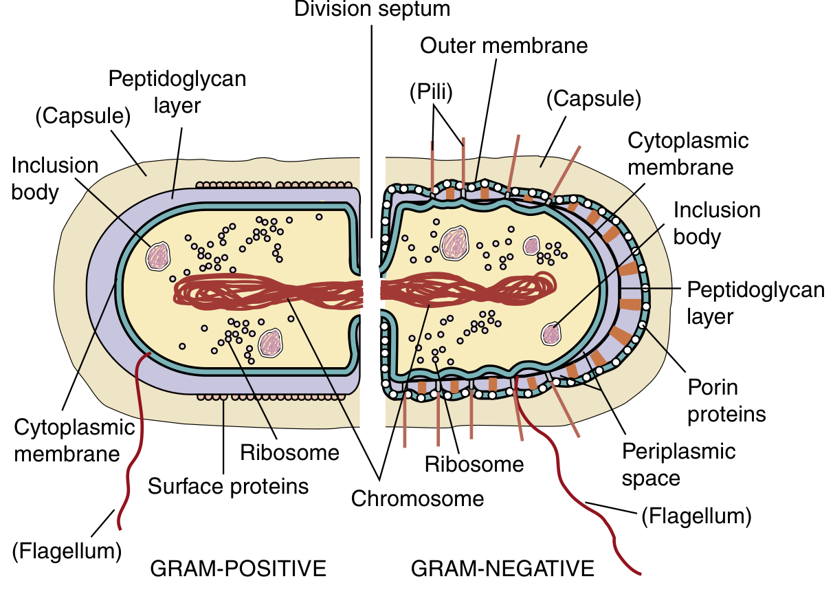
Fig. 12.4 - Gram-positive (left) and Gram-negative (right) bacterial cell structures showing all major labeled components. (Medical Microbiology 9e)
Structures and Their Functions
1. Capsule (Glycocalyx / Slime Layer)
- Structure: A loose polysaccharide or polypeptide layer surrounding the cell wall. When compact and well-defined, it is called a capsule; when loose and diffuse, it is a slime layer.
- Functions:
- Protects the bacterium from phagocytosis by host immune cells (major virulence factor)
- Helps bacteria adhere to surfaces and host tissues
- Prevents desiccation (drying out)
- Confers resistance to some antimicrobial agents
- Examples: Streptococcus pneumoniae, Klebsiella pneumoniae
2. Cell Wall
- Structure: A rigid layer made primarily of peptidoglycan (murein) - a polymer of N-acetylglucosamine (NAG) and N-acetylmuramic acid (NAM) cross-linked by peptide bridges.
- Gram-positive: thick peptidoglycan (up to 40 layers, comprising ~50% of the cell wall)
- Gram-negative: thin peptidoglycan (1-2 layers) + outer membrane
- Functions:
- Maintains the shape of the bacterium
- Provides rigid structural support and prevents osmotic lysis
- Determines Gram staining reaction
- Contains surface antigens involved in immune recognition
- Is the target of beta-lactam antibiotics (penicillins, cephalosporins) and vancomycin
3. Cytoplasmic (Plasma) Membrane
- Structure: A phospholipid bilayer with embedded proteins. Unlike eukaryotes, it contains no sterols (except Mycoplasma).
- Functions:
- Selective permeability - controls entry and exit of nutrients and waste
- Site of electron transport chain and ATP (energy) production (performs functions of mitochondria)
- Contains transport proteins (permeases) for active transport of metabolites
- Contains enzymes for biosynthesis (e.g., lipid synthesis)
- Site of DNA attachment during replication and cell division
- Houses ion pumps that maintain membrane potential
4. Nucleoid (Bacterial Chromosome)
- Structure: A single, circular, double-stranded DNA molecule (haploid genome) located in the nucleoid region of the cytoplasm. There is no nuclear membrane enclosing it.
- Functions:
- Contains all genetic information for bacterial growth, reproduction, and metabolism
- Since transcription and translation are coupled (no nuclear membrane), ribosomes bind mRNA while it is still being synthesized
5. Plasmids
- Structure: Small, circular, extrachromosomal double-stranded DNA molecules that replicate independently.
- Functions:
- Carry genes for antibiotic resistance, toxin production, virulence factors, and metabolic enzymes
- Can be transferred between bacteria by conjugation, contributing to horizontal gene transfer
- Important in the spread of antibiotic resistance
6. Ribosomes
- Structure: 70S ribosomes made of a 30S small subunit and a 50S large subunit (unlike eukaryotic 80S ribosomes).
- Functions:
- Site of protein synthesis
- The structural differences from eukaryotic ribosomes make them targets for many antibiotics (aminoglycosides, macrolides, tetracyclines, chloramphenicol)
7. Flagella
- Structure: Long, whip-like filamentous appendages made of the protein flagellin. May be arranged as:
- Monotrichous (single polar flagellum)
- Lophotrichous (tuft at one end)
- Amphitrichous (flagella at both ends)
- Peritrichous (flagella all around)
- Functions:
- Primary organ of motility - enables bacteria to swim toward nutrients (chemotaxis) and away from harmful substances
- Act as virulence factors (help bacteria reach and colonize host tissues)
8. Pili (Fimbriae)
- Structure: Short, hair-like, protein filaments (made of pilin). Two main types:
- Common pili (fimbriae): numerous, short, involved in adhesion
- Sex pili (F-pili): longer, fewer in number, involved in conjugation
- Functions:
- Mediate adherence of bacteria to host cell surfaces and mucosal membranes (critical first step in infection)
- Sex pili form conjugation bridges for transfer of plasmids and DNA between bacteria (conjugation)
9. Inclusion Bodies (Granules)
- Structure: Storage granules within the cytoplasm; may include glycogen granules, polyphosphate granules (volutin), lipid granules, sulfur granules.
- Functions:
- Storage of nutrient reserves (carbon, energy, phosphate) used during periods of starvation or stress
10. Endospores (in select bacteria only)
- Structure: Highly resistant dormant structures formed within the cell by bacteria such as Bacillus and Clostridium. Contain dipicolinic acid and have multiple protective layers.
- Functions:
- Allow the bacterium to survive extreme conditions - heat, desiccation, UV radiation, disinfectants, and lack of nutrients
- Germinate back into vegetative cells when conditions improve
11. Outer Membrane (Gram-negative bacteria only)
- Structure: An additional lipid bilayer outside the thin peptidoglycan layer, containing lipopolysaccharide (LPS) on its outer leaflet and porin proteins that form channels.
- Functions:
- Acts as a permeability barrier
- LPS (endotoxin) triggers a strong immune response and septic shock in the host
- Porin proteins control passage of small molecules across the outer membrane
- Confers resistance to many antibiotics and lysozyme
12. Periplasmic Space (Gram-negative bacteria)
- Structure: The space between the inner cytoplasmic membrane and the outer membrane, containing the thin peptidoglycan layer and periplasmic proteins.
- Functions:
- Contains enzymes for nutrient processing, detoxification (beta-lactamases), and transport
Question 2: What Are Bacteria? Classification Based on Shape (10 Marks)
Definition of Bacteria
Bacteria are unicellular prokaryotic microorganisms - they are living cells that lack a true membrane-bound nucleus and membrane-enclosed organelles. Key characteristics:
- Size: approximately 0.5-3.0 µm
- Single, circular DNA (haploid genome) in a nucleoid region
- 70S ribosomes (30S + 50S subunits)
- Cell wall containing peptidoglycan (murein)
- Reproduce by binary fission
- Can be free-living, commensal, or pathogenic
- Found in virtually every environment on Earth - soil, water, air, and within the bodies of all living organisms
Bacteria belong to the domain Prokaryota (or Bacteria), distinct from Archaea (another prokaryotic domain) and Eukarya. Unlike eukaryotes (fungi, protozoa, plants, animals), bacteria do not have mitochondria, Golgi bodies, or an endoplasmic reticulum.
Classification of Bacteria Based on Shape (Morphology)
Shape-based classification (morphological classification) is one of the most fundamental ways to classify bacteria. As seen in the diagram below, bacteria exist in several distinct shapes:
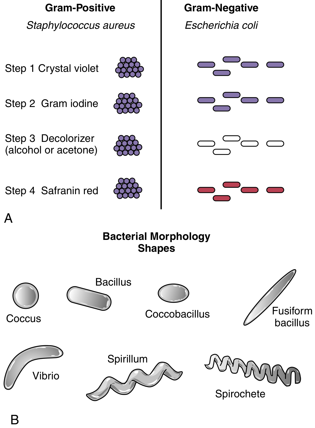
Fig. 12.3B - Bacterial Morphology Shapes (Medical Microbiology 9e)
1. Cocci (Spherical Bacteria)
- Shape: Round or spherical, approximately 1 µm in diameter
- Arrangements:
- Diplococci: pairs of cocci - e.g., Streptococcus pneumoniae, Neisseria gonorrhoeae
- Streptococci: chains of cocci - e.g., Streptococcus pyogenes (Group A Streptococcus)
- Staphylococci: grape-like irregular clusters - e.g., Staphylococcus aureus
- Tetrads: groups of four cocci - e.g., Micrococcus species
- Sarcinae: cubical packets of eight cocci
- Clinical significance: Cause pneumonia, meningitis, gonorrhoea, skin infections, toxic shock syndrome
2. Bacilli (Rod-shaped Bacteria)
- Shape: Cylindrical/rod-shaped bacteria, varying in length
- Sub-types:
- Bacillus (true rod): straight rods, e.g., Escherichia coli, Bacillus anthracis
- Coccobacillus: short, oval rods intermediate between cocci and bacilli - e.g., Haemophilus influenzae, Bordetella pertussis
- Fusiform bacillus: spindle-shaped, tapered at both ends - e.g., Fusobacterium species
- Palisade/Chinese letter arrangement: seen in Corynebacterium diphtheriae
- Pairs (diplobacilli): two rods end-to-end
- Chains (streptobacilli): e.g., Streptobacillus moniliformis
- Clinical significance: Cause tuberculosis, diphtheria, typhoid, E. coli infections, anthrax, gas gangrene
3. Spiral Bacteria
Bacteria with a helical or curved shape. Three main sub-types:
a) Vibrio (Comma-shaped)
- Shape: Short, curved rods resembling a comma; incomplete spiral (less than one full turn)
- Example: Vibrio cholerae (cholera), Campylobacter jejuni
- Note: Highly motile with a single polar flagellum
b) Spirillum (Rigid Spiral)
- Shape: Rigid, helical (corkscrew-like) rods with multiple complete turns; have a stiff cell wall
- Example: Spirillum minus (rat-bite fever)
- Motility: By flagella
c) Spirochete (Flexible Spiral)
- Shape: Thin, flexible, tightly coiled spiral bacteria; move by means of internal axial filaments (endoflagella) wound around the cell body
- Examples:
- Treponema pallidum (syphilis)
- Borrelia burgdorferi (Lyme disease)
- Leptospira interrogans (leptospirosis)
- Distinguishing feature: Unique internal locomotion by axial filaments; do not stain well with Gram stain
Summary Table: Classification by Shape
| Shape | Term | Arrangement | Example |
|---|---|---|---|
| Spherical | Coccus | Clusters, chains, pairs | S. aureus, S. pneumoniae |
| Rod/Cylinder | Bacillus | Solitary, chains, palisades | E. coli, B. anthracis |
| Short oval rod | Coccobacillus | Variable | H. influenzae |
| Comma-shaped | Vibrio | Solitary | V. cholerae |
| Rigid spiral | Spirillum | Solitary | Spirillum minus |
| Flexible spiral | Spirochete | Solitary | T. pallidum |
| Spindle | Fusiform bacillus | Often in clusters | Fusobacterium |
Question 3: Study of Bacteria, and Difference Between Gram-Positive and Gram-Negative Bacteria (10 Marks)
What is the Study of Bacteria Called?
The scientific study of bacteria is called Bacteriology.
- Bacteriology is a branch of Microbiology - the study of all microorganisms
- The term comes from the Greek: bakterion (small rod/staff) + logos (study)
- Hans Christian Gram (1884) developed the Gram staining technique, a cornerstone of bacteriology
- Key related sub-disciplines:
- Clinical Bacteriology: study of bacteria causing human diseases
- Medical Microbiology: encompasses bacteriology in the context of human health and disease
- Environmental Bacteriology: study of bacteria in nature
- Industrial Microbiology: uses bacteria for biotechnology, fermentation, etc.
Gram Staining - The Key Differentiating Test
The Gram stain, developed by Hans Christian Gram in 1884, is a differential staining technique that divides bacteria into two major groups based on differences in their cell wall structure. It is the single most important staining technique in clinical microbiology.
Steps of the Gram Stain:
| Step | Reagent | Gram-Positive Result | Gram-Negative Result |
|---|---|---|---|
| 1 | Crystal violet (primary stain) | Purple | Purple |
| 2 | Gram's iodine (mordant) | Purple (iodine-dye complex forms) | Purple |
| 3 | Acetone/alcohol (decolorizer) | Retain purple (thick PG layer traps dye) | Decolorized (thin PG, dye washes out) |
| 4 | Safranin (counterstain) | Remain purple | Stain red/pink |
Fig. 12.3A - Gram stain process with Staphylococcus aureus (Gram-positive) and Escherichia coli (Gram-negative) (Medical Microbiology 9e)
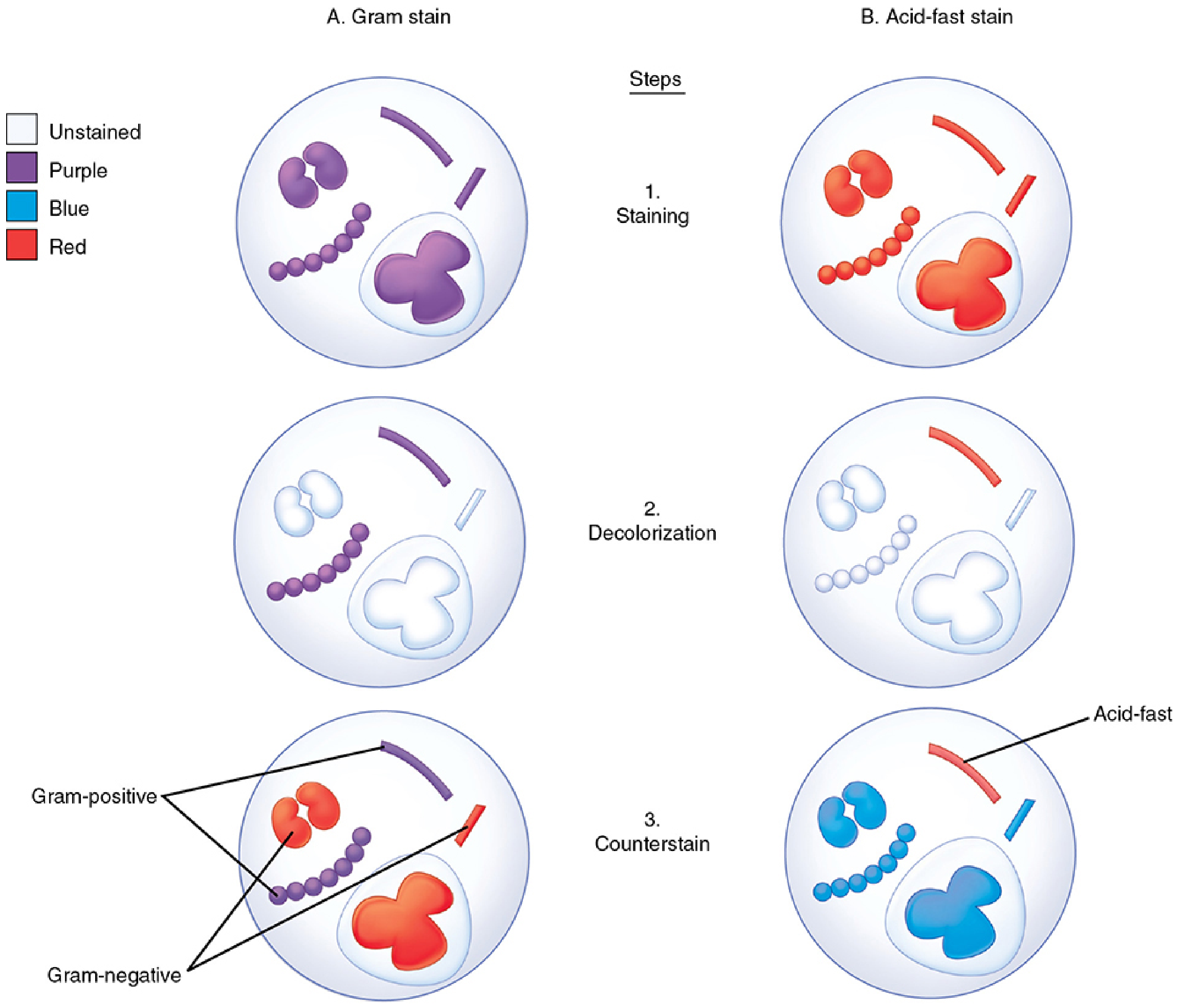
FIGURE 4-2. Gram stain procedure showing all stages of staining (Sherris & Ryan's Medical Microbiology, 8e)
Differences Between Gram-Positive and Gram-Negative Bacteria
Structural Diagram:
Fig. 12.4 - Structural differences between Gram-positive and Gram-negative bacteria (Medical Microbiology 9e)
Detailed Comparison Table:
| Feature | Gram-Positive | Gram-Negative |
|---|---|---|
| Gram stain result | Purple/violet | Pink/red |
| Cell wall thickness | Thick (20-80 nm) | Thin (5-10 nm) |
| Peptidoglycan content | Abundant (up to 40 layers, ~50% of cell wall) | Scanty (1-2 layers, ~10% of cell wall) |
| Outer membrane | Absent | Present (contains LPS) |
| Lipopolysaccharide (LPS / Endotoxin) | Absent | Present (on outer membrane) |
| Teichoic acids | Present (wall teichoic acid + lipoteichoic acid) | Absent |
| Periplasmic space | Narrow/absent | Present (well-defined) |
| Porin proteins | Absent | Present (in outer membrane) |
| Lipid content | Low (~2-4%) | High (~15-20%) |
| Sensitivity to penicillin | More sensitive (thick PG = more target) | Less sensitive (outer membrane is a barrier) |
| Lysozyme sensitivity | More sensitive | Less sensitive (outer membrane protects) |
| Exotoxin production | Many produce exotoxins | Some produce exotoxins |
| Endotoxin (LPS) | Not produced | Produced; causes fever, septic shock |
| Flagella structure | 2 rings in basal body | 4 rings in basal body |
| Examples | S. aureus, S. pneumoniae, B. anthracis, C. perfringens, C. diphtheriae | E. coli, Salmonella, Pseudomonas, Klebsiella, N. gonorrhoeae |
Key Differences Explained:
1. Cell Wall:
The fundamental difference is in the cell wall. Gram-positive bacteria have a thick peptidoglycan layer (up to 40 sheets) with teichoic acids embedded in it. This thick layer traps the crystal violet-iodine complex during Gram staining, keeping the cells purple.
Gram-negative bacteria have a thin peptidoglycan layer (1-2 sheets) plus an outer membrane outside it. The outer membrane contains LPS (lipopolysaccharide), porin proteins, and phospholipids. During Gram staining, the acetone-alcohol decolorizer disrupts the outer membrane and washes the crystal violet from the thin peptidoglycan layer, leaving cells colorless until counterstained red by safranin. (Sherris & Ryan's Medical Microbiology, 8e)
2. Teichoic Acids (Gram-positive only):
- Wall teichoic acids (covalently attached to peptidoglycan) and lipoteichoic acids (anchored in cytoplasmic membrane) are unique to Gram-positive bacteria
- Function in cell wall integrity, regulation of cell division, and act as adhesins
- Are important antigens for immune recognition
3. LPS/Endotoxin (Gram-negative only):
- Lipopolysaccharide (LPS) is composed of lipid A (toxic component), core polysaccharide, and O-antigen
- Lipid A = endotoxin - released when bacteria lyse; triggers fever, hypotension, disseminated intravascular coagulation (DIC), and septic shock
- Gram-positive bacteria do NOT produce endotoxin
4. Outer Membrane (Gram-negative only):
- Forms an additional permeability barrier - makes Gram-negative bacteria intrinsically more resistant to antibiotics, detergents, and lysozyme
- Porin proteins in the outer membrane create size-selective channels for small hydrophilic molecules
Clinical Relevance:
- The Gram stain result guides antibiotic selection BEFORE culture results are available
- Gram-positive infections are often treated with beta-lactams, vancomycin, or clindamycin
- Gram-negative infections often require broader-spectrum antibiotics (aminoglycosides, fluoroquinolones, carbapenems) because of the outer membrane barrier
- Gram-negative sepsis is particularly dangerous due to endotoxin (LPS) release
Sources: Medical Microbiology 9e (Murray); Sherris & Ryan's Medical Microbiology, 8th Edition; Jawetz, Melnick & Adelberg's Medical Microbiology, 28e
This is a shared conversation. Sign in to Orris to start your own chat.