
Congenital sensory neural hearing loss
Reading File
Now I'll search the medical library for congenital sensorineural hearing loss content in parallel.
Finding Sources
Finding Sources
Searching PubMed
"congenital sensorineural hearing loss"[MeSH Terms]
Excellent sources found. Now reading the most comprehensive sections in parallel.
Reading File
Reading File
Reading File
Now reading more content - the detailed syndromic genetics, workup, and management sections:
Reading File
Reading File
Reading File
Reading File
Reading File
Reading File
I now have comprehensive information from the textbooks. Let me compile the full response.
Congenital Sensorineural Hearing Loss (SNHL)
Overview
Congenital SNHL affects 1-3 per 1,000 live births and is one of the most common congenital disabilities. The causes are broadly split 50:50 between genetic and acquired origins. Of genetic cases, 75% are non-syndromic and 25% are syndromic. - K.J. Lee's Essential Otolaryngology, p.1001; Bailey and Love's Surgery, p.781
1. Genetic Causes
A. Nonsyndromic (75% of genetic cases)
Autosomal Recessive (most common)
| Gene/Mutation | Mechanism | Key Features |
|---|---|---|
| GJB2 (Connexin 26) | Dysfunctional gap junctions impair K+ recycling in stria vascularis | Most common genetic cause; 35delG accounts for 80% of GJB2 mutations |
| GJB6 (Connexin 30) | Same pathway as above | Often co-inherits with GJB2 mutations |
| SLC26A4 (EVA) | Enlarged vestibular aqueduct | Mild-to-profound SNHL, can worsen with head trauma; flat or downsloping audiogram |
| OTOF (Otoferlin) | Vesicle membrane fusion defect | Auditory dyssynchrony: present OAEs, absent ABR |
| Mitochondrial A1555G | Mutation in mitochondrial 12S rRNA (maternally inherited) | 15% of US patients with aminoglycoside-induced SNHL; more prevalent in Mongolia |
Autosomal Dominant
- Wolfram syndrome: Most common cause of dominantly inherited low-frequency SNHL; progressive course.
B. Syndromic Genetic Causes (>400 syndromes)
Autosomal Recessive Syndromes
1. Pendred Syndrome
- Accounts for ~2% of profound congenital SNHL
- Mutation in SLC26A4 (iodine-chloride transporter defect)
- Bilateral/unilateral SNHL + Mondini malformation + euthyroid goiter
- Work-up: SLC26A4 testing, TSH, T3, T4
2. Usher Syndrome
- Affects 3% of the deaf population; 50% of the deaf-blind population
- Mutations in MYO7A, USH2A, and others (tip-link/usherin protein dysfunction)
- Three types:
- USH1 (most severe): profound SNHL, vestibular dysfunction, early retinitis pigmentosa
- USH2 (most common): moderate-severe SNHL, normal vestibular function, later retinitis pigmentosa
- USH3 (~3%, mostly Norwegian): progressive SNHL, variable vestibular function
3. Jervell and Lange-Nielsen
- 1 in 1,000 profoundly deaf patients
- Mutation in KVLQT1 - dysregulated potassium channel
- Severe/profound SNHL + prolonged QT interval + sudden death in young adults
- Treatment: beta-blockers; screen first-degree relatives for Romano-Ward syndrome
4. Biotinidase Deficiency
- Incidence: 1/60,000; screened in neonatal programs
- SNHL + hair loss + seizures + hypotonia
- Preventable/treatable with dietary biotin replacement
Autosomal Dominant Syndromes
1. Waardenburg Syndrome - Most common inherited congenital deafness (~2%)
- Mutation in neural crest cells → defective intermediate layer of stria vascularis
- Features: white forelock, dystopia canthorum, synophrys, heterochromia iridis, SNHL
- Four types: WS1 (PAX3), WS2A/Tietz (MITF), WS3 (Klein), WS4 (SOX10 + Hirschsprung)
2. Branchio-Oto-Renal (BOR) Syndrome
- EYA1 mutation (only 40% confirmed by testing)
- Major features: branchial clefts/fistulas, outer/middle/inner ear anomalies
- Temporal bone findings pathognomonic: fused malleoincudal complex, funnel-shaped IAC, hypoplastic cochlear apex
3. CHARGE Syndrome
- Sporadic microdeletion in CHD7 (chr 8) - only 50% have confirmed mutation
- Coloboma, Heart defects, Atresia choanae, Retardation, Genitourinary anomalies, Ear anomalies
- Inner ear: absent oval window, Mondini malformation, absent/hypoplastic semicircular canals
4. NF2 (Neurofibromatosis Type 2)
- Mutation in schwannomin (chr 22) → abnormal merlin protein
- Bilateral acoustic neuromas, hearing loss ~age 20, tinnitus, imbalance
5. Treacher-Collins (Mandibulofacial Dysostosis)
- Mutation in chr 5q → abnormal treacle protein
- Bilateral: microtia, aural atresia, mid-face hypoplasia, downsloping palpebral fissures, coloboma of lower eyelid, micrognathia
6. Stickler Syndrome
- Progressive mixed hearing loss (80%)
- Myopia, retinal detachment, cataracts, Robin sequence, hypermobile joints
X-Linked
1. Nonsyndromic - POU3F4 Mutation (Xq21)
- Most common X-linked deafness
- Males: stable hearing at birth → progressive mixed/SNHL (bilateral in 75%)
- Large IAC, absent lamina cribrosa; risk of perilymphatic gusher during surgery
2. Alport Syndrome
- COL4A5 mutation (X-linked) or COL4A3/COL4A4 (AR, 15%)
- Progressive high-frequency SNHL + glomerulonephritis + hematuria + anterior lenticonus
Chromosomal
- Down Syndrome (Trisomy 21): Stenotic EAC, middle ear disease, SNHL (common)
- Turner Syndrome (45,XO): Mixed conductive and SNHL
- Fetal Alcohol Syndrome: Microcephaly, mental retardation; conductive and SNHL
2. Acquired Causes
TORCH Infections
| Organism | Key Features |
|---|---|
| CMV (most common acquired cause in developed world) | Affects ~1% of pregnancies; 40% vertical transmission. IUGR, microcephaly, intracerebral calcification, SNHL (may be delayed onset, can be unilateral progressing to bilateral). Diagnose with PCR on neonatal dried blood spot |
| Rubella | CRS when maternal infection in first trimester; deafness + cataracts/glaucoma + PDA/pulmonary stenosis + microcephaly. Nearly eliminated in countries with vaccination |
| Toxoplasmosis | Part of TORCH screen |
| Syphilis (T. pallidum) | Vertical transmission 70-100% (primary stage); hearing loss is a late feature; congenital syphilis SNHL |
| Herpes | Part of TORCH spectrum |
Perinatal Causes
- Hypoxia / birth asphyxia
- Neonatal jaundice (kernicterus)
- Prematurity
Postnatal Causes
- Bacterial meningitis (Streptococcus most common organism)
- Mumps
3. Investigations / Diagnosis
| Test | Use |
|---|---|
| Newborn hearing screening | OAE (otoacoustic emissions) as first-pass screen; ABR (auditory brainstem response) for confirmation and thresholds |
| OAE | Tests outer hair cell function; present in auditory dyssynchrony (OTOF mutations) |
| ABR | Absent in otoferlin/auditory dyssynchrony despite present OAEs |
| Audiogram | Flat/downsloping in EVA; high-frequency loss in Alport |
| CT temporal bone | Mondini malformation, EVA, absent semicircular canals, funnel-shaped IAC |
| MRI | Cochlear nerve aplasia, CNS anomalies (CMV) |
| Genetic testing | GJB2/GJB6 first-line; SLC26A4 (Pendred/EVA); EYA1 (BOR); CHD7 (CHARGE); panel testing available |
| TORCH serology / PCR | CMV (PCR on dried blood spot preferred); rubella, syphilis serology |
| ECG | Prolonged QT for Jervell and Lange-Nielsen |
| Ophthalmology | Retinitis pigmentosa (Usher), lenticonus (Alport), coloboma (CHARGE) |
| Renal function / urinalysis | Hematuria in Alport syndrome |
| TSH, T3, T4 | Pendred (euthyroid goiter) |
4. Management
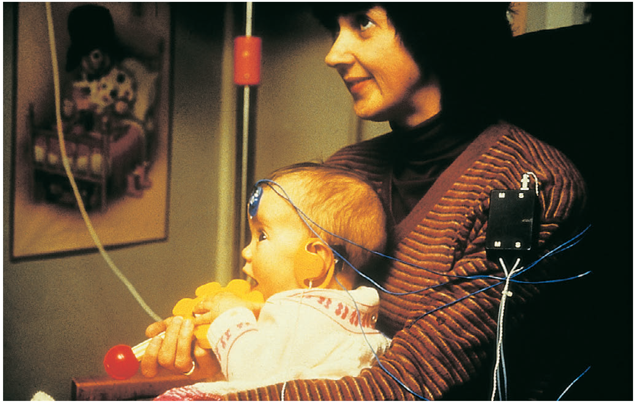
- Early identification - Universal newborn hearing screening programs are now standard (UK: OAE at birth; ABR if failed)
- Hearing aids - Fitting as early as possible to exploit neural plasticity of the developing auditory cortex
- Cochlear implantation - For profound SNHL; early intervention essential; stimulates cochlear neurones (which usually remain intact even when hair cells are lost); insertion through round window
- Auditory brainstem implant (ABI) - In cases of absent cochlear nerve (e.g., NF2, cochlear aplasia)
- Sign language / habilitation - Complementary to amplification
- Genetic counselling - Essential for families; test first-degree relatives
- Condition-specific management:
- Jervell-Lange-Nielsen: beta-blockers, screen relatives for Romano-Ward
- Pendred: monitor thyroid; avoid unnecessary thyroid surgery
- CMV: antiviral therapy (valganciclovir) may reduce progression of CMV-related SNHL
- Biotinidase deficiency: dietary biotin replacement
- K.J. Lee's Essential Otolaryngology, pp.1001-1006
- Bailey and Love's Short Practice of Surgery (28th ed.), p.781
- Scott-Brown's Otorhinolaryngology Head & Neck Surgery (Vol 2)
Bilateral abductor palsy
Bilateral abductor palsy refers to paralysis of both vocal cord abductors (posterior cricoarytenoid muscles), leaving both cords in the paramedian/median position. It's a life-threatening emergency due to airway obstruction, presenting with stridor and dyspnea but a relatively preserved voice. Causes include thyroid surgery, bilateral thyroid malignancy, and neurological conditions.
Searching sources...
Finding Sources
Finding Sources
Searching PubMed
"bilateral vocal cord paralysis" AND management
Finding Sources
Reading File
Finding Sources
Finding Sources
Finding Sources
Reading File
Reading File
Reading File
Finding Sources
Excellent - Scott-Brown's has dedicated sections. Reading both in parallel:
Reading File
Reading File
Reading File
Reading File
I now have comprehensive textbook content. Let me also fetch the laryngoscopy image for illustration:
Loading Image
I have all the information needed for a comprehensive answer. Here is the full response:
Bilateral Abductor Palsy (Bilateral Vocal Cord Paralysis)
Definition & Anatomy
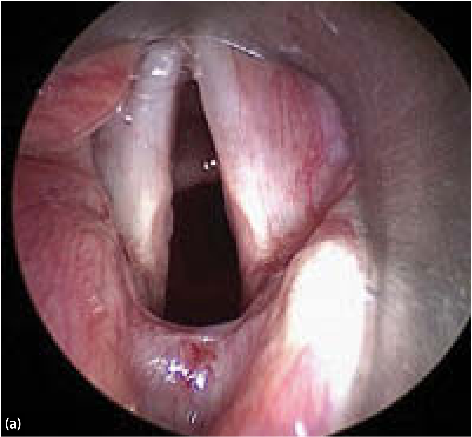
Bilateral abductor palsy is the failure of both vocal cords to abduct (open), due to paralysis or mechanical dysfunction of the posterior cricoarytenoid (PCA) muscles - the only intrinsic laryngeal abductors, innervated by the recurrent laryngeal nerve (RLN). Both cords assume the median or paramedian position, severely narrowing the glottic airway.
The key physiological paradox: because adductor muscle tone is intact (or unopposed), the voice may be relatively preserved while the airway is dangerously compromised.
Causes
Three Principal Mechanisms
| Mechanism | Common Causes |
|---|---|
| Bilateral laryngeal denervation | Thyroid surgery (most common), neck/chest malignancy (thyroid, tracheal, esophageal), esophagectomy, thymectomy, mediastinal procedures, thoracic aortic aneurysm |
| Bilateral cricoarytenoid joint fixation | Trauma, rheumatoid arthritis, post-intubation ankylosis |
| Inter-arytenoid scarring | Prolonged endotracheal intubation (common ICU cause), posterior glottic scar |
Neurological Causes
- Amyotrophic lateral sclerosis (ALS)
- Post-polio syndrome
- Charcot-Marie-Tooth neuropathy
- Arnold-Chiari malformation
- Guillain-Barré syndrome
- Idiopathic (same infectious agents as unilateral paralysis)
Important distinction: Bilateral vocal fold immobility following intubation may represent mechanical fixation (joint/scar) rather than true denervation. This matters because mechanical causes may be surgically correctable without destructive procedures.
Clinical Features
| Feature | Explanation |
|---|---|
| Inspiratory stridor | Dominant symptom - negative intrathoracic pressure during inspiration passively pulls denervated folds into closer approximation |
| Dyspnoea / exercise intolerance | Severity inversely proportional to residual glottic aperture |
| Voice relatively preserved | Adductors are intact; cords can still approximate and vibrate |
| Swallowing usually unaffected | Unlike unilateral palsy, aspiration is not a primary concern |
| Acute vs. progressive onset | Post-thyroidectomy extubation = acute emergency; neurological causes = insidious compensation |
The typical scenario is unexpected respiratory distress after extubation from thyroid surgery - in this setting, securing the airway is the only immediate consideration.
In progressive neurological disease, patients may compensate as the paralysis gradually worsens, tolerating unexpectedly small glottic airways with minimal symptoms until a threshold is crossed.
Investigations
| Investigation | Purpose |
|---|---|
| Flexible nasolaryngoscopy | Visualise cord position and mobility; assess for posterior glottic scar or arytenoid fixation |
| Operative laryngoscopy ± EMG (LEMG) | Distinguish true denervation from mechanical fixation |
| Flow-volume loop | Shows variable extra-thoracic obstruction - cords passively adduct on inspiration and passively abduct on expiration (vs. fixed obstruction in subglottic stenosis) |
| CT neck and mediastinum | Identify malignancy, aneurysm, or other compressive lesions along the RLN course |
| MRI brain/brainstem | If neurological cause suspected (Arnold-Chiari, ALS, etc.) |
| Laryngeal EMG | Confirm denervation; assess for spontaneous reinnervation potential |
Management
Management is guided by the degree of airway limitation and the likelihood of spontaneous recovery. The patient may be the best judge of functional handicap in non-acute cases.
Acute Phase
- Immediate airway security - re-intubation or emergency tracheotomy if post-operative stridor after extubation
- Do NOT proceed to destructive surgery until recovery potential is excluded
Long-term Options
1. Tracheotomy
- Guarantees the airway
- Minimal compromise of phonation or swallowing
- May be a long-term option for some patients, especially the elderly or those who are poor surgical candidates
2. Reversible Glottic Enlargement Procedures (when recovery potential exists)
- Vocal cord suture lateralization (endo-extralaryngeal suture) - lateralizes one cord, reversible, buys time
- Arytenoid abduction suture - mimics PCA action; pulls the vocal process posterolaterally; does NOT abolish residual adductor activity during phonation
3. Definitive Glottic Enlargement (when no recovery expected)
These are destructive and irreversible - only after excluding spontaneous recovery AND confirming a safe swallow:
- Endoscopic CO2 laser partial posterior cordectomy + partial arytenoidectomy - unilateral or bilateral; most widely used. Voice becomes breathier but remains functional. Shorter hospitalization; higher compliance.
- Open arytenoidectomy - rarely performed now with laser available
- Restenosis of the glottis is the main complication requiring reoperation
4. Reinnervation (Selective Reinnervation)
- Indicated for stridor due to bilateral abductor paralysis
- Selective reinnervation procedure: C3 root of phrenic nerve → cable graft (great auricular nerve, Y-shaped) → both PCA muscles; PLUS ansa hypoglossi anastomosed to RLN bilaterally
- Technically demanding; performed via extended anterior neck incision
- Aim: selectively restore abductor function while adductors are separately reinnervated
5. Laryngeal Pacing (Experimental)
- Electrodes implanted into both PCA muscles → automatic abduction/adduction
- Connected to subcutaneous pacemaker device (similar to cardiac pacemaker)
- Currently in clinical trials; not yet standard of care
Summary Table
| Parameter | Bilateral Abductor Palsy |
|---|---|
| Cord position | Median / paramedian |
| Voice | Relatively preserved |
| Swallowing | Usually unaffected |
| Airway | COMPROMISED (primary problem) |
| Stridor | Inspiratory (worsens with exertion) |
| Most common cause | Thyroid surgery |
| Emergency management | Tracheotomy / re-intubation |
| Definitive surgery | CO2 laser cordectomy ± arytenoidectomy |
- Scott-Brown's Otorhinolaryngology Head & Neck Surgery, Vol 3, pp.1134, 1152
- Cummings Otolaryngology Head and Neck Surgery, Ch. 63
- Fishman's Pulmonary Diseases and Disorders, p.436
- Goldman-Cecil Medicine, p.3979
Bilateral adductor palsy
Bilateral adductor palsy means both vocal cords are paralyzed in the abducted (open) position - the opposite of abductor palsy. The airway is patent, so there is no stridor or dyspnea, but phonation and airway protection are severely impaired, causing a weak/breathy voice, aphonia, and aspiration.
Searching sources...
Finding Sources
Finding Sources
Finding Sources
Finding Sources
Finding Sources
Finding Sources
Reading File
Finding Sources
Finding Sources
Finding Sources
Reading File
Reading File
Reading File
Reading File
Reading File
I now have comprehensive material. Here is the full, structured response:
Bilateral Adductor Palsy
Definition & Physiology
Bilateral adductor palsy is paralysis of the adductor muscles of both vocal cords, leaving both cords fixed in the abducted (open) cadaveric position. Since both cords cannot close, the glottis remains permanently wide open.
The key physiological consequences flow directly from this:
- Airway is patent - no obstruction, no stridor
- Glottis cannot close - phonation fails, cough fails, airway protection fails
This is the direct clinical inverse of bilateral abductor palsy. The two conditions are compared below.
Anatomy of Adduction
The following muscles are responsible for adduction (closing) of the vocal cords, all innervated by the recurrent laryngeal nerve (RLN):
- Lateral cricoarytenoid (LCA) - primary adductor; rotates arytenoid to bring vocal process medially
- Transverse and oblique arytenoids (interarytenoids) - close the posterior glottis
- Thyroarytenoid (TA/vocalis) - tenses and shortens the cord
In complete RLN palsy (flaccid), all these are paralysed. The cord assumes a cadaveric/lateral position with a wide posterior glottic gap. This is bilateral adductor palsy when it affects both sides.
Causes
The causes of bilateral adductor palsy are the same as bilateral vocal cord paralysis in general - since the RLN innervates adductors predominantly:
Surgical / Iatrogenic (most common)
| Cervical | Thoracic | Other |
|---|---|---|
| Thyroidectomy / parathyroidectomy | Oesophagectomy | Anterior cervical spine surgery |
| Carotid endarterectomy | Thymectomy | Vagal nerve stimulator implantation |
| Cricopharyngeal myotomy | Mediastinal dissection | Skull base surgery |
Malignancy
- Thyroid carcinoma invading both RLNs
- Tracheal, oesophageal, or lung malignancy
- Mediastinal lymphadenopathy compressing both nerves
Neurological
- Amyotrophic lateral sclerosis (ALS)
- Post-polio syndrome
- Guillain-Barré syndrome
- Charcot-Marie-Tooth neuropathy
- Arnold-Chiari malformation
- Brainstem stroke or tumour (nucleus ambiguus)
High Vagal Injury (more severe syndrome)
When injury is above the origin of the superior laryngeal nerve (e.g., jugular foramen syndrome, skull base tumour, stroke), the full picture includes:
- Glottic insufficiency (RLN component)
- Hemi-laryngeal anaesthesia (internal branch of SLN)
- Pharyngeal constrictor atony
- Cricopharyngeal hyperfunction This combination produces far greater aspiration risk than RLN palsy alone.
Idiopathic / Infectious
- Viral neuropathy (same agents as unilateral palsy)
- Post-intubation cricoarytenoid joint injury (mechanical, not true palsy - important to distinguish)
Clinical Features
| Feature | Detail |
|---|---|
| Voice | Hoarse, breathy, weak to near-total aphonia; vocal fatigue; patients may report phonation-induced breathlessness from excessive air escape |
| Cough | Ineffective - cannot build subglottic pressure; predisposes to retained secretions and chest infections |
| Aspiration | Major risk - cords cannot protect the airway during swallowing; worse if SLN also injured (absent laryngeal sensation) |
| Dysphagia | Common, especially with high vagal lesions |
| Airway | Patent - no stridor, no dyspnoea at rest (contrast with bilateral abductor palsy) |
| Wet voice | Pooled secretions in the laryngeal introitus cause a 'wet' vocal quality; sign of aspiration penetration |
| Laryngospasm | Can occur paradoxically from reaction to unexpected aspiration or penetration |
In complete flaccid bilateral paralysis, the cords lie in the laterally displaced cadaveric position with a wide posterior glottic gap - severe glottal incompetence results and injection laryngoplasty alone cannot close this gap.
Important: The degree of glottic insufficiency depends on cord position. Residual or regenerated innervation of adductors may hold cords near the midline (paramedian), allowing some phonation and aspiration protection. Truly cadaveric (lateralized) cords indicate complete flaccid denervation.
Comparison: Bilateral Adductor vs. Abductor Palsy
| Feature | Bilateral ADDUCTOR Palsy | Bilateral ABDUCTOR Palsy |
|---|---|---|
| Cord position | Abducted (open) - cadaveric | Adducted (closed) - median/paramedian |
| Airway | Patent | Obstructed |
| Stridor | Absent | Present (inspiratory) |
| Voice | Aphonic / severely breathy | Relatively preserved |
| Cough | Ineffective | Preserved |
| Aspiration | Major problem | Not a problem |
| Primary danger | Aspiration pneumonia | Respiratory failure |
| Emergency | Aspiration / nutrition failure | Tracheotomy |
Investigations
| Investigation | Purpose |
|---|---|
| Flexible nasolaryngoscopy | Visualise cord position; assess posterior glottic gap; note pooling of secretions |
| Laryngeal EMG (LEMG) | Confirm denervation; assess prognosis for recovery; distinguish palsy from mechanical fixation |
| Videostroboscopy | Assess mucosal wave asymmetry and cord tension in paresis |
| Swallowing assessment (VFSS / FEES) | Quantify aspiration risk; direct management of diet and feeding |
| CT neck, chest, mediastinum | Identify malignancy, aneurysm, or compressive lesion along RLN course |
| MRI brain/brainstem | If central/neurological cause suspected |
| Pulmonary function / flow-volume loop | May show reduced expiratory peak flow (weak cough); non-obstructive pattern |
| Chest X-ray | Screen for aspiration pneumonia |
Management
The goal is to restore glottic competence - for phonation, cough, and airway protection.
Principle: Medialization
Moving the paralysed cord(s) to the midline to allow the opposite cord (or the other paralysed cord) to approximate during phonation and swallowing.
1. Vocal Fold Injection (Injection Laryngoplasty)
- Injection of bulking agent (collagen, fat, hydroxylapatite, carboxymethylcellulose) lateral to the thyroarytenoid muscle
- Ideal when recovery is anticipated within weeks (use temporary/absorbable material)
- Can be done under local or general anaesthesia
- For bilateral palsy: bilateral injections described for aspiration, though they do not prevent all aspiration and carry risk of causing airway compromise - must be done with care
- Used as a test of medialization suitability before permanent thyroplasty
2. Medialization Thyroplasty (Isshiki Type I)
- Permanent implant (Silastic, Gore-Tex, titanium, hydroxylapatite/VoCoM) inserted through a window in the thyroid lamina to push the cord medially
- Done under local anaesthesia so the patient can phonate during the procedure - allows real-time optimisation
- For bilateral adductor palsy with aspiration: bilateral medialization thyroplasty has been described
- Posterior edge of implant must sit anterior to the vocal process to allow abduction in non-aspiration cases (presbyphonia/bowing)
- For aspiration: adduction of the entire vocal cord (including vocal process) is required
- Works in carefully selected cases of mild aspiration; does not prevent all aspiration; risk of converting to airway obstruction requiring tracheostomy
- Indicated when likelihood of recovery is negligible
- Can be considered early (even before recovery excluded) if aspiration or severe dysphonia requires urgent intervention
3. Arytenoid Adduction
- Suture placed in the muscular process of the arytenoid; traction applied anteriorly (mimicking lateral cricoarytenoid action) to internally rotate the arytenoid → medial displacement of the vocal process
- Closes the posterior glottic gap which thyroplasty alone cannot reliably close
- Combined with thyroplasty for superior acoustic and aerodynamic results
- Most valuable when vocal process is significantly laterally displaced (cadaveric position)
- More technically demanding than thyroplasty alone
4. Reinnervation (Non-selective)
- Ansa cervicalis to RLN anastomosis - restores bulk and tone to the thyroarytenoid muscle without restoring purposeful motion; provides neuromuscular medialization without an implant
- Does not restore voluntary adduction but prevents progressive cord atrophy
- Indicated in unilateral adductor palsy (hoarseness from unilateral palsy)
5. Laryngeal Closure Procedures (Severe Intractable Aspiration)
When all other measures fail and aspiration is life-threatening:
- Montgomery-type glottic closure - median laryngofissure, cord mucosa stripped, cords sutured together; patient becomes tracheostomy-dependent and loses all voice; not easily reversible
- Supraglottic laryngeal closure - two-layer closure of the supraglottis via pharyngotomy; similarly renders patient voiceless and tracheostomy-dependent
- Epiglottoplasty / tracheoesophageal diversion - reserved for the most severe cases
6. Non-surgical / Adjunctive
- Speech and swallowing therapy - compensatory swallowing techniques, postural manoeuvres (chin tuck, supraglottic swallow)
- Dietary modification - thickened fluids, texture-modified food
- Nasogastric / PEG feeding - if aspiration risk prohibits safe oral intake
- Antibiotics - for aspiration pneumonia episodes
- Cricopharyngeal myotomy - combined with medialization for high vagal injuries where UOS dysfunction compounds the aspiration
Summary
Bilateral adductor palsy produces the clinical triad of aphonia, ineffective cough, and aspiration on a background of a patent airway. It is a condition of glottic incompetence, not obstruction. The dominant therapeutic challenge is protecting the airway and lungs from chronic aspiration while restoring as much voice as possible. Management is staged from reversible (injection) through permanent (thyroplasty ± arytenoid adduction) to irreversible (laryngeal closure) depending on severity and likelihood of recovery.
- Scott-Brown's Otorhinolaryngology Head & Neck Surgery, Vol 3, pp.554-578, 2156-2205
- Cummings Otolaryngology Head and Neck Surgery, Ch. 62-63, pp.1152-1164
- Schwartz's Principles of Surgery (11th ed.), p.1656
- Goldman-Cecil Medicine, p.3973
U/l rln palsy
Unilateral RLN palsy causes ipsilateral vocal cord paralysis in the paramedian or cadaveric position. The main symptoms are hoarseness, breathy voice, weak cough, and variable aspiration. The airway is usually not compromised because the contralateral cord compensates. Causes differ by side - the left RLN has a longer intrathoracic course making it more vulnerable to mediastinal pathology.
Searching sources...
Finding Sources
Finding Sources
Searching PubMed
"vocal fold paralysis" AND "recurrent laryngeal nerve"
Reading File
Finding Sources
Reading File
Reading File
Reading File
Reading File
Reading File
Reading File
Reading File
Reading File
I now have very rich content. Let me fetch the key clinical images and also read the reinnervation/prognosis section:
Reading File
Loading Image
Excellent - I have all the material needed. Here is the complete, well-structured response:
Unilateral Recurrent Laryngeal Nerve (RLN) Palsy
Anatomy Review - Why the RLN is Vulnerable
The RLN is a branch of the vagus nerve (CN X) that innervates all intrinsic laryngeal muscles except the cricothyroid (innervated by the external branch of the superior laryngeal nerve).
Left RLN: Arises at the aortic arch, loops around the ligamentum arteriosum, and ascends in the tracheo-oesophageal groove - a longer intrathoracic course, making it more susceptible to mediastinal pathology (lung cancer, aortic aneurysm, mediastinal nodes).
Right RLN: Loops around the right subclavian artery, more oblique course - at risk from thyroid and cervical pathology.
Both nerves may branch in the neck, pass anterior/posterior/interdigitate with branches of the inferior thyroid artery, and are vulnerable near the ligament of Berry (25% of individuals have nerve branches traversing it).
Causes
Surgical / Iatrogenic (most common overall)
| Cervical | Thoracic | Other |
|---|---|---|
| Thyroidectomy / parathyroidectomy (most common) | Oesophagectomy | Anterior cervical spine surgery |
| Carotid endarterectomy | Thymectomy | Skull base surgery |
| Cricopharyngeal myotomy | Mediastinal dissection | Vagal nerve stimulator implant |
| Neck dissection | Cardiac surgery | Endotracheal intubation |
Malignancy
- Thyroid carcinoma (most common on right)
- Lung carcinoma / apical tumour (most common non-surgical cause on left - Pancoast)
- Oesophageal carcinoma
- Mediastinal lymphadenopathy (lymphoma, metastatic nodes)
Neurological / Central
- Brainstem lesions (stroke, tumour, demyelination) - affect nucleus ambiguus
- Guillain-Barré syndrome, ALS, post-polio syndrome
- Skull base tumours or jugular foramen syndrome (co-involves CN IX, X, XI)
Vascular
- Thoracic aortic aneurysm (classically left RLN)
- Aortic arch anomalies
Idiopathic / Viral
- Accounts for 10-40% of cases in large series
- Presumed viral neuropathy (similar to Bell's palsy)
- Generally good prognosis for recovery
Iatrogenic - Post-intubation
- Arytenoid dislocation or cricoarytenoid joint injury
- Cuff-pressure neuropathy
- Accounts for ~5-11% of cases in large series
Key epidemiological data from pooled studies: Across series, causes break down approximately as - surgical trauma 12-75%, tumour 7-52%, idiopathic 10-41%, intubation 5-11%. The wide variation reflects referral patterns and era.
Pathophysiology - Cord Position
The paralysed cord may assume one of several positions, and this carries important clinical implications:
| Position | Mechanism | Clinical significance |
|---|---|---|
| Paramedian | Residual adductor tone or partial reinnervation | Near-normal voice; less aspiration risk; good compensation by opposite cord |
| Cadaveric / lateral | Complete flaccid denervation | Breathy aphonia; large posterior glottic gap; significant aspiration; medialization alone often inadequate |
| Intermediate | Partial denervation | Variable symptoms |
Important: Cord position does NOT reliably indicate the site of injury or prognosis. Position is determined by the balance of residual innervation, not by where the nerve is damaged.
Prolapsed Arytenoid
In profound denervation, the arytenoid cartilage may fall forward into the laryngeal introitus ("prolapsed arytenoid"), creating height and tension mismatch. This predicts a need for arytenoid repositioning during surgical rehabilitation, as simple medialization will be insufficient.
Clinical Features
Symptoms
- Hoarseness / dysphonia - the cardinal complaint; ranges from subtle vocal fatigue to near-total aphonia
- Breathy voice - excessive air escape through the incompetent glottis
- Weak/ineffective cough - inability to generate adequate subglottic pressure
- Dysphagia and aspiration - especially in 'high vagal' injury (see below)
- Phonation-induced dyspnoea - breathlessness from air escape rather than obstruction; often mistaken for asthma
- 'Wet' voice quality - secretions pooling in the laryngeal introitus; sign of penetration/aspiration
- Laryngospasm - reactive to unexpected aspiration
- 'Paralytic falsetto' - patient uses intact cricothyroid (SLN intact) to increase pitch and raise vocal fold resistance; characteristic strained high-pitched voice
Signs on Laryngoscopy
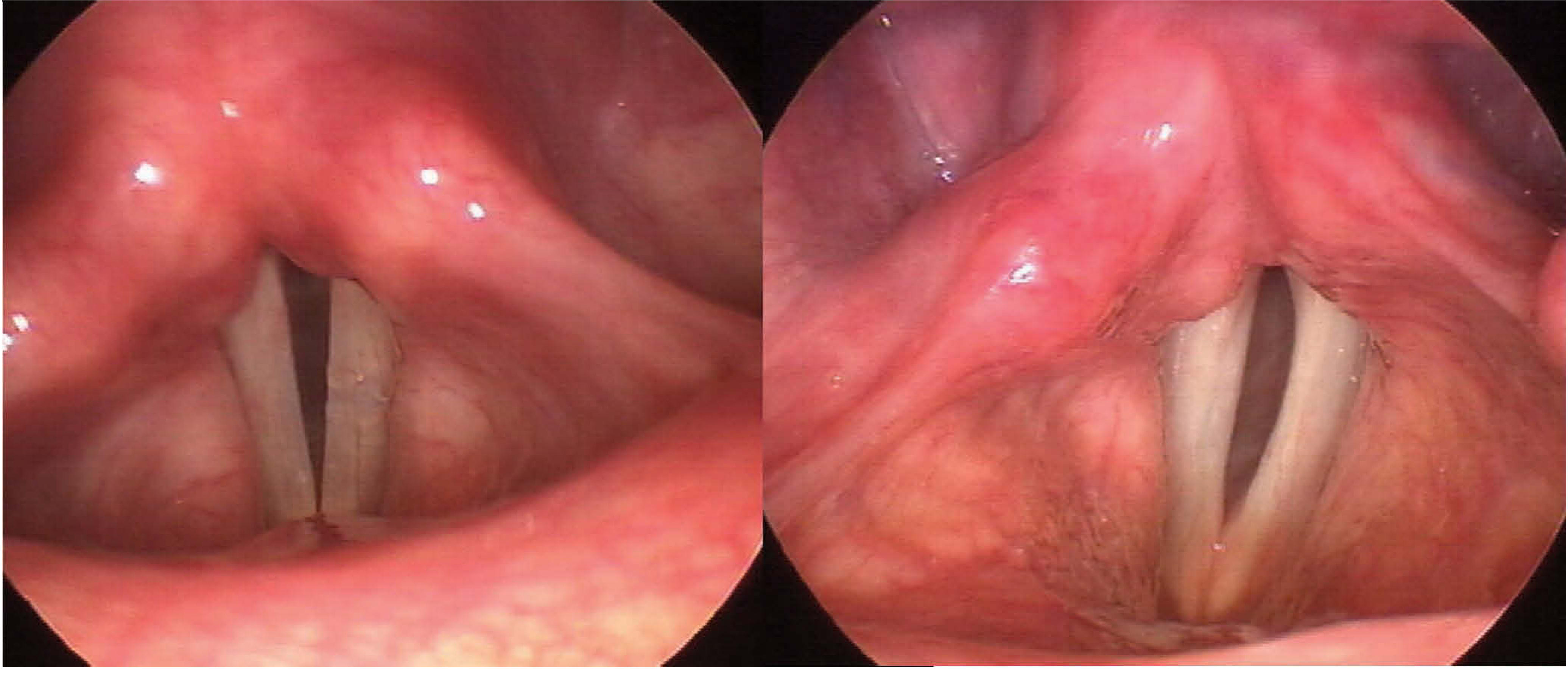
- Immobile vocal fold on the affected side
- Use the 'eee-sniff' manoeuvre to exaggerate cord asymmetry on examination
- Spindle-shaped gap: membranous portion fails to close, cartilaginous portion approximates (relatively common)
- Posterior gap: wide distance between vocal processes; indicates complete denervation; associated with prolapsed arytenoid; responds poorly to injection/thyroplasty alone
- Supraglottic hyperfunction (false cord squeezing) - compensatory mechanism for glottic insufficiency
- Jostle sign (positive in mechanical fixation): the paralysed arytenoid is passively pushed laterally by the contralateral arytenoid during phonation; negative in true denervation where the arytenoid is flaccid
'High Vagal' Injury (above origin of SLN)
When the vagus or its superior laryngeal branch is also affected (jugular foramen syndrome, skull base surgery, high neck dissection), additional features emerge:
- Hemi-laryngeal anaesthesia - absent sensation, greatly increases silent aspiration
- Pharyngeal constrictor atony - pooling in pyriform fossa
- Cricopharyngeal hyperfunction - UOS dysfunction worsening dysphagia
- Associated CN deficits (IX, XI) if jugular foramen involved; Horner syndrome if cervical sympathetics involved
Voice Assessment Tools
| Tool | Description |
|---|---|
| Voice Handicap Index (VHI) | Patient-rated validated scale of vocal disability |
| V-RQOL | Voice-related quality of life scale |
| CAPE-V | Clinician-rated perceptual voice assessment |
| GRBAS scale | Grade, Roughness, Breathiness, Asthenia, Strain |
| Maximum Phonation Time (MPT) | Duration of sustained vowel /i/; inversely proportional to glottic insufficiency |
| s/z ratio | Ratio of MPT of unvoiced /s/ to voiced /z/; normally large (s>>z); approaches 1.0 with glottic insufficiency |
Investigations
| Investigation | Purpose |
|---|---|
| Flexible nasolaryngoscopy | First-line; visualise cord position, gap configuration, arytenoid position, pooling |
| Videostroboscopy | Assess mucosal wave; detect subtle paresis; evaluate tension mismatch |
| CT neck + chest (skull base to aortic arch / right subclavian) | Mandatory to image full nerve course; exclude malignancy |
| MRI skull base / brain | If high vagal injury suspected; central cause; jugular foramen lesion |
| Laryngeal EMG (LEMG) | Distinguishes true denervation from mechanical fixation; useful prognostic tool especially <6 months from onset; better at predicting poor recovery than good recovery |
| Modified barium swallow / FEES | If aspiration suspected; guides dietary modification |
| Serology | Yield is essentially 0% without clinical suspicion; not routine |
LEMG Prognostic Accuracy (Table 78.3, Scott-Brown's)
| Study | N | Predicted Recovery | Predicted No Recovery |
|---|---|---|---|
| Munin et al. | 31 | 80% | 80% |
| Sittel et al. | 111 | 13% | 94% |
| Parnes & Satya-Murti | 18 | 80% | 100% |
LEMG is most reliable as a predictor of poor outcome - fibrillations and positive sharp waves indicate absent reinnervation. Presence of reinnervation on EMG does not always correlate with return of function (synkinesis).
Management
Decision Framework
Unilateral RLN Palsy diagnosed
├── Cause identified post-surgically? → No further workup for cause needed
├── Unknown cause → CT skull base to aortic arch (mandatory)
│
└── Treatment decision guided by:
    ├── Severity of aspiration/dysphagia (trumps all other factors)
    ├── Degree of vocal disability
    ├── Prognosis for recovery
    └── Glottic gap configuration
1. Observation + Voice Therapy
Favourable factors for observation:
- No aspiration
- Intact nerve (structurally) with recovery potential
- Minimal vocal disability or demand
- Significant comorbidities
Voice therapy does not alter the course of paralysis but offers insight and compensatory strategies. Natural improvement occurs over time in many cases (especially idiopathic/viral).
However - severe dysphagia, aspiration pneumonia, or frank aspiration on evaluation overrides all other factors and demands intervention.
2. Injection Laryngoplasty (Temporary / Early)
Best for:
- Recovery anticipated (use absorbable material)
- Small glottic gap (2-3 mm)
- No posterior glottic gap
- Aspiration / high vocal demand
- Short life expectancy (avoids open surgery)
- As test of medialization suitability
Injectables used:
- Hyaluronic acid preparations (shortest duration)
- Micronized human dermis (Cymetra)
- Autologous fat
- Carboxymethylcellulose-glycerine gel
- Calcium hydroxylapatite (CaHA) - most durable injectable; ~1 year effect
Routes: Direct laryngoscopy (GA), or percutaneous/peroral under topical LA in office
Limitations:
- Cannot correct posterior glottic gap or height discrepancy
- Requires over-injection (allowing for reabsorption) - fine tuning impossible
- Superficial injection into lamina propria stiffens the mucosa and impairs vibration
- All available materials stiffen the lamina propria
3. Medialization Thyroplasty (Laryngeal Framework Surgery)
Isshiki Type I thyroplasty - permanent procedure; reserved for cases where recovery is unlikely.
Favourable factors:
- Poor prognosis for recovery
- Large glottic gap or posterior gap
- High degree of vocal disability
- Dysphagia / aspiration
Technique: Under local anaesthesia (patient phonates to guide implant placement in real-time), a window is cut in the thyroid lamina and a biologically inert implant (silicone, Gore-Tex, hydroxylapatite/VoCoM, titanium) is inserted into the paraglottic space to push the cord medially.
Advantages over injection: Precise, predictable, durable, adjustable, can be done safely in high-risk patients
Complications:
- Airway obstruction (post-op oedema + medialization narrows airway; some surgeons keep overnight)
- Lumen perforation → infection/extrusion
- Suboptimal voice (revision rate 5.4-33%) - causes: persistent posterior gap, under-medialization, superior/anterior implant malposition
4. Arytenoid Adduction (± Thyroplasty)
Added when:
- Significant posterior glottic gap
- Prolapsed / poorly supported arytenoid
- Height mismatch between cord levels
Technique: Suture through the muscular process of the arytenoid, secured to the thyroid lamina with anterolateral traction → internal rotation of arytenoid → vocal process medializes and descends.
More effective than thyroplasty alone for closing the posterior gap (thyroplasty pushes the arytenoid body but biomechanics of the cricoarytenoid joint mean it cannot reliably rotate the vocal process medially).
More technically demanding, higher risk of oedema/bleeding causing airway obstruction. Combined thyroplasty + arytenoid adduction is the gold standard for cadaveric cord with posterior gap.
5. Reinnervation
Ansa cervicalis to RLN anastomosis (non-selective)
- Restores muscle bulk and tone to the thyroarytenoid; prevents atrophy
- Does not restore purposeful cord motion (due to synkinesis from complex innervation)
- Provides neuromuscular medialization without an implant
- Ideal if RLN was knowingly sectioned during surgery - immediate re-anastomosis (or reinnervation if tension-free anastomosis impossible) is the treatment of choice when the cut is recognized intraoperatively
- If considering later in the disease course, must confirm patient is not depriving the cord of spontaneous reinnervation
- Improvement takes weeks to months; sometimes combined with other procedures
6. Prognosis for Spontaneous Recovery
- Good prognosis: Post-intubation, idiopathic/viral (Ramsay Hunt, Bell's equivalent), blunt trauma
- Poor prognosis: Complete nerve section during tumour resection, nerve invasion by tumour, thoracic aortic aneurysm, progressive neurological disease
Summary
| Feature | Unilateral RLN Palsy |
|---|---|
| Cord position | Paramedian or cadaveric (lateral) |
| Airway | Patent (no stridor) |
| Voice | Hoarse, breathy, weak - up to aphonia |
| Cough | Weak / ineffective |
| Aspiration | Variable (worst with high vagal or cadaveric position) |
| Most common cause | Thyroid surgery |
| First investigation | Flexible nasolaryngoscopy + CT neck/chest |
| Temporary treatment | Injection laryngoplasty |
| Definitive treatment | Medialization thyroplasty ± arytenoid adduction |
| If nerve sectioned intra-op | Immediate re-anastomosis |
- Scott-Brown's Otorhinolaryngology Head & Neck Surgery (Vol 3), pp.2114-2563
- Cummings Otolaryngology Head and Neck Surgery, Ch. 62-63
- Schwartz's Principles of Surgery (11th ed.), p.1656
- Goldman-Cecil Medicine, p.3973
B/l sln and rln palsy
Bilateral SLN + RLN palsy means complete bilateral vagal/laryngeal denervation - both the sensory-motor superior laryngeal nerves and both recurrent laryngeal nerves are non-functional. This is the most severe laryngeal paralysis syndrome: the airway is obstructed (cords in median position), there is aphonia, and crucially, all laryngeal protective sensation is lost, making silent aspiration inevitable and catastrophic.
Searching sources...
Finding Sources
Finding Sources
Finding Sources
Reading File
Reading File
Reading File
Finding Sources
Finding Sources
Reading File
Reading File
Reading File
Finding Sources
Finding Sources
Reading File
Reading File
Finding Sources
Finding Sources
Your credits are exhausted. You can purchase a top-up or subscribe to a plan to continue using Orris. If you're already subscribed, your daily credits will reset tomorrow.
Bells palsy
Your credits are exhausted. You can purchase a top-up or subscribe to a plan to continue using Orris. If you're already subscribed, your daily credits will reset tomorrow.
This is a shared conversation. Sign in to Orris to start your own chat.