
Tremors in young causes and management
tremor young adults causes management
tremor classification types neurological diagram
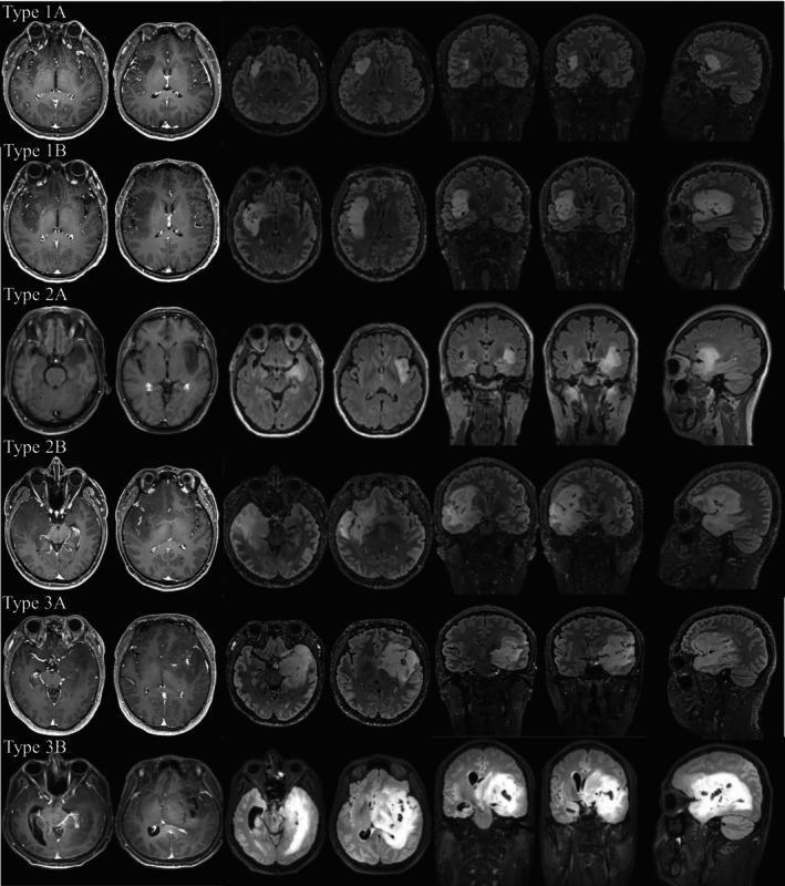
This diagnostic image composite presents a neurological classification system for gliomas based on tumor spread and anatomical involvement, organized into six types: 1A, 1B, 2A, 2B, 3A, and 3B. Each row displays axial, coronal, and sagittal MRI views, primarily utilizing T1-weighted and T2/FLAIR sequences to demonstrate lesion morphology. Type 1A shows a localized hyperintense lesion restricted to the insular cortex. Type 1B displays extension into the orbitofrontal cortex or temporal pole. The Type 2 category focuses on medial temporal involvement: 2A involves peri-hippocampal structures (amygdala and parahippocampal gyrus), while 2B shows direct invasion of the hippocampus. Type 3 represents deep structure involvement: 3A demonstrates invasion into the internal capsule, and 3B shows the most extensive spread, involving both the internal capsule and the thalamus. The images highlight the progression from localized cortical lesions to deep white matter and nuclear gray matter invasion, serving as a radiological tool for preoperative staging and surgical planning.
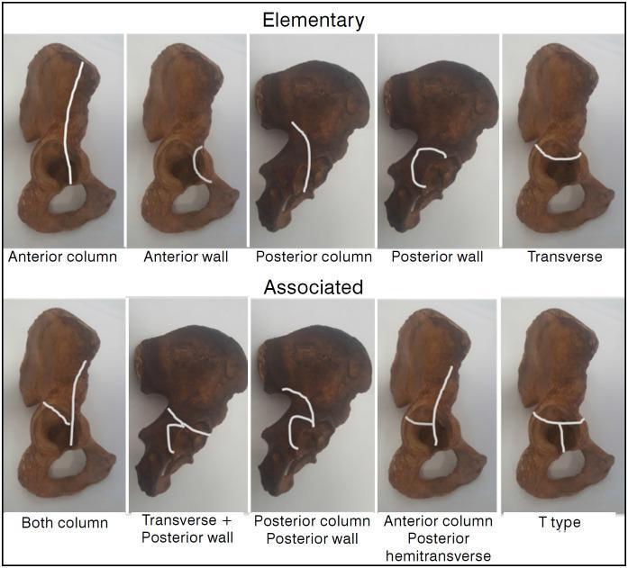
Educational diagram illustrating the Judet-Letournel classification of acetabular fractures using anatomical specimens of the hemipelvis. The visual is divided into two rows: 'Elementary' and 'Associated' fracture types. White lines superimposed on the specimens delineate specific fracture patterns. The 'Elementary' row features five simple patterns: Anterior column (vertical line through the pelvic brim), Anterior wall (localized articular segment), Posterior column (ischiadic notch through obturator foramen), Posterior wall (localized posterior rim), and Transverse (horizontal line bisecting the acetabulum). The 'Associated' row demonstrates complex configurations: Both column, Transverse plus posterior wall, Posterior column plus posterior wall, Anterior column with posterior hemitransverse (a vertical anterior column line joined by a horizontal posterior component), and T-type (a transverse fracture with an additional vertical stem extending through the obturator foramen). This orthopedic diagram serves as a clinical reference for orthopedic surgeons and medical students to understand pelvic trauma morphology and surgical planning.
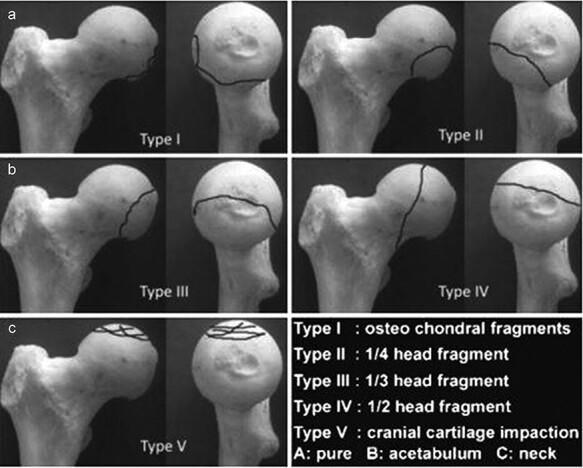
This orthopaedic anatomical diagram illustrates the Chiron classification (2004) for femoral head fractures, shown across five categorized types (Type I to Type V). Each type is presented using a dry bone model of the proximal femur from two perspectives: a lateral/oblique view and a frontal view of the femoral head. Fracture lines are demarcated by solid black lines to indicate the morphology and size of the fracture fragments. Type I displays small osteochondral fragments at the superior-lateral aspect. Type II shows a 1/4 head fragment on the anterior-superior surface. Type III demonstrates a 1/3 head fragment extending from superior to posterior. Type IV depicts a vertical fracture dividing approximately 1/2 of the femoral head. Type V illustrates cranial cartilage impaction with multiple intersecting lines across the superior articular surface. The classification is clinically relevant for orthopedic surgeons and residents for determining the severity of femoral head trauma and informing surgical decisions, such as fragment fixation versus excision based on size and location.
"essential tremor" OR "Wilson disease" tremor young onset
Tremors in Young Patients: Causes and Management
What is Tremor?
- Bradley and Daroff's Neurology in Clinical Practice
Classification by Activation Condition
| Type | Description | Common Causes in Young |
|---|---|---|
| Rest tremor | Present when body part is fully relaxed; disappears with action | Parkinsonian (juvenile PD, Wilson's disease) |
| Postural tremor | Appears when limb is held against gravity | Essential tremor, enhanced physiological tremor |
| Kinetic/Intention tremor | Worsens as limb approaches a target | Cerebellar disorders (MS, SCA, trauma) |
| Task-specific | Only during one activity | Primary writing tremor |
| Isometric tremor | During sustained muscle contraction without movement | -- |
| Dystonic tremor | Slow, irregular, in body part affected by dystonia | Wilson's disease, DYT-1 dystonia |
Causes in Young Patients
1. Essential Tremor (ET)
-
Characteristics: Bilateral postural/kinetic tremor, 5-8 Hz frequency, involves hands/arms most commonly; also head, voice, and tongue
-
Family history: Autosomal dominant inheritance in many cases; sporadic cases occur
-
Typical complaint: Difficulty holding a glass of water without spilling; problems with handwriting
-
Key features: Tremor increases with stress; temporarily reduced by alcohol ingestion
-
Pathophysiology: Associated with pathological changes in the cerebellum; cerebellar-thalamic pathway dysfunction
-
Neuroanatomy through Clinical Cases, 3rd Edition
2. Wilson's Disease (Hepatolenticular Degeneration)
- Onset: Typically age 5-35 years
- Neurologic signs: Tremor (including the characteristic "wing-beating" tremor - arms abducted, elbows flexed), choreiform movements, rigidity, parkinsonism, ataxia, dysarthria, dysphagia, cognitive changes
- Hepatic signs: Liver disease (can be the presenting feature in children under age 10)
- Psychiatric features: Personality change, psychosis
- Diagnosis:
- Kayser-Fleischer rings on slit-lamp exam (present in virtually all with neurologic involvement)
- Serum ceruloplasmin < 20 mg/dL
- Elevated 24-hour urine copper
- Liver biopsy in equivocal cases
"Wilson's disease should be considered and formally excluded in all young adults with new-onset psychiatric symptoms, especially if liver function tests are abnormal" - Goldman-Cecil Medicine
3. Enhanced Physiological Tremor
-
Caffeine excess
-
Hyperthyroidism (thyrotoxicosis)
-
Alcohol withdrawal
-
Anxiety and intense fear
-
Drugs: lithium, valproate, beta-agonists (salbutamol), steroids, SSRIs, tricyclics, amiodarone
-
Metabolic derangements (hypoglycemia, hypomagnesemia)
-
Peripheral neuromuscular disorders
-
Neuroanatomy through Clinical Cases, 3rd Edition
4. Cerebellar Tremor (Intention Tremor)
- Frequency: 2-4 Hz; irregular oscillations in multiple planes that worsen as target is approached ("terminal tremor")
- Young causes:
- Multiple sclerosis (MS) - very common cause in young adults; appendicular tremor limiting daily activities
- Spinocerebellar ataxias (SCAs) - hereditary
- Posterior fossa tumors or trauma
- Friedreich's ataxia
5. Juvenile / Young-Onset Parkinson's Disease
- Onset before age 40 (young-onset PD) or before age 21 (juvenile PD)
- Rest tremor: 4-7 Hz, "pill-rolling" character, asymmetric, suppressed by voluntary movement, re-emerges with sustained posture ("reemergent tremor")
- Accentuated by stress and mental activity (asking patient to perform mental calculations)
- Associated features: bradykinesia, rigidity, postural instability
- Causes: PARK2 (parkin), PARK7 (DJ-1), PINK1 mutations
6. Dystonic Tremor
- Slow, irregular, jerky oscillation in a body part affected by dystonia
- Important in young patients: DYT-1 (TOR1A gene deletion) - autosomal dominant early-onset generalized dystonia; often starts in lower limbs
- Dopa-responsive dystonia (DRD) - an essential diagnosis not to miss; responds dramatically to low-dose levodopa
- Also seen in Wilson's disease, tardive dyskinesia (from antipsychotic/antiemetic drugs)
7. Drug-Induced Tremor
| Drug Class | Examples |
|---|---|
| Antipsychotics/antiemetics | Haloperidol, metoclopramide (tardive tremor/parkinsonism) |
| Mood stabilizers | Lithium, valproate |
| Stimulants | Caffeine, cocaine, amphetamines |
| Bronchodilators | Salbutamol (beta-2 agonist) |
| Antidepressants | SSRIs, TCAs |
| Immunosuppressants | Cyclosporine, tacrolimus |
| Thyroid hormone | Levothyroxine (excessive dosing) |
8. Functional/Psychogenic Tremor
-
Variable frequency - the tremor starts and stops
-
Entrainment test: tremor in affected limb stops OR entrains to the frequency being tapped by the unaffected hand
-
Ballistic movements: functional tremor stops briefly when the unaffected hand makes sudden movements
-
Loading paradox: adding weights makes functional tremor worse (organic tremor improves)
-
Coactivation sign: co-contraction of agonist and antagonist muscles
-
Coherence analysis: if bilateral, functional tremor has the same frequency in both limbs (organic tremors have different frequencies)
-
Bradley and Daroff's Neurology in Clinical Practice
9. Other Causes in Young Patients
| Condition | Key Features |
|---|---|
| Hyperthyroidism | Postural tremor + tachycardia, weight loss, exophthalmos |
| Alcohol withdrawal | Coarse postural tremor + autonomic hyperactivity (12-48 hours after last drink) |
| Multiple sclerosis | Intention tremor; demyelinating plaques on MRI |
| Spinocerebellar ataxias | Hereditary; cerebellar signs + family history |
| Fragile X tremor/ataxia syndrome (FXTAS) | In older carriers; rare in young |
| Neuropathic tremor | Associated with peripheral neuropathy (Charcot-Marie-Tooth) |
| Primary writing tremor | Task-specific; only on writing |
Diagnostic Approach
| Test | Condition screened |
|---|---|
| Serum ceruloplasmin + 24h urine copper | Wilson's disease (mandatory in all young tremor cases) |
| Thyroid function tests (TFTs) | Hyperthyroidism |
| LFTs + slit-lamp exam | Wilson's disease |
| MRI brain | MS, cerebellar pathology, structural lesions |
| Genetic testing | SCA panel, PARK2/PINK1, DYT-1, Fragile X |
| DAT-SPECT scan | Differentiates PD (abnormal) from ET/functional (normal) |
| Nerve conduction studies | Neuropathic tremor |
Management
Essential Tremor
- First line: Propranolol (beta-blocker) or Primidone (anticonvulsant)
- Second line: Topiramate, gabapentin
- Others: Phenobarbital, diazepam, atenolol
-
VIM nucleus thalamic Deep Brain Stimulation (DBS) - most effective; improves tremor, quality of life, and ADLs
-
Focused ultrasound thalamotomy
-
Stereotactic radiosurgery (gamma knife thalamotomy)
-
Neuroanatomy through Clinical Cases, 3rd Edition; Bradley and Daroff's Neurology
Wilson's Disease
- Dietary restriction: Keep copper intake < 2 mg/day (avoid shellfish, nuts, chocolate, mushrooms)
- Copper chelation:
- Penicillamine (500 mg 3-4x daily in adults) - first-line historically; neurologic worsening occurs in ~10%; co-administer pyridoxine 25 mg/day
- Trientine hydrochloride (1-1.5 g/day) - preferred over penicillamine due to fewer side effects and less neurologic worsening
- Tetrathiomolybdate - promising for neurologic Wilson's; in clinical trials
- Zinc (50 mg acetate 3x daily) - blocks GI copper absorption; low toxicity; used for maintenance or mild cases
- Liver transplantation - for refractory hepatic failure
- Katzung's Basic and Clinical Pharmacology, 16th Edition
Parkinsonian Tremor (Juvenile/Young-Onset PD)
- Levodopa/carbidopa - gold standard
- Dopamine agonists (ropinirole, pramipexole) - often preferred first-line in young patients to delay dyskinesias
- MAO-B inhibitors (selegiline, rasagiline)
- VIM-DBS or subthalamic nucleus DBS for refractory cases
Cerebellar (Intention) Tremor - MS
-
Weighted wrist braces and adapted utensils (non-pharmacological)
-
Carbamazepine (up to 800 mg/day), gabapentin (up to 3600 mg/day), topiramate
-
Primidone 125-250 mg BID-TID
-
Clonazepam 0.5-2 mg 1-4x daily
-
Surgical thalamotomy or DBS for refractory cases
-
Bradley and Daroff's Neurology in Clinical Practice
Enhanced Physiological / Drug-Induced Tremor
- Remove the offending drug or trigger
- Treat underlying cause (e.g., antithyroid therapy for hyperthyroidism; benzodiazepines for alcohol withdrawal)
- If drug cannot be stopped: dose reduction, switch to alternative, or add low-dose propranolol
Functional/Psychogenic Tremor
- Positive diagnosis with above clinical tests (entrainment, loading, coherence)
- Psychoeducation - explain the diagnosis clearly and positively
- Physiotherapy with distraction-based movement retraining
- Cognitive behavioral therapy (CBT) and treatment of underlying psychiatric comorbidity
- Avoid unnecessary medications
Dopa-Responsive Dystonia (DRD)
- Small doses of levodopa produce dramatic, sustained benefit - this diagnosis must not be missed
- Trial of levodopa is recommended in any young patient with dystonia
Summary Table: Tremors in Young Patients
| Cause | Type | Age Group | Key Diagnostic Feature | Treatment |
|---|---|---|---|---|
| Essential tremor | Postural, 5-8 Hz | Any age, young adult | Family history, alcohol-responsive | Propranolol, primidone, DBS |
| Wilson's disease | Rest + wing-beating | 5-35 years | Kayser-Fleischer rings, low ceruloplasmin | Trientine/penicillamine + zinc |
| Enhanced physiological | Postural, 8-12 Hz | Any age | Drug/caffeine/thyroid history | Remove cause |
| Juvenile PD | Rest, 4-7 Hz | < 40 years | Asymmetric, bradykinesia, DAT-SPECT | Levodopa, DA agonists, DBS |
| Cerebellar (MS, SCA) | Intention, 2-4 Hz | Young adult | Worsens toward target, MRI lesions | Gabapentin, DBS |
| Functional | Variable frequency | Young adult | Entrainment, loading paradox | Physiotherapy, CBT |
| Drug-induced | Postural | Any | Drug history | Withdraw offending drug |
| Dopa-responsive dystonia | Dystonic | Childhood/teen | Dramatic levodopa response | Low-dose levodopa |
Important: Wilson's disease must be actively excluded in every young patient presenting with tremor, as it is treatable and progressive if missed.