Pectoralis major insertion

Reading File
Finding Sources
Finding Sources
Finding Sources
Reading File
Reading File
Reading File
Reading File
Loading Image
Here is a complete summary of the pectoralis major insertion from the textbooks:

Pectoralis Major - Insertion

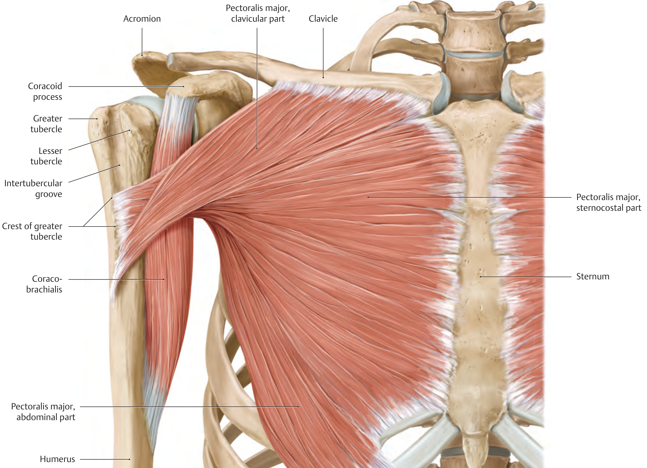
Insertion site: The lateral lip of the intertubercular sulcus (bicipital groove) of the humerus - also described as the crest of the greater tubercle.
The tendon is broad and has a horseshoe-shaped cross section. A key anatomical feature is that the tendon fibers are twisted on themselves before inserting:
PartOriginInserts...
Clavicular headMedial half of clavicleLower on the lateral lip (more superficially)
Sternocostal headSternum + upper 6 ribsHigher / deeper on the lateral lip
Abdominal partAnterior rectus sheathDeepest / highest on the humerus
The twist explained: The parts with a more superior origin (clavicular) end up inserting lower on the humerus; the parts with a more inferior origin (sternocostal/abdominal) end up inserting higher. This reversal unfolds as the arm is elevated, which progressively stretches the fibers and increases the force the muscle can generate at higher angles of arm elevation.
Pectoralis major muscle - all three parts converging to insert on the crest of the greater tubercle/intertubercular groove
Actions at insertion:
  • Adduction + medial (internal) rotation of the arm (entire muscle)
  • Flexion (clavicular + sternocostal parts)
  • Extension from a flexed position against resistance (sternocostal head)
Innervation: Medial and lateral pectoral nerves (C5-T1)

Sources: Gray's Anatomy for Students, THIEME Atlas of General Anatomy & Musculoskeletal System, Rockwood & Green's Fractures in Adults 10th ed, Imaging Anatomy Vol. 3

What is the vascular supply to pectoralis major?

Finding Sources
Reading File
Reading File
Finding Sources
Reading File
Reading File
Loading Image

Vascular Supply to Pectoralis Major

The pectoralis major has three arterial sources, with one dominant pedicle:

1. Thoracoacromial Artery (Dominant - Pedicle Type I)

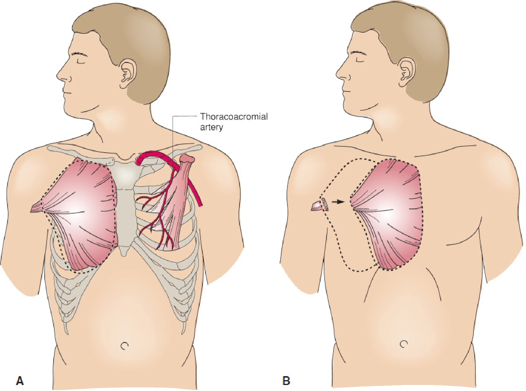
This is the dominant blood supply. It arises from the 1st or 2nd segment of the axillary artery, proximal to the coracoid attachment of the pectoralis minor. Its pectoral branch is the key vessel - it runs between the pectoralis major and pectoralis minor, distributing to the deep surface of the muscle.
The thoracoacromial artery has four named branches:
  • Pectoral branch (to pectoralis major and minor)
  • Acromial branch
  • Clavicular branch
  • Deltoid branch (runs laterally with the cephalic vein in the deltopectoral groove)
Each part of the muscle has a distinct vascular supply running in the epimysium on its posterior surface.

2. Lateral Thoracic Artery

Also a branch of the axillary artery (though with significant origin variation - most commonly from the thoracoacromial in 67% of cases, and directly from the axillary artery in only 17%). It travels along the lateral chest wall and supplies the pectoralis major, serratus anterior, and subscapularis. It also sends branches to the axilla and breast.

3. Perforating Branches of the Internal Thoracic (Mammary) Artery

These intercostal perforators enter the medial part of the muscle and provide additional supply, particularly relevant when using the muscle as a pedicled flap based on these vessels (turning it into a reverse-pedicle or "turnover" flap).

Pectoralis major rotation flap showing the dominant thoracoacromial artery pedicle (A) and medial transposition preserving the neurovascular bundle (B)

Clinical Relevance - Muscle Flap

Because the thoracoacromial artery is such a reliable dominant pedicle, the pectoralis major is classified as a Mathes and Nahai Type V muscle flap (one dominant pedicle + secondary segmental pedicles). It is widely used in head and neck reconstruction, chest wall reconstruction, and sternal wound coverage - the flap is rotated medially while preserving the thoracoacromial neurovascular bundle.

Sources: Imaging Anatomy Vol. 3 (Bones, Joints, Vessels & Nerves), Rockwood & Green's Fractures in Adults 10th ed, Mulholland & Greenfield's Surgery 7th ed, Fischer's Mastery of Surgery 8th ed

Explain brachial plexus and axillary artery

Finding Sources
Finding Sources
Reading File
Reading File
Reading File
Reading File
Reading File
Loading Image
Loading Image
Loading Image
Excellent - all the content I need is gathered. Here is the full explanation:

Brachial Plexus

Formation

The brachial plexus is formed by the anterior rami of C5, C6, C7, C8, and T1. It originates in the neck between the anterior and middle scalene muscles, passes over rib I, and enters the axilla.
Brachial plexus schematic - roots to terminal nerves with cord relationships to 2nd part of axillary artery

Structure: Roots - Trunks - Divisions - Cords - Branches

Roots (anterior rami)

The roots receive gray rami communicantes from the sympathetic trunk (postganglionic sympathetic fibers). They lie between the anterior and middle scalene muscles, posterior to the subclavian artery.

Trunks (3)

TrunkFormed by
SuperiorC5 + C6
MiddleC7 alone
InferiorC8 + T1
The inferior trunk lies on rib I posterior to the subclavian artery.

Divisions (6)

Each trunk splits into an anterior and a posterior division:
  • Anterior divisions - supply anterior (flexor) compartments
  • Posterior divisions - supply posterior (extensor) compartments
  • No peripheral nerves arise directly from divisions

Cords (3) - named by position relative to the 2nd part of the axillary artery

CordFormed byPositionContributions
LateralAnterior div. of superior + middle trunksLateralC5-C7
MedialAnterior div. of inferior trunkMedialC8, T1
PosteriorAll three posterior divisionsPosteriorC5-T1

Branches

Brachial plexus - all branches with relationships to the axillary artery

From the Roots

NerveRootsSupplies
Dorsal scapularC5Rhomboids major & minor
Long thoracicC5, C6, C7Serratus anterior
Contribution to phrenicC5Diaphragm (partial)

From the Trunks (Superior trunk only)

NerveRootsSupplies
SuprascapularC5, C6Supraspinatus, infraspinatus
Nerve to subclaviusC5, C6Subclavius muscle

From the Lateral Cord

NerveSupplies
Lateral pectoral nervePectoralis major (clavicular part)
Musculocutaneous nerveCoracobrachialis, biceps brachii, brachialis + lateral cutaneous nerve of forearm
Lateral root of median nerve(joins medial root to form median nerve)

From the Medial Cord

NerveSupplies
Medial pectoral nervePectoralis major and minor
Medial cutaneous nerve of armSkin, medial arm
Medial cutaneous nerve of forearmSkin, medial forearm
Medial root of median nerve(joins lateral root)
Ulnar nerveMost intrinsic hand muscles, flexor carpi ulnaris, medial FDP

From the Posterior Cord

NerveSupplies
Superior subscapularSubscapularis (upper)
ThoracodorsalLatissimus dorsi
Inferior subscapularSubscapularis (lower) + teres major
Axillary nerveDeltoid, teres minor + lateral cutaneous nerve of arm
Radial nerveAll posterior compartment muscles of arm and forearm
The 5 terminal nerves: Musculocutaneous, Median, Ulnar, Radial, Axillary (Mnemonic: "My Mate Likes Real Ale")


Axillary Artery

Course

The subclavian artery becomes the axillary artery at the lateral margin of rib I, and ends at the inferior border of teres major, becoming the brachial artery.
The pectoralis minor crosses it anteriorly and divides it into 3 parts:
PartPositionNumber of branches
1stProximal to pectoralis minor1
2ndPosterior to pectoralis minor2
3rdDistal to pectoralis minor3
Mnemonic: 1-2-3 (one branch, two branches, three branches)
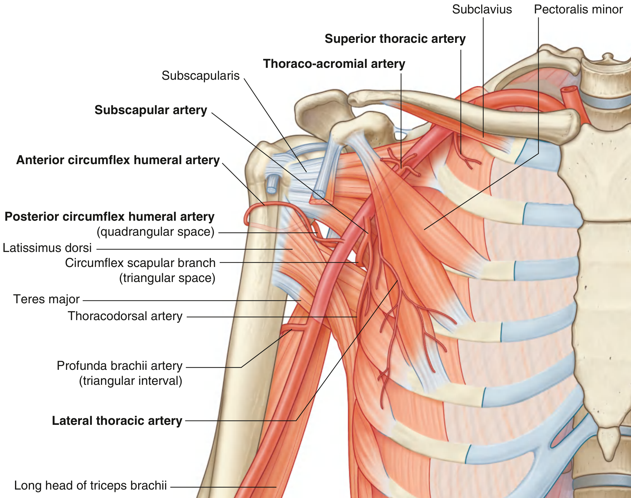
Branches of the axillary artery - all 6 branches labeled with surrounding muscles

The 6 Branches

1st Part (1 branch)

Superior thoracic artery - small; supplies upper medial and anterior axillary walls

2nd Part (2 branches)

Thoraco-acromial artery - arises from anterior surface, curves around superior margin of pectoralis minor, pierces clavipectoral fascia, divides into 4 branches:
  • Pectoral - supplies pectoralis major & minor, contributes to breast
  • Deltoid - runs with cephalic vein in deltopectoral groove
  • Clavicular
  • Acromial
Lateral thoracic artery - arises from anterior surface, follows lateral margin of pectoralis minor; supplies medial/anterior axillary walls and contributes to breast supply in women

3rd Part (3 branches)

Subscapular artery - largest branch; arises from posterior surface; divides into:
  • Circumflex scapular artery (through triangular space → infraspinous fossa)
  • Thoracodorsal artery (follows lateral border of scapula)
Anterior circumflex humeral artery - small; passes anterior to surgical neck of humerus; supplies glenohumeral joint and humeral head
Posterior circumflex humeral artery - larger; exits through quadrangular space with the axillary nerve; curves around surgical neck of humerus; supplies surrounding muscles, glenohumeral joint

Key Relationships

  • The cords of the brachial plexus are named by their relationship to the 2nd part of the axillary artery (lateral, medial, posterior)
  • The axillary vein lies medial and anterior to the axillary artery throughout its course
  • The posterior circumflex humeral artery travels with the axillary nerve through the quadrangular space - both are at risk in surgical neck of humerus fractures

Sources: Gray's Anatomy for Students (block 4), THIEME Atlas General Anatomy & Musculoskeletal System

Make flowchart for subclavian artery

Finding Sources
Reading File
Reading File
Finding Sources
Reading File
Reading File
Generating Image

A clean medical anatomy flowchart of the subclavian artery and all its branches. The flowchart should be structured top to bottom with clear boxes and connecting arrows. At the top: "SUBCLAVIAN ARTERY" in a large header box. Below it show the origin: "Right: from Brachiocephalic trunk | Left: from Arch of Aorta directly". The artery is divided into 3 parts by the Anterior Scalene muscle, so show 3 clearly labeled columns/sections: PART 1 (Medial to anterior scalene): - Vertebral Artery → branches: Spinal branches, Basilar artery (forms circle of Willis) - Internal Thoracic Artery (Internal Mammary) → branches: Pericardiophrenic artery, Anterior intercostal arteries, Superior epigastric artery, Musculophrenic artery - Thyrocervical Trunk → sub-branches: Inferior Thyroid Artery (→ ascending cervical artery), Suprascapular Artery, Transverse Cervical Artery (→ dorsal scapular artery) PART 2 (Posterior to anterior scalene): - Costocervical Trunk → branches: Supreme (Highest) Intercostal Artery (→ 1st & 2nd posterior intercostal arteries), Deep Cervical Artery Note: On LEFT side, costocervical trunk comes from Part 1 PART 3 (Lateral to anterior scalene): - Dorsal Scapular Artery (may arise here or from Part 2) - No branches on the RIGHT side in Part 3 At the bottom: arrow pointing to "AXILLARY ARTERY (at lateral border of 1st rib)" Use a clean white background, blue header boxes, light blue branch boxes, yellow terminal boxes. Include a mnemonic at the bottom: "VITamin C & D" = Vertebral, Internal thoracic, Thyrocervical trunk, Costocervical trunk, Dorsal scapular. Professional medical textbook diagram style with clear hierarchy and all text legible.

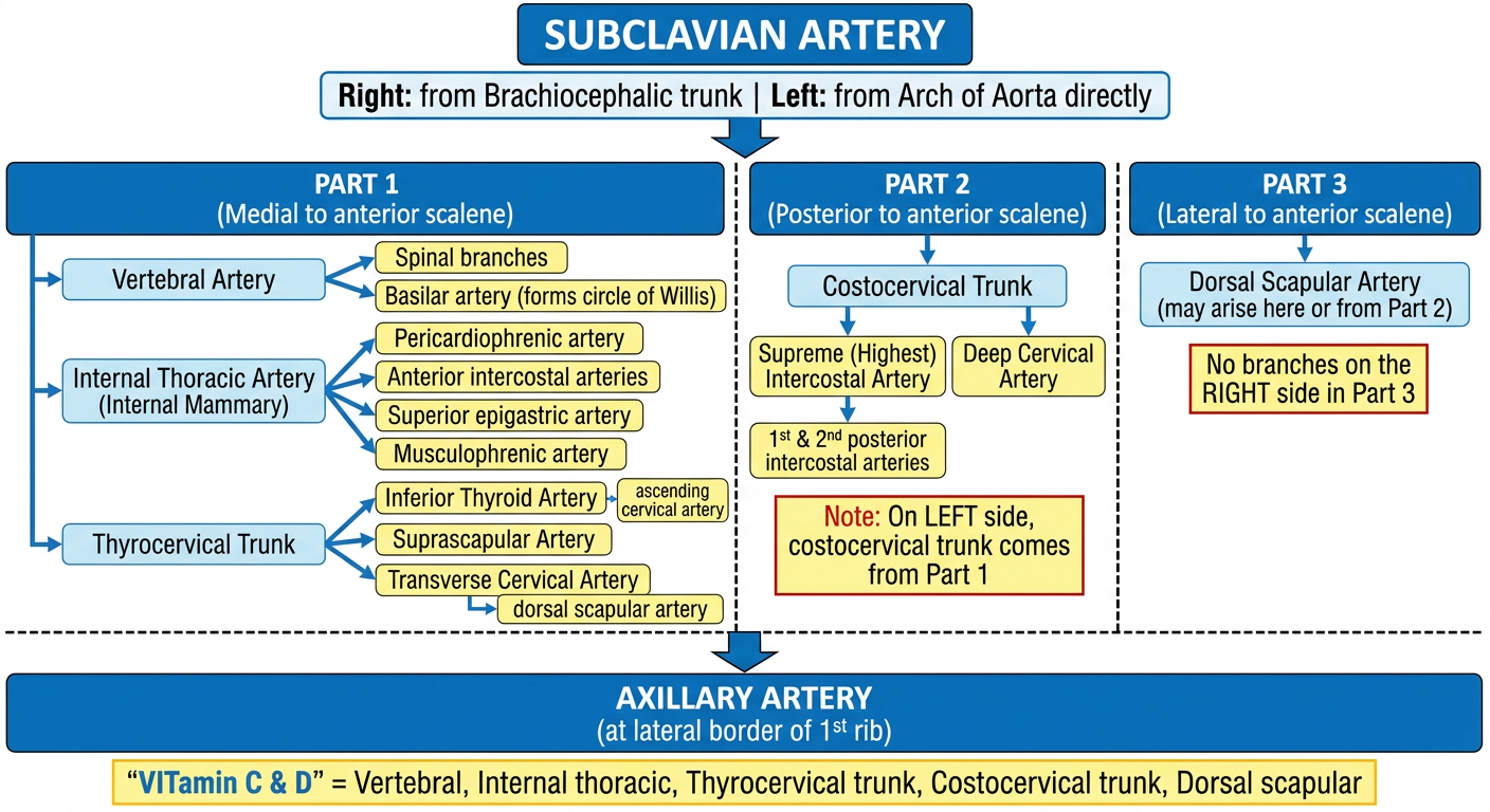
Subclavian Artery Flowchart

Here is a summary of the complete structure shown in the flowchart:

Subclavian Artery - Branches by Part

The artery is divided into 3 parts by the anterior scalene muscle:

Part 1 - Medial to anterior scalene (3 branches)

BranchSub-branches
Vertebral arterySpinal branches → ascends through transverse foramina C6-C1 → basilar artery → circle of Willis
Internal thoracic artery (internal mammary)Pericardiophrenic, anterior intercostals → divides into: superior epigastric + musculophrenic
Thyrocervical trunkInferior thyroid artery (+ ascending cervical), suprascapular artery, transverse cervical artery (+ dorsal scapular)

Part 2 - Posterior to anterior scalene (1 branch)

BranchSub-branches
Costocervical trunkSupreme intercostal artery (→ 1st & 2nd posterior intercostals) + Deep cervical artery
Note: On the left side, the costocervical trunk arises from Part 1. On the right, it arises from Part 2.

Part 3 - Lateral to anterior scalene (1 branch - variable)

BranchNotes
Dorsal scapular arteryMay arise here or from Part 2, or from the transverse cervical artery
Part 3 has no branches in many descriptions (the dorsal scapular is highly variable in origin).

Continues as → Axillary Artery (at lateral border of rib I)


Mnemonic: "VITamin C & D"
  • Vertebral
  • Internal thoracic
  • Thyrocervical trunk
  • Costocervical trunk
  • Dorsal scapular
Sources: Scott-Brown's Otolaryngology, Imaging Anatomy Vol. 3, THIEME Atlas of General Anatomy
This is a shared conversation. Sign in to Orris to start your own chat.