
Varicose vein
Reading File
Now let me search the medical library for varicose veins content in parallel.
Finding Sources
Searching PubMed
"varicose veins"[MeSH Terms] AND treatment
Good. Now let me do a broader paragraph-level search to find comprehensive content on varicose veins across the library.
Finding Sources
Excellent. Multiple rich sources found. Let me read the key sections in parallel - Harrison's (pathophysiology + clinical), Bailey & Love (epidemiology + main section), and Schwartz's (varicose veins section).
Reading File
Reading File
Reading File
Excellent content from Schwartz's and Bailey & Love. Let me now read more from Harrison's (pathophysiology + treatment) and Bailey & Love (investigations + treatment), and also grab the image from Gray's Anatomy.
Reading File
Reading File
Loading Image
Good - rich content obtained. Let me read more on treatment from Bailey & Love and Harrison's, and also the CEAP classification content.
Reading File
Reading File
I now have comprehensive content from multiple authoritative sources. Let me read the treatment section from Harrison's and the remaining treatment options from Bailey & Love.
Reading File
Reading File
I now have sufficient content from multiple authoritative textbooks. Let me also get the surgical treatment section and complications from Bailey & Love.
Reading File
I have comprehensive information from all the textbooks. Now let me compile the full response.
Varicose Veins
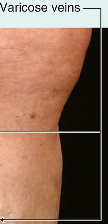
Fig. 1.29 - Photograph demonstrating varicose veins (Gray's Anatomy for Students)
Definition
Varicose veins are dilated, tortuous, bulging superficial veins measuring at least 3 mm in diameter. They are part of the spectrum of chronic venous disease (CVD), which ranges from telangiectasias and reticular veins at the mild end, through varicose veins, to chronic venous insufficiency with edema, skin changes, and ulceration at the severe end. - Harrison's Principles of Internal Medicine 22E, p. 2224
Epidemiology
-
Adult prevalence of visible varicose veins: 30-50% (Bailey & Love) / ~15% in men and ~30% in women in the US (Harrison's)
-
Prevalence increases sharply with age - the Edinburgh Vein Study showed prevalence rising from 11.5% (age 18-24) to 55.7% (age 55-64)
-
Risk factors:
- Female sex - higher prevalence in women across most studies
- Age - progressive increase with aging
- Pregnancy - significantly increases risk
- Family history - strong familial susceptibility; ~50% of primary cases have a positive family history
- Obesity and increased height - BMI and height correlate with prevalence
- Prolonged standing - inconclusive but commonly cited
- Ethnicity - influences prevalence
-
Bailey and Love's Short Practice of Surgery 28th Ed., p. 1052
Anatomy Relevant to Varicose Veins
The superficial venous system of the leg consists of:
- Great saphenous vein (GSV) - originates on the medial foot, ascends anterior to the medial malleolus, runs along the medial calf and thigh, drains into the common femoral vein at the saphenofemoral junction (SFJ)
- Small saphenous vein (SSV) - originates on the dorsolateral foot, ascends posterior to the lateral malleolus, drains into the popliteal vein
- Perforating veins - connect superficial to deep systems; normally direct flow superficial → deep
Varicosity distribution reflects which system is incompetent:
-
Medial thigh/calf → GSV incompetence
-
Posterolateral calf → SSV incompetence
-
Anterolateral thigh/calf → Anterior accessory GSV (AAGSV) incompetence
-
Harrison's Principles of Internal Medicine 22E; Bailey and Love 28th Ed.
Classification
Primary vs. Secondary
| Type | Mechanism |
|---|---|
| Primary | Intrinsic abnormalities of the venous wall, defective valve structure/function, wall weakness; no underlying deep vein disease |
| Secondary | Result of deep-vein thrombosis, venous hypertension, incompetent perforating veins, AV fistulas, or congenital anomalies (e.g., Klippel-Trénaunay syndrome) |
CEAP Classification (Clinical, Etiologic, Anatomic, Pathophysiologic)
The standard staging system for chronic venous disease:
| CEAP Class | Description |
|---|---|
| C0 | No visible/palpable signs |
| C1 | Telangiectasias or reticular veins |
| C2 | Varicose veins |
| C2r | Recurrent varicose veins |
| C3 | Edema |
| C4a | Pigmentation or eczema |
| C4b | Lipodermatosclerosis or atrophie blanche |
| C4c | Corona phlebecta |
| C5 | Healed venous ulcer |
| C6 | Active venous ulcer |
| C6r | Recurrent active venous ulcer |
- Harrison's Principles of Internal Medicine 22E, Table 293-1; Bailey and Love 28th Ed.
Pathophysiology
-
Valve incompetence - Normally, one-way venous valves prevent reflux. When valve leaflets fail (primary degeneration or post-thrombotic damage), blood refluxes downward under gravity, raising hydrostatic pressure in the superficial system.
-
Venous wall weakness - Intrinsic structural defects in smooth muscle and connective tissue of the vein wall allow progressive dilatation.
-
Chronic venous hypertension - Sustained high pressure leads to extravasation of fluid and blood elements (including hemosiderin, fibrin cuffs) into surrounding tissue, causing the chronic skin changes of venous insufficiency.
-
Calf muscle pump failure - Normal venous return relies on the calf muscle pump; dysfunction worsens venous hypertension.
- Harrison's Principles of Internal Medicine 22E, p. 2224-2225
Clinical Features
Symptoms
- Aching, heaviness, throbbing, burning or "bursting" sensation in the affected leg
- Symptoms worsen with prolonged standing and improve with elevation or compression hosiery
- Pruritus (itching) - especially with complications
- Ankle swelling - more common with complications
- Symptoms can severely impact daily activities and quality of life, independent of the degree of incompetence
Signs
- Tortuous, dilated, bulging subcutaneous veins (clinically obvious in standing position)
- Saphena varix - a large, dilated vein at the SFJ presenting as a painless groin lump that disappears on lying down; may be confused with a femoral hernia; may show a cough impulse
- Skin changes in advanced disease: hyperpigmentation, stasis dermatitis (eczema), lipodermatosclerosis, atrophie blanche
- Venous ulcer - typically in the gaiter area (medial/lateral malleolus)
Acute Complications
- Superficial vein thrombosis (thrombophlebitis) - painful, tender, inflamed cord along a varicosity
- Bleeding - rupture of an attenuated vein cluster, can be significant
Chronic Complications
-
Lipodermatosclerosis, hyperpigmentation, eczema, venous ulceration (~20% of chronic venous insufficiency patients)
-
Bailey and Love's Short Practice of Surgery 28th Ed., pp. 1052-1053; Schwartz's Principles of Surgery 11th Ed.
Investigations
Duplex Ultrasound Scanning (Gold Standard)
Hand-held Doppler and tourniquet tests have been abandoned in favour of duplex ultrasound for all patients prior to intervention. Duplex scan establishes:
- Presence and distribution of reflux in deep and superficial systems
- Exact extent of reflux, including affected junctions and perforators
- Presence of deep-venous obstruction
- Suitability of veins for the different treatment modalities (diameter, tortuosity, depth)
A high-frequency linear array transducer (7.5-13 MHz) is used. Reflux is defined as reversal of flow >0.5 seconds on colour/spectral Doppler after calf compression and release.
Other Investigations (rarely needed)
-
CT venography / MR venography / conventional venography - reserved for suspected proximal obstruction, May-Thurner syndrome, or IVC filter occlusion
-
Plethysmography - can quantify severity of reflux and calf pump function
-
Bailey and Love's Short Practice of Surgery 28th Ed., pp. 1053-1054; Harrison's 22E, p. 2225
Treatment
1. Conservative (Supportive) Measures
- Leg elevation - periodic elevation reduces symptoms
- Avoid prolonged standing/sitting
- Compression hosiery - graduated compression stockings:
- 20-30 mmHg for simple varicose veins (CEAP C2)
- 30-40 mmHg for CVI with edema/ulcers (CEAP C3-C6)
- British classification: Class 1 (14-17 mmHg), Class 2 (18-24 mmHg), Class 3 (25-35 mmHg)
- Improves symptoms but does not prevent progression; compliance is poor
- Level 1 evidence shows interventional treatment offers superior quality of life improvement
- Weight loss in overweight/obese patients
- Exercise - leg strengthening improves calf pump function
2. Endothermal Ablation (First-line Interventional Treatment)
Replaced surgical ligation/stripping as the gold standard after RCTs showed superior or equivalent outcomes with faster recovery.
Principle: A catheter is inserted percutaneously into the incompetent vein. Tumescent local anaesthetic is infiltrated around the vein (compresses vein, hydro-dissects nearby nerves, acts as heat sink). The device destroys the vein wall causing permanent occlusion.
Endovenous Laser Ablation (EVLA)
- A flexible glass fibre is inserted; laser energy (~1470 nm wavelength) generates thermal energy at the tip
- Energy delivery ~60-80 J/cm used to achieve durable closure
- Efficacy: >95% closure rate
Radiofrequency Ablation (RFA)
- ClosureFast™ device (Medtronic) is most widely used
- Electromagnetic current heats a wire coil to 120°C for 20-second cycles
- Automatic treatment cycle reduces operator-dependent error
- Efficacy: >95% closure rate
- Recent meta-analysis (Jiang et al., 2024, PMID 38316290) found both RFA and EVLA are effective with no significant difference in outcomes; choice is often personal preference
EVLA vs. RFA: Evidence is generally equivocal. RFA has a simpler user interface (single button), freeing the surgeon to perform concurrent phlebectomy.
3. Foam Sclerotherapy
- Sclerosing agents include hypertonic saline, sodium tetradecyl sulfate (STS), and polidocanol
- Mechanism: destroys venous endothelium, causing inflammatory occlusion
- Used for telangiectasias, reticular veins, and larger varicose veins (higher concentrations for larger veins)
- Can be used alone or adjunctively after thermal ablation for tributaries
- After injection, elastic bandages are worn continuously for 3-5 days, then compression stockings for ≥2 weeks
- Complications: allergic reaction, local hyperpigmentation, matting, superficial thrombophlebitis, and rarely, skin necrosis or DVT
- A multicenter RCT confirmed significant symptom relief vs. placebo
4. Surgery
Traditional surgical treatment includes:
- Saphenofemoral/saphenopopliteal junction ligation (high tie)
- Stripping of the GSV or SSV
- Multiple avulsion phlebectomy (stab avulsion) of visible tributaries
Surgery remains an option where endothermal ablation is not suitable (very tortuous veins, lack of facilities) and is still employed for recurrent disease. The systematic review by Bontinis et al. (2024) compared treatments for recurrent varicose veins arising from the saphenofemoral junction and GSV, supporting modern endovenous approaches even in recurrent disease.
5. NICE/Guideline Recommendations
NICE (UK) guideline recommends offering endothermal ablation (EVLA or RFA) as first-line treatment; if not suitable, ultrasound-guided foam sclerotherapy; and if not suitable, surgical ligation/stripping.
- Bailey and Love's Short Practice of Surgery 28th Ed., pp. 1054-1057; Harrison's Principles of Internal Medicine 22E, pp. 2225-2226; Schwartz's Principles of Surgery 11th Ed.
Complications of Varicose Vein Surgery/Procedures
-
Bruising and haematoma
-
Wound infection
-
DVT and pulmonary embolism (rare)
-
Nerve injury - saphenous nerve (medial calf paraesthesia with GSV surgery), sural nerve (lateral calf/foot with SSV surgery)
-
Lymphatic injury - lymphocele, lymphoedema
-
Recurrence - neovascularisation at the junction is a common cause
-
Skin burns - with thermal ablation if tumescent anaesthesia is inadequate
-
Bailey and Love's Short Practice of Surgery 28th Ed., p. 1057
Summary Table
| Feature | Detail |
|---|---|
| Definition | Dilated, tortuous superficial veins ≥3 mm |
| CEAP Class | C2 (varicose veins) |
| Key mechanism | Valvular incompetence → venous reflux → hypertension |
| Most common system | GSV (60%), SSV (20%) |
| Key investigation | Duplex ultrasound (mandatory before intervention) |
| First-line treatment | Endothermal ablation (EVLA or RFA) |
| Sclerotherapy agents | Sodium tetradecyl sulfate, polidocanol, hypertonic saline |
| Ulcer prevalence | ~20% of chronic venous insufficiency patients |
Recent evidence: A 2024 meta-analysis (PMID 38316290) found RFA and EVLA have comparable outcomes for lower extremity varicose veins. A 2024 systematic review and meta-analysis (PMID 39559844) examined interventions for recurrent varicose veins, supporting endovenous approaches even in recurrence. No major updates from these contradict the textbook guidance above.
This is a shared conversation. Sign in to Orris to start your own chat.