
Eustachian tube anatomy and Eustachian tuboplasty in detail according to standard ENT textbook in simplified manner
Eustachian tube anatomy diagram cross section
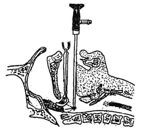
A sagittal cross-sectional anatomical diagram illustrating the technique for endoscopically assisted adenoidectomy. The diagram depicts the head and neck anatomy, including the nasal cavity, hard palate, soft palate, tongue, and cervical vertebrae. Two surgical instruments are shown inserted transorally into the nasopharynx: an adenoid curette and a 70-degree nasal endoscope. The curette, identifiable by its loop-shaped distal tip, is positioned superiorly to engage the adenoid tissue mass on the posterior nasopharyngeal wall. Simultaneously, a rigid endoscope is introduced posterior to the curette, providing direct visualization of the surgical field to ensure complete resection and avoid injury to adjacent structures like the Eustachian tube orifices. The clinical significance of this illustration is to demonstrate the spatial relationship between the instruments during a combined endoscopic-curettage procedure for treating adenoid hypertrophy. This material is relevant for otolaryngology training, specifically for surgical planning and understanding the anatomical approach to the nasopharynx.
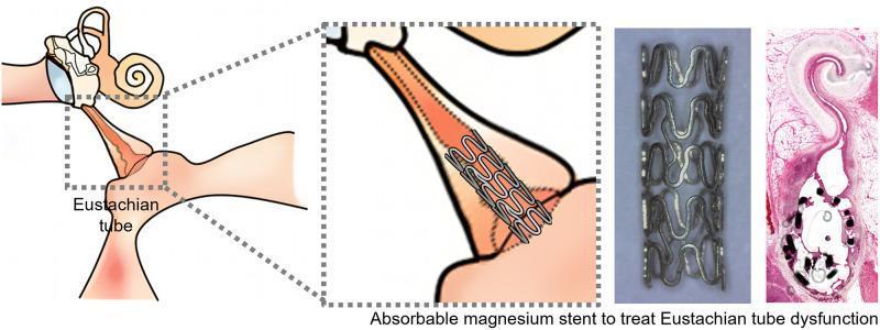
A multi-panel educational graphic illustrating the use of an absorbable magnesium (Mg) stent for managing Eustachian tube dysfunction (ETD). The leftmost section is an anatomical schematic of the ear and nasopharynx, highlighting the Eustachian tube (ET) with a dotted box. A magnified inset shows the stent positioned within the ET lumen, distal to the middle ear space, maintaining patency. The third panel features a high-resolution photograph of the standalone magnesium stent, revealing a metallic, cylindrical lattice structure composed of interconnected sinusoidal or wave-like struts. The final panel is a histological cross-section (H&E stain) of the ET lumen containing the stent, demonstrating the relationship between the stent struts and the surrounding submucosal tissue. The visual material focuses on the clinical application of biodegradable materials in otolaryngology to prevent stent-induced tissue hyperplasia while providing temporary mechanical support to the Eustachian tube.
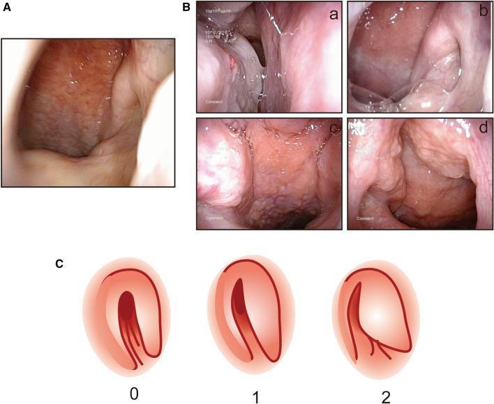
This composite educational image illustrates nasal endoscopic findings and scoring systems used to evaluate allergic rhinitis and Eustachian tube dysfunction. Section A shows a clinical photograph of a normal posterior nostril area with smooth, healthy mucosa. Section B contains four panels (a-d) demonstrating specific pathologies: (a) posterior turbinate swelling, (b) viscous posterior nostril secretions, (c) torus tubarius swelling, and (d) nasopharyngeal lymphatic hyperplasia characterized by a cobblestone mucosal appearance. Section C provides a schematic diagram for scoring the shape of the pharyngeal orifice of the Eustachian tube under a 30° endoscope: Score 0 represents a normal round or oval opening; Score 1 shows a narrow, fissure-like orifice; and Score 2 indicates a completely blocked or non-visualized orifice. This material is designed for Otolaryngology (ENT) training to standardize endoscopic assessment of the nasopharynx and its impact on Eustachian tube patency.
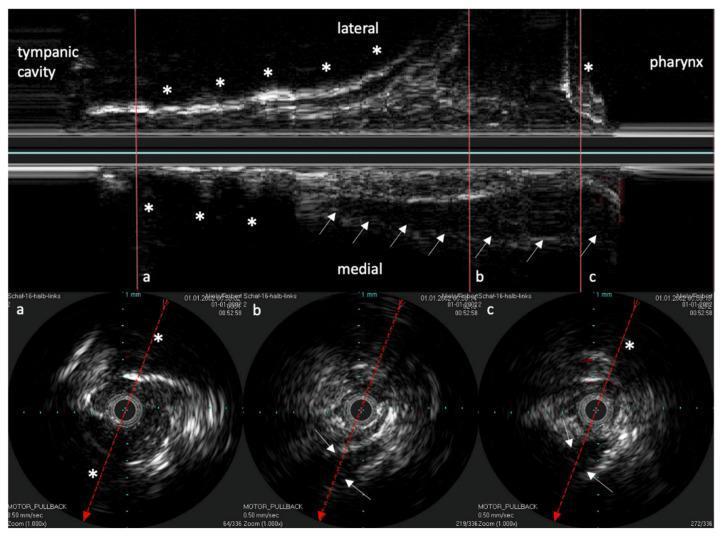
This diagnostic image displays Intravascular Ultrasound (IVUS) scans of the human Eustachian Tube (ET), featuring a longitudinal reconstruction and three corresponding circular cross-sections (a, b, and c). The longitudinal view (top) illustrates the ET's path from the tympanic cavity to the pharynx. Highly echogenic areas marked with asterisks (*) identify bony structures, which serve as consistent anatomical landmarks. The ET cartilage is characterized by low echogenicity (hypoechoic), indicated by white arrows on the medial aspect. The circular cross-sections correspond to specific anatomical locations indicated by vertical red lines in the longitudinal view: (a) represents the ET isthmus, showing surrounding bone; (b) represents the middle cartilaginous section; and (c) represents the area near the pharyngeal ostium. A dashed red line in each cross-section indicates the reconstruction axis, while the arrow tips indicate the orientation relative to the longitudinal view. This educational material demonstrates the use of high-resolution endoluminal ultrasound to distinguish between bony, cartilaginous, and soft tissue components of the ET for diagnostic or interventional planning.
Eustachian tube dysfunction balloon dilatation tuboplasty endoscopic
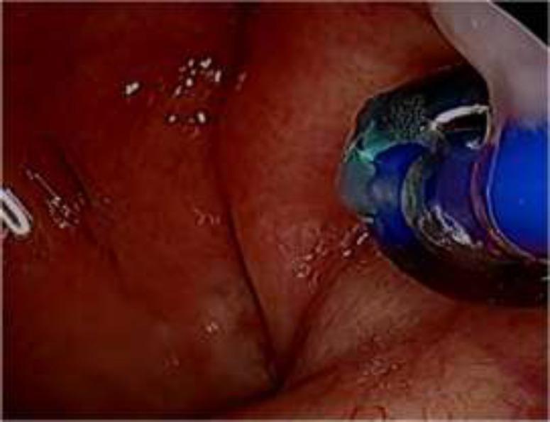
An endoscopic clinical photograph demonstrating a Balloon Eustachian Tuboplasty (BET) procedure within the nasopharynx. The image shows a dark blue, inflated balloon catheter positioned within the pharyngeal orifice of the Eustachian tube. The balloon is inserted into the cartilaginous portion of the tube to perform dilation. The surrounding mucosa appears erythematous and highly vascularized, characteristic of the nasopharyngeal lining. Significant anatomical landmarks include the torus tubarius and the folds of the Eustachian tube opening. The procedure is performed under transnasal endoscopic guidance to alleviate Eustachian tube dysfunction by widening the narrowest portion of the tube. This visual highlights the surgical intervention for middle ear aeration disorders, illustrating the precise placement of the dilation device relative to the surrounding soft tissue structures.
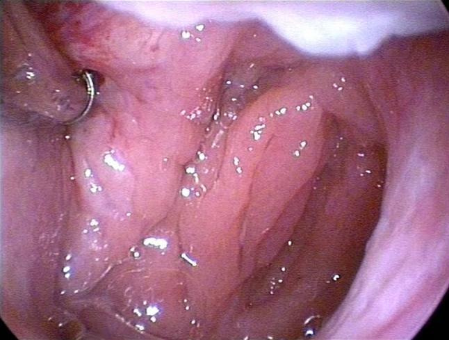
This endoscopic clinical photograph demonstrates a Balloon Eustachian Tuboplasty (BET) procedure. The image provides an internal view of the nasopharynx, focusing on the pharyngeal orifice of the Eustachian tube. The surrounding mucosa is pinkish-red, moist, and glistening, with natural anatomical folds visible. A specialized 600 µm balloon catheter is seen positioned at the entrance of the Eustachian tube. On the left side of the field, a metallic, semi-circular instrument or guide is visible, characterized by its silver, reflective surface. The educational focus of this image is the visualization of the nasopharyngeal anatomy during the insertion phase of a balloon dilation procedure intended to treat Eustachian tube dysfunction. The procedure typically involves advancing the catheter 2 cm into the tube and inflating the balloon to a pressure of 10 bars using saline solution to facilitate dilation and improve middle ear ventilation.
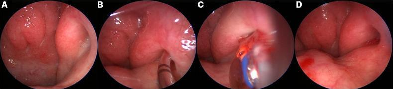
This set of four endoscopic images (A-D) demonstrates the intraoperative sequence of a Balloon Eustachian Tuboplasty (BET). Image A shows the baseline endonasal view of the nasopharyngeal orifice of a grade II Eustachian tube, characterized by pink, healthy mucosa and the torus tubarius. In image B, a semi-rigid guide catheter is introduced and positioned at the orifice to facilitate the entry of the dilation device into the cartilaginous portion of the tube. Image C displays the active dilation phase, where a blue-colored balloon catheter is inflated within the Eustachian tube lumen to achieve mechanical widening. Image D depicts the post-procedural state immediately following balloon deflation and removal; the orifice appears patent with minimal traumatic bleeding, indicating a successful dilation. This clinical sequence illustrates a minimally invasive otolaryngological intervention for chronic Eustachian tube dysfunction, specifically highlighting the navigation from the nasopharynx into the tubal lumen.
Eustachian Tube: Anatomy and Tuboplasty
PART 1: EUSTACHIAN TUBE ANATOMY
1. Overview
2. Dimensions and Orientation
| Feature | Newborn | Adult |
|---|---|---|
| Length | 17-18 mm | ~35 mm |
| Angle with horizontal | Near horizontal (10°) | 45° inclined |
| Pharyngeal vs tympanic end | Level | Pharyngeal orifice is ~15 mm LOWER than tympanic orifice |
- The tube grows progressively and becomes more angled with age.
- In infants, the near-horizontal position predisposes them to middle ear infections (pathogens ascend easily from nasopharynx).
3. Structural Divisions
[Tympanic orifice] ---BONY PART (11 mm)--- ISTHMUS ---CARTILAGINOUS PART (24 mm)--- [Pharyngeal orifice]
                    posterolateral                        anteromedial
A. Bony (Osseous) Portion - 11 mm
- Situated in the temporal bone (petrous part)
- Posterolateral in position
- Always OPEN (patent); does not collapse
- Tympanic ostium diameter: 3-5 mm (located on the anterior wall of the tympanic cavity, about 4 mm above the floor)
- Lined by ciliated cuboidal epithelium near the tympanic end
B. Cartilaginous Portion - 24 mm
- Anteromedial in position
- Normally CLOSED at rest; opens only during swallowing, yawning, or sneezing
- The cartilage is J-shaped or hook-shaped in cross section:
- Medial lamina (larger): forms the posterior cushion / torus tubarius
- Lateral lamina (smaller): forms the anterolateral wall
- Lined by pseudostratified columnar ciliated epithelium (respiratory type)
- Ostmann's fat pad lies between the dilator tubae muscle and the lateral wall - its bulk helps keep the tube closed at rest
C. Isthmus - the Narrowest Point
- Junction of the bony and cartilaginous portions
- Functionally the most important segment ("valve" mechanism is here)
- When obstructed, this is the site of ET dysfunction
4. Openings (Ostia)
| Ostium | Location | Size |
|---|---|---|
| Tympanic ostium | Anterior wall of middle ear, ~4 mm above floor | 3-5 mm diameter |
| Pharyngeal ostium | Posterior lateral nasopharynx | 3-10 mm vertical, 2-5 mm horizontal |
5. Muscles of the Eustachian Tube
A. Tensor Veli Palatini (TVP) - PRIMARY DILATOR
- Innervation: V3 (mandibular branch of trigeminal nerve)
- Its medial bundle is called the "dilator tubae" - this is the actual dilator of the ET lumen
- Acts by pulling the lateral cartilaginous wall anterolaterally, widening the lumen
- The only functional dilator in infants and young children
- Dilation begins at the nasopharyngeal end and propagates toward the isthmus
B. Levator Veli Palatini (LVP) - ACCESSORY OPENER
- Innervation: Vagus nerve (CN X) via pharyngeal plexus
- Elevates the soft palate and medially rotates the torus tubarius
- Creates a scaffold against which the TVP can act
- Becomes functionally relevant only in older children/adults (in children it is too far from ET cartilage)
C. Tensor Tympani
- Associated with the bony portion
- Does not directly contribute to tubal opening
D. Salpingopharyngeus
- May assist in opening by pulling the posterior ET wall inferiorly
- LVP contracts first → soft palate rises, torus tubarius rotates medially
- Lateral pharyngeal wall medializes (protecting ET during opening)
- TVP (dilator tubae) contracts → lumen visibly dilates from nasopharyngeal end toward isthmus
- Valve closes proximally toward nasopharynx, expelling secretions outward
6. Epithelial Lining
| Segment | Epithelium |
|---|---|
| Pharyngeal end (cartilaginous) | Pseudostratified columnar ciliated + goblet cells |
| Near tympanic end | Ciliated cuboidal epithelium |
| Lymphoid tissue within tube | "Tonsil of Gerlach" |
7. Blood Supply and Nerve Supply
- Arterial supply: Ascending pharyngeal artery, middle meningeal artery, artery of pterygoid canal
- Venous drainage: Pterygoid plexus
- Nerve supply: Tympanic plexus (CN IX), pharyngeal branch of CN IX and X
8. Functions of the Eustachian Tube (The Three Ps)
| Function | Details |
|---|---|
| Pressure equalization | Equalizes atmospheric pressure with middle ear pressure |
| Protection | Keeps nasopharyngeal secretions, pathogens, and sound out of middle ear |
| Patency / Drainage | Clears mucus and debris from middle ear into nasopharynx |
- A pressure difference of 200-300 mm H₂O is needed for airflow through the tube
- A Valsalva maneuver generates ~20-40 mm Hg
- Negative pressure of -30 mm Hg for 15 minutes can produce a middle ear transudate
- Critical pressure difference = 90 mm Hg - at this point the tube "locks" and cannot be opened by the TVP
- Tympanic membrane rupture risk above 100 mm Hg
- Air is expelled from the middle ear more easily than it enters (explains why airplane descent causes more symptoms than ascent)
9. Differences: Child vs Adult ET
| Feature | Child | Adult |
|---|---|---|
| Length | 17-18 mm | ~35 mm |
| Angle | Near horizontal | 45° |
| Active dilator | TVP only | TVP + LVP |
| ET dysfunction risk | Higher (OME, AOM common) | Lower |
PART 2: EUSTACHIAN TUBOPLASTY
1. Why Tuboplasty? - The Problem It Solves
- Otitis media with effusion (OME / "glue ear")
- Middle ear atelectasis
- Negative middle ear pressure
- Recurrent AOM
- Failed tympanoplasty
- Repeatedly need new tympanostomy tubes
- Have persistent OME or atelectasis despite multiple tubes
- Show slow-motion endoscopic evidence of obstructive mucosal disease causing inadequate tubal dilation
2. Types of Eustachian Tuboplasty
A. Laser Eustachian Tuboplasty (ETP)
- Refractory OME with multiple prior tympanostomy tubes (2 or more)
- Middle ear atelectasis despite medical therapy
- Difficulty with airplane flights (pressure changes)
- Underlying mucosal disease controlled (allergy, LPR treated)
- Slow-motion ET video endoscopy showing obstructive dysfunction
- Uncontrolled inflammatory disease (active allergy, uncontrolled LPR)
- Primary middle ear disease unrelated to ET dysfunction (e.g., thick proteinaceous "glue ear" repeatedly blocking tubes)
- Prior nasopharyngeal radiation therapy
- Extensive uncontrolled nasopharyngeal mucosal disease
- Treat underlying causes (allergy, LPR, rhinitis) for at least 6 weeks
- 6-week course of nasal steroid sprays
- High-resolution CT scan of nasopharynx and temporal bones
- Video endoscopy review to assess ET pathology extent
- Anesthesia: General anesthesia with endotracheal intubation; patient supine
- Setup: Tonsil mouth gag placed; 30-degree, 4-mm Hopkins rod nasal endoscope via nasal cavity; procedure monitored on surgical video monitor
- Myringotomy: May be done to aspirate middle ear effusion
- Local infiltration: Lidocaine 1% with epinephrine 1:100,000 injected into nasopharyngeal orifice via curved endosinus needle through the mouth
- Dilation: ET orifice dilated with 2 mm Merocel sponge (soaked in epinephrine 1:50,000) placed into lumen for 5 minutes, then removed
- Laser ablation:
- Fiber-delivered diode-pumped KTP laser (settings: 2,500 mW, 1-second continuous mode)
- Handpiece gently bent to 60-degree arc and passed through the mouth
- Ablation begins on mucosa overlying the leading edge of the medial cartilaginous lamina within the posterior cushion
- A triangular defect is created: ablating all mucosa and submucosa down to exposed cartilage, extending proximally up to the valve
- No more than 40% of the circumference of the lumen is ablated (to prevent synechiae)
- Advanced disease: Additional cartilage resection (medial cartilaginous lamina thinning) to reduce the "spring" of the cartilage; ablation may extend into the valve (with great care to protect the opposite wall)
- Packing: Merogel soaked in sulfacetamide/prednisolone ophthalmic solution applied to the defect at end of procedure
- Day surgery; discharged same day
- Light activities for 10 days
- Saline nasal spray 3 times daily for 2 weeks
- Proton pump inhibitors for 6 weeks (if LPR suspected)
- Nasal steroid sprays for 6 weeks (if allergic disease)
- Follow-up at 1, 6, 12, 24, and 36 months
- In a 2-year follow-up study using KTP laser: 37% of refractory OME patients had complete remission; additional patients reported substantial improvement
- 3/13 had continuing intermittent effusions but were substantially improved
- Microdebrider variant: 70% improvement in combined OME + sinus surgery patients (by subjective score, tympanogram, and audiogram)
- Failure correlated with: active LPR, allergic disease, high CT sinus staging, high eosinophil count in biopsy
- Synechiae (adhesions between anterior and posterior walls) - most feared complication, worsens obstruction
- Small granuloma at posterior cushion (resolves with nasal steroid sprays)
- Minor nasal synechia (rare)
- No cases of significant bleeding or worsened dysfunction reported in published series
- Postoperative pain is minimal
B. Balloon Dilation Eustachian Tuboplasty (BET)
- Uncomfortable ear pressure with atmospheric changes (airplane travel)
- Inability to perform Valsalva maneuver
- Chronic OME
- Middle ear atelectasis
- Recurrent middle ear disease (perforation, cholesteatoma)
- Failed tympanoplasty (e.g., protruding middle ear prosthesis)
- General anesthesia (usually); local anesthesia also reported (outpatient technique)
- Transnasal 30-degree endoscope visualization of nasopharynx
- Semi-rigid guide catheter positioned at ET orifice
- Balloon catheter (600 µm diameter) advanced 2 cm into the ET to reach the cartilaginous segment
- Balloon inflated with saline to 10 bars pressure
- Pressure maintained for 2 minutes
- Balloon deflated and removed
- Can be performed bilaterally in the same session
- In a series of >400 patients: ~80% reported subjective benefit with significant improvement in ET score
- Meta-analysis (1155 patients): short-term benefit in majority; medium-to-long-term benefit in a smaller number
- Post-procedure: virtually all patients able to perform Valsalva maneuver
- Safe in adults and children ≥7 years
- Nasopharyngeal scarring (post-radiation for nasopharyngeal cancer)
- Injury to torus tubarius from prior adenoidectomy (technically challenging)
- Cleft palate patients - underlying pathology is muscular (TVP dysfunction), NOT structural obstruction; BET does not help
3. Comparison: Laser ETP vs Balloon BET
| Feature | Laser Tuboplasty | Balloon Dilation (BET) |
|---|---|---|
| Mechanism | Tissue ablation (mucosal debulking) | Mechanical dilation |
| Target site | Posterior cushion / medial cartilaginous lamina | Cartilaginous portion (2 cm in) |
| Anesthesia | General | GA or local |
| Success rate | ~70% (KTP laser) | ~80% subjective benefit |
| Key risk | Synechiae if anterior wall damaged | Minimal scarring risk |
| Recurrence | Yes, if underlying disease (allergy, LPR) uncontrolled | Medium-long term benefit in fewer cases |
| Available for children? | Adults (mainly) | Children ≥7 years |
| Not suitable for | Uncontrolled mucosal inflammation | Cleft palate, post-radiation |
4. Key Points to Remember
- The ET is normally closed at rest (cartilaginous part) and opens only actively with muscular effort - different from the bony part which is always open.
- The isthmus is the narrowest point and the functional "valve."
- TVP is the primary dilator; its medial bundle (dilator tubae) is the actual opener.
- In children, only the TVP functions; the LVP becomes important in adulthood.
- Cleft palate disrupts TVP function → ET dysfunction → recurrent OME.
- Tuboplasty targets the cartilaginous portion (the obstructed/dysfunctional segment), not the bony part.
- Underlying conditions (allergy, LPR) must be controlled before and after tuboplasty or results will be poor.
- The Tonsil of Gerlach = lymphoid tissue within the tube - can contribute to obstruction.
- Critical pressure = 90 mm Hg locks the ET and cannot be overcome by muscle action alone.
- K.J. Lee's Essential Otolaryngology, 11th edition - Chapter 13
- Cummings Otolaryngology Head & Neck Surgery - Chapters 130-131
- Shambaugh Surgery of the Ear - Chapter 12
- Scott-Brown's Otorhinolaryngology Head & Neck Surgery Vol. 2 - Chapters 46, 86