
Osteomeatal complex , what are the structure involved , what are the anatomical pathology involved . Complete overview of the omc and its anatomical parts
osteomeatal complex anatomy diagram paranasal sinuses
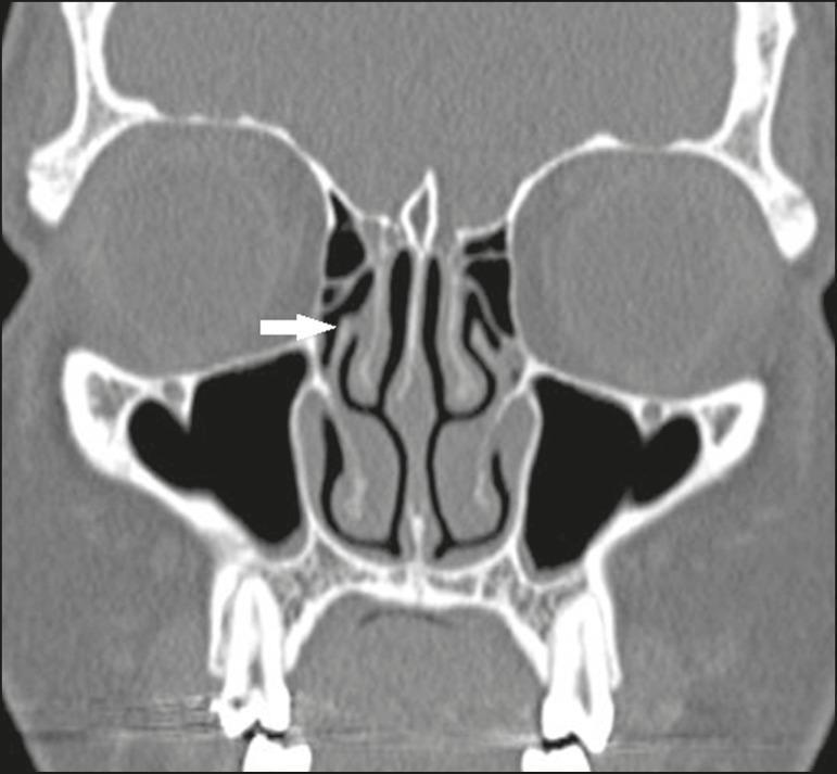
This diagnostic image is a coronal CT scan of the paranasal sinuses using bone window settings, highlighting the complex anatomy of the sinonasal cavities. The focus is on the right osteomeatal complex, where a white arrow points to a Landsberg and Friedman type 6 anatomical variation: the superior portion of the right uncinate process inserts directly into the medial surface of the middle turbinate. Surrounding structures clearly visible in high contrast include the nasal septum, bilateral middle and inferior turbinates, ethmoid air cells, and the air-filled maxillary sinuses. The orbits and orbital floors are visualized superior to the maxillary sinuses, and the frontal sinuses are seen at the top of the frame. This educational visual is critical for otorhinolaryngologists and radiologists to identify anatomical variants of the uncinate process, which determines the drainage pathway of the frontal recess and influences surgical planning for functional endoscopic sinus surgery (FESS).
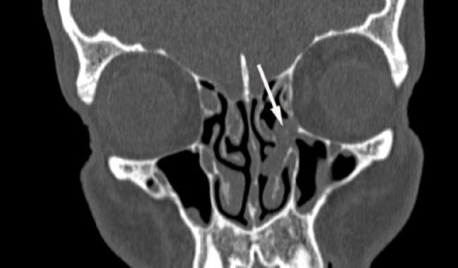
This diagnostic image is a coronal CT scan of the paranasal sinuses focusing on the sinonasal anatomy in the context of pre-transplant screening. The image displays the frontal sinuses, ethmoid air cells, maxillary sinuses, and the nasal cavity with its turbinates. A white arrow points to a distinct soft tissue density mass, identified as a nasal polyp, located within the left osteomeatal complex (right side of the image). This mass is causing mechanical obstruction of the drainage pathway. The contralateral (right) osteomeatal complex remains patent and air-filled. The surrounding bony structures, including the orbital floors and nasal septum, appear intact. Such imaging is critical in hematology-oncology patients, specifically those with acute myeloid leukemia undergoing hematopoietic stem cell transplantation, to identify and manage potential foci of infection or obstructive sinonasal disease prior to immunosuppression.
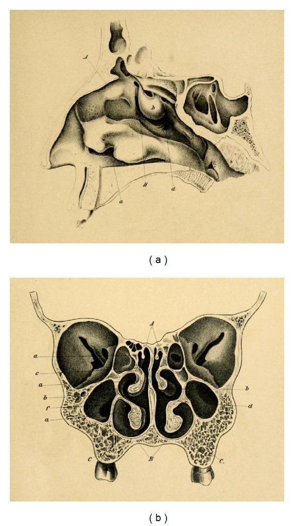
This historical anatomical diagram by Emil Zuckerkandl (1882) illustrates the paranasal sinuses and nasal anatomy in two planes. Image (a) is a sagittal section of the nasal cavity, detailing the complex arrangement of the superior, middle, and inferior turbinates (conchae) along the lateral wall. It visualizes the frontal sinus superiorly, the sphenoid sinus posteriorly, and the relationship between the nasal cavity and the hard palate. Image (b) presents a frontal (coronal) section, showcasing the bilateral symmetry of the mid-facial anatomy. Key features include the central nasal septum, the convoluted turbinates projecting into the nasal passages, and the expansive maxillary sinuses situated laterally within the maxilla bones. Small ethmoid air cells are visible medially to the orbits and superior to the maxillary sinuses. This anatomical illustration serves as a foundational reference for otorhinolaryngology and neurosurgical approaches, particularly regarding the development of transsphenoidal surgery for pituitary lesions.
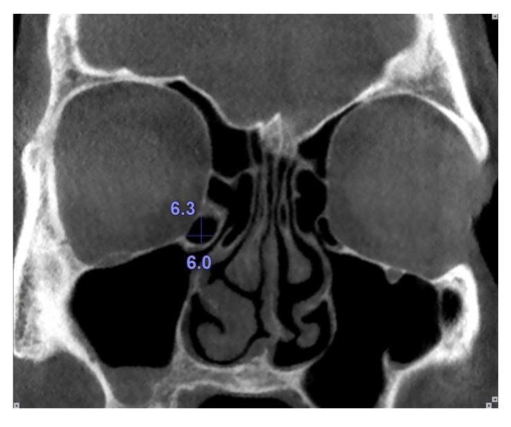
This diagnostic image is a coronal computed tomography (CT) scan of the paranasal sinuses focusing on the osteomeatal complex. The scan reveals a clear view of the nasal cavity divided by the nasal septum, flanked by bilateral air-filled maxillary sinuses and ethmoid air cells. Centrally, the inferior and middle nasal turbinates are visible. An anatomical variant known as a Haller cell (infraorbital ethmoid cell) is identified on the patient's right side (visualized on the left side of the image), located along the medial orbital floor and the superior-lateral aspect of the maxillary sinus ostium. Blue annotations and a crosshair indicate quantitative measurements of this Haller cell, with a vertical dimension of 6.3 mm and a horizontal dimension of 6.0 mm. The image serves as an educational example of sinonasal anatomy and anatomical variations that can contribute to the narrowing of the infundibulum and potential predisposition to recurrent sinusitis. The clinical focus is on preoperative mapping and diagnostic radiology in otolaryngology.
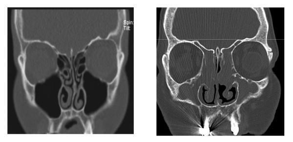
This diagnostic image displays a side-by-side comparison of two coronal CT scans of the paranasal sinuses. The left image represents a 'Normal' anatomy, characterized by clear, air-filled (black) maxillary and ethmoid sinuses, well-defined middle and inferior turbinates, and a patent osteomeatal complex. The nasal septum is midline and the nasal passages are open. In contrast, the right image depicts a patient with Chronic Rhinosinusitis with Nasal Polyps (CRSwNP). This scan reveals significant bilateral opacification of the ethmoid and maxillary sinuses, indicating mucosal thickening, fluid, or soft tissue masses. Large, irregular soft-tissue densities (nasal polyps) fill and obstruct the nasal cavities, obscuring the normal anatomy of the turbinates and significantly narrowing the airway passages. This comparison illustrates the typical radiographic presentation of sinonasal inflammatory disease and its impact on sinus aeration and nasal patency, commonly used in otolaryngology for diagnostic and surgical planning.
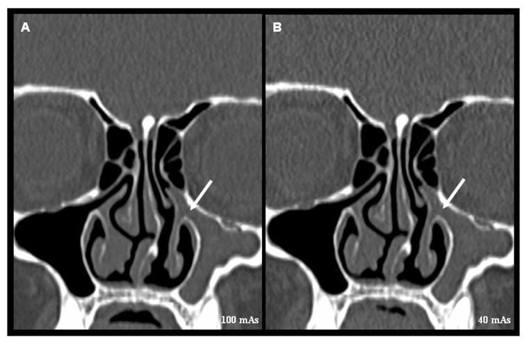
This diagnostic image is a side-by-side comparison of two coronal reformatted Multidetector Computed Tomography (MDCT) scans of the paranasal sinuses. The images illustrate the clinical pathology of the osteomeatal complex (OMC) and demonstrate the impact of radiation dose optimization on image quality. Image A is captured at 100 mAs, showing high resolution and sharp detail of the ethmoid air cells and turbinates. Image B is captured at a lower dose of 40 mAs, resulting in a subtle increase in image noise but maintaining diagnostic utility. On both scans, the right osteomeatal complex is patent with clear aeration. In contrast, the left osteomeatal complex is indicated by a white arrow, revealing opacification and obstruction by soft tissue or fluid density, consistent with sinusitis or mechanical blockage. Key anatomical structures visible include the nasal septum, middle turbinates, maxillary sinuses, and the ethmoid bulla. This educational comparison highlights the balance between radiation dose (ALARA principle) and the visualization of critical diagnostic features in rhinology and otorhinolaryngology.
Osteomeatal Complex (OMC) - Complete Overview
Definition
- Posterior: Basal lamella of the middle turbinate
- Medial: Middle turbinate
- Lateral: Lamina papyracea
- Superior: Fovea ethmoidalis (ethmoid roof)
"The ostiomeatal unit is not a discrete anatomic structure but rather is a functional designation that describes the drainage pathway of the maxillary, frontal, and anterior ethmoid sinuses. Collectively it also encompasses the middle meatus, uncinate process, and ethmoid infundibulum." - K.J. Lee's Essential Otolaryngology
Ostiomeatal Channels: Two Units
| Channel | Structures Involved | Sinuses Drained |
|---|---|---|
| Anterior OMU | Frontal sinus ostium, frontal recess, maxillary sinus ostium, ethmoidal infundibulum, middle meatus | Frontal, anterior ethmoid, maxillary |
| Posterior OMU | Sphenoid ostium, sphenoethmoidal recess, superior meatus | Sphenoid, posterior ethmoid |
- The frontal recess
- The ethmoidal infundibulum
- The sphenoethmoidal recess
Structures of the OMC - Detailed Anatomy
1. Uncinate Process
- A thin, sickle-shaped bony projection of the ethmoid bone forming the medial wall of the ethmoidal infundibulum
- Its free posterior edge forms the medial boundary of the hiatus semilunaris
- Its superior attachment has 3 important variations (Cummings):
- Type 1: Attaches laterally to the lamina papyracea or ethmoid bulla → frontal recess opens directly to middle meatus (terminal recess formed)
- Type 2: Attaches medially to the lateral surface of the middle turbinate → frontal recess drains to infundibulum
- Type 3: Attaches medially and superiorly to the skull base → frontal recess drains to infundibulum
- Its inferior aspect forms part of the medial wall of the maxillary sinus
- The maxillary sinus natural ostium lies lateral and superior to this part
2. Ethmoidal Bulla (Bulla Ethmoidalis)
- The largest and most constant of the anterior ethmoid air cells
- Forms the posterior boundary of the hiatus semilunaris
- Forms the posterior boundary of the frontal recess (if it extends to skull base)
- The space posterior to the bulla (between it and the basal lamella) is the retrobullar recess (sinus lateralis)
- Its anterior wall can extend to the skull base and limit the frontal recess posteriorly
3. Hiatus Semilunaris
- A two-dimensional, crescent-shaped gap between:
- The posterior free margin of the uncinate process (medially)
- The anterior wall of the ethmoid bulla (posteriorly)
- Through this gap, the middle meatus communicates with the ethmoidal infundibulum
- It is the "entrance" to the infundibulum
4. Ethmoidal Infundibulum
| Direction | Structure |
|---|---|
| Medial | Uncinate process |
| Lateral | Lamina papyracea |
| Posterior | Anterior wall of ethmoid bulla |
| Anterior/Superior | Frontal process of the maxilla |
| Superior/Lateral | Lacrimal bone |
5. Middle Meatus
- The space lateral to the middle turbinate in the nasal cavity
- Receives drainage from the anterior OMU via the hiatus semilunaris and infundibulum
- The agger nasi cell sits anteriorly, the uncinate process and ethmoid bulla lie laterally
6. Middle Turbinate
- A projection of the ethmoid bone with three segments:
- Vertical (sagittal) portion: Attaches to the lateral edge of the cribriform plate
- Horizontal (axial) portion (Basal lamella): Attaches to the lamina papyracea - divides anterior from posterior ethmoid
- Oblique posterior portion: Attaches to the perpendicular process of the palatine bone
- The basal lamella is the posterior boundary of the OMC
7. Frontal Recess
| Direction | Structure |
|---|---|
| Lateral | Lamina papyracea |
| Medial | Middle turbinate |
| Anterior | Posterior superior wall of agger nasi cell |
| Posterior | Anterior wall of ethmoid bulla (if it reaches skull base) |
8. Agger Nasi Cell
- An ethmoturbinal remnant present in most individuals
- The most anterior ethmoid air cell
- Lies anterior to the frontal recess
- If large, it directly narrows the frontal recess and anterior middle meatus
9. Lamina Papyracea
- The very thin paper-like orbital plate of the ethmoid bone
- Forms the lateral wall of the ethmoid sinuses and the lateral boundary of the ethmoidal infundibulum
- A key surgical landmark - dehiscence risks orbital fat herniation and orbital injury during FESS
CT Anatomy of the OMC
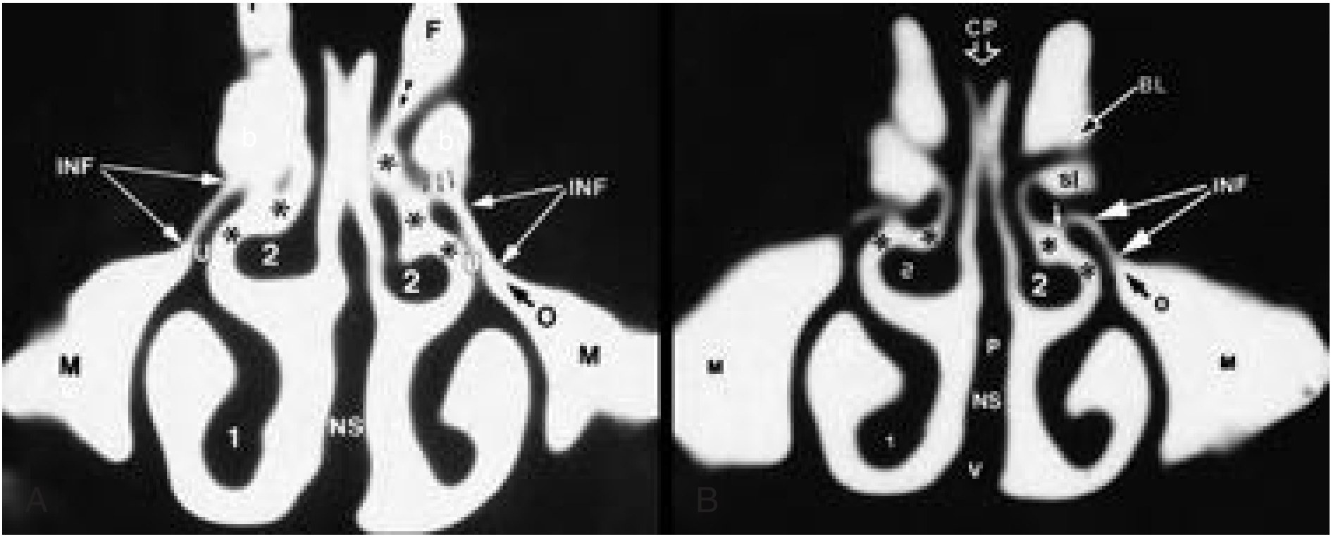
Anatomical Variants and Pathological Anatomy of the OMC
1. Concha Bullosa
- Pneumatization of the middle turbinate head = concha bullosa
- Pneumatization of the vertical (intralaminar) portion = intralaminar cell
- A large concha bullosa compresses the uncinate process laterally, narrows the middle meatus and infundibulum
- The most common anatomical variant of the middle turbinate
2. Paradoxical Middle Turbinate
- Normal curvature projects medially toward the septum
- In the paradoxical variant, the major curvature projects laterally
- This narrows or obstructs the middle meatus and infundibulum directly
- Can cause recurrent sinusitis
3. Variations in the Uncinate Process (Superior Attachment)
- Atelectatic uncinate process: Free edge adheres to the orbital floor or lamina papyracea → occluded infundibulum → hypoplastic ipsilateral maxillary sinus (risk of orbital injury during surgery)
- Pneumatized uncinate - rare but causes infundibular narrowing
- Bifid uncinate - can obstruct drainage
4. Agger Nasi Hyperpneumatization
- An overly pneumatized agger nasi cell encroaches on the frontal recess from anterior
- Most common cause of frontal recess obstruction
5. Haller Cells (Infraorbital Ethmoid Cells)
- Ethmoid air cells that extend laterally over the medial roof of the maxillary sinus
- Lie lateral to the infundibulum along the orbital floor
- If large, they narrow the infundibulum → impaired maxillary sinus drainage
- Present in approximately 10-45% of individuals
- Radiographically identifiable on coronal CT as cells below the orbit along the maxillary sinus roof
6. Giant Ethmoid Bulla
- Enlargement of the ethmoid bulla
- Can narrow or obstruct the middle meatus and infundibulum directly
7. Nasal Septal Deviation
- High septal deviation can compress the middle turbinate laterally and narrow the middle meatus
- May mechanically obstruct the anterior OMC
- High-angle deviation is particularly significant
8. Frontal Cells (Types I-IV, Kuhn Classification)
- Ethmoid cells that pneumatize into the frontal recess or sinus
- Can obstruct the frontal recess depending on their size and location
9. Onodi (Sphenoethmoidal) Cells
- Lateral and posterior extensions of posterior ethmoid air cells
- Can surround the optic nerve - major surgical risk
- Not directly OMC pathology but relevant in posterior sinus disease
10. Keros Classification (Ethmoid Roof Height) - Surgical Risk Variant
| Type | Olfactory Fossa Depth | Risk |
|---|---|---|
| Keros I | 1-3 mm | Low |
| Keros II | 4-7 mm | Moderate |
| Keros III | 8-16 mm | Highest (long thin lateral lamella) |
Pathophysiology: Why the OMC Matters
- Viral URTI → mucosal edema → apposition of mucosal surfaces in the narrow OMC structures
- OMC obstruction → impaired mucociliary clearance → mucus stasis in the draining sinuses
- Reduced oxygen tension → bacterial superinfection (most commonly Streptococcus pneumoniae, Haemophilus influenzae, Moraxella catarrhalis)
- Resulting in acute bacterial rhinosinusitis (ABRS) or, if persistent, chronic rhinosinusitis (CRS)
Clinical and Surgical Significance
- FESS (Functional Endoscopic Sinus Surgery) targets the OMC - the Messerklinger technique begins with uncinectomy (removal of the uncinate process) which exposes the ethmoidal infundibulum and allows visualization and opening of the ethmoid bulla and frontal recess
- Pre-operative coronal CT is mandatory for OMC assessment and surgical planning
- OMC variants must be identified pre-operatively to avoid orbital injury (atelectatic uncinate, Haller cells), optic nerve injury (Onodi cells), carotid injury (extensive sphenoid pneumatization), and CSF leak (Keros III)
- Coronal CT is the gold standard for OMC imaging - plain radiographs cannot capture the structural detail needed
Quick Reference: OMC Structures Summary
| Structure | Role in OMC |
|---|---|
| Uncinate process | Medial wall of infundibulum; separates infundibulum from middle meatus |
| Ethmoid bulla | Posterior wall of hiatus semilunaris; posterior limit of frontal recess |
| Hiatus semilunaris | 2D crescent gap; entrance to infundibulum from middle meatus |
| Ethmoidal infundibulum | Funnel collecting drainage from maxillary + anterior ethmoid (± frontal) |
| Middle meatus | Final common drainage pathway into nasal cavity |
| Frontal recess | Most anterior ethmoid; conduit for frontal sinus drainage |
| Agger nasi cell | Most anterior ethmoid cell; anterior wall of frontal recess |
| Basal lamella | Posterior boundary of OMC; separates anterior from posterior ethmoid |
| Lamina papyracea | Lateral wall of infundibulum and ethmoid labyrinth |
- K.J. Lee's Essential Otolaryngology, 11th Ed. - Chapters on Nasal Anatomy and Rhinosinusitis
- Cummings Otolaryngology Head and Neck Surgery - Chapter 38 (Imaging of the Paranasal Sinuses and Nasal Cavity)
- Scott-Brown's Otorhinolaryngology Head & Neck Surgery Vol. 1 & 2 - Rhinology chapters