Microscopy Superficial luminal part of fibrous cap → composed of smooth muscle cells and collagen. Cellular area under fibrous cap (shoulder) comprised by a mixture of macrophages, foam cells, lymphocytes and lipid laden smooth muscle cells Deeper central soft core consists of extracellular lipid material, cholesterol clefts "
Microscopy Superficial luminal part of fibrous cap → composed of smooth muscle cells and collagen. Cellular area under fibrous cap (shoulder) comprised by a mixture of macrophages, foam cells, lymphocytes and lipid laden smooth muscle cells Deeper central soft core consists of extracellular lipid material, cholesterol clefts " create histological image as per given text.
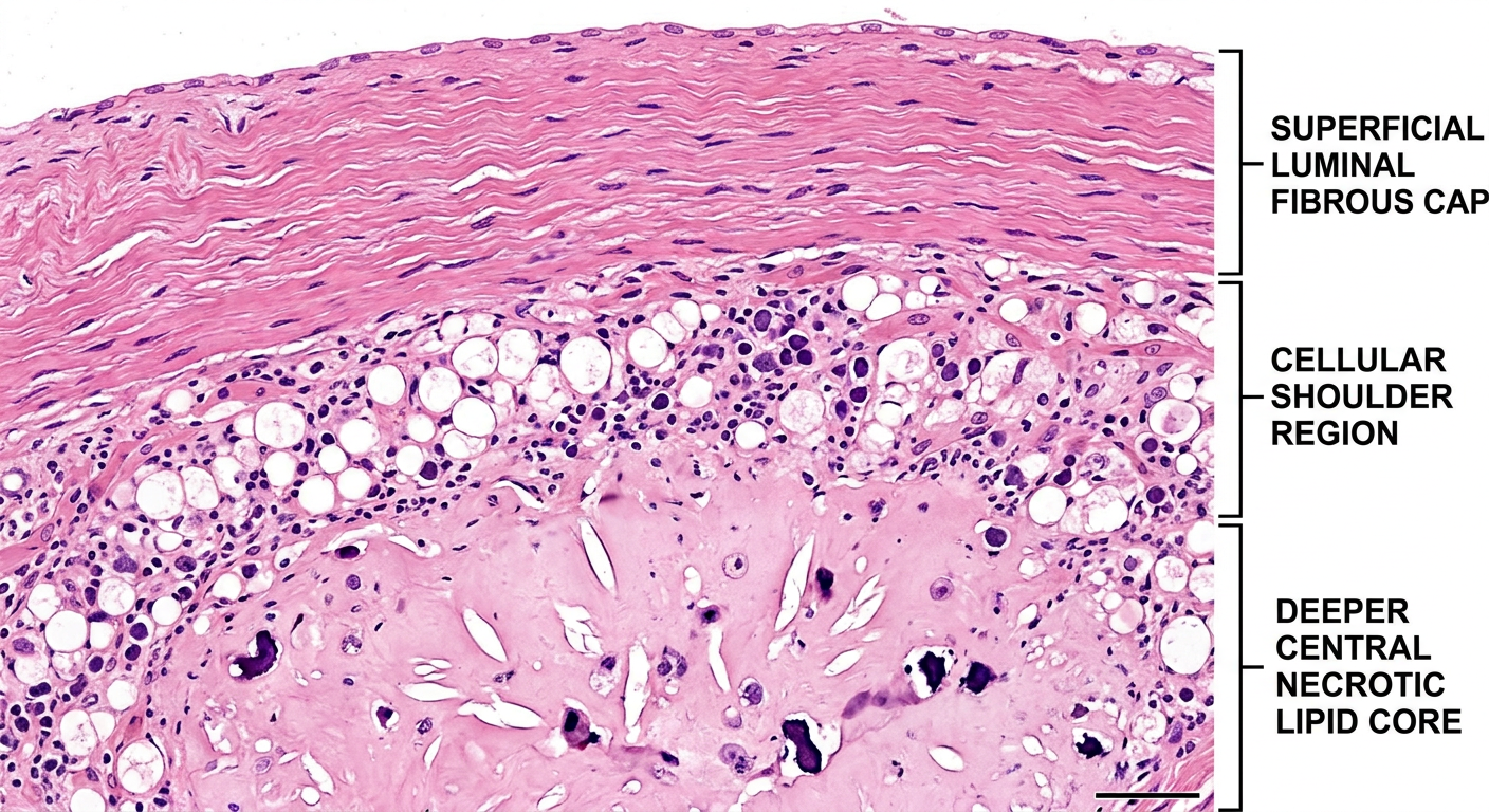
- Dense pink collagen bundles
- Elongated smooth muscle cells with cigar-shaped nuclei embedded in the matrix
- Endothelial lining facing the lumen
- Macrophages and foam cells with vacuolated cytoplasm (lipid-filled)
- Lymphocytes with dark compact nuclei
- Lipid-laden smooth muscle cells with pale, foamy cytoplasm
- This is the most vulnerable region - prone to plaque rupture
- Acellular amorphous material (extracellular lipid)
- Characteristic cholesterol clefts - needle-shaped empty slits (cholesterol dissolves during tissue processing, leaving ghost spaces)
- Necrotic debris
Microscopy Superficial luminal part of fibrous cap → composed of smooth muscle cells and collagen. Cellular area under fibrous cap (shoulder) comprised by a mixture of macrophages, foam cells, lymphocytes and lipid laden smooth muscle cells Deeper central soft core consists of extracellular lipid material, cholesterol clefts " create schematic habd scatched H & E histological image as per the given text.
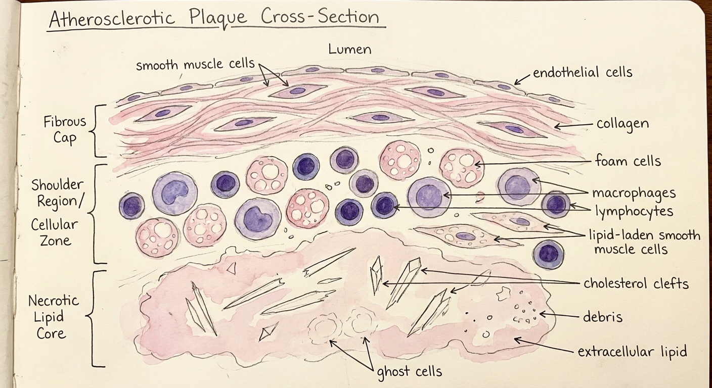
- Wavy pink collagen bundles (eosinophilic)
- Spindle-shaped smooth muscle cells with elongated purple nuclei
- Thin endothelial lining at the lumen
- Foam cells - large round cells with bubbly vacuolated cytoplasm (lipid-filled)
- Macrophages with round/kidney-shaped nuclei
- Lymphocytes - small dark cells with compact nuclei
- Lipid-laden smooth muscle cells with pale foamy cytoplasm
- Pale acellular amorphous extracellular lipid material
- Cholesterol clefts - needle-like empty slits (cholesterol dissolved during processing)
- Necrotic cellular debris