
Give comprehensive notes on tuberculosis, especially in the context of the Pakistani population and it's guidelines followed in Pakistan and also compare with International guidelines: - its diagnosis - treatment and management - what its different types are - how to diagnose each - how to give information on each type - what different protocols and guidelines there are for Pakistani populations - and also give recent updates regarding diagnsosis and management.. What are the different types of emergencies in a tuberculosis case? How to diagnose and treat each? What are the protocols for a latent case of tuberculosis? What PACT questions about history of tuberculosis are to be taken? What examination findings are to be expected in a case of tuberculosis? Based on each case scenario, also give algorithms, illustrations, diagrams, and images from the latest guidelines. Textbooks from Davidson, Harrison, Kumar, and Clark textbook of medicine, and the latest Pakistani local tuberculosis guidelines. Also give memorable mnemonics in an easy way to remember different information wherever necessary. Don't make unnecessary mnemonics
Pakistan NTP tuberculosis guidelines 2024 2025 diagnosis treatment protocols
tuberculosis Pakistan diagnosis treatment guidelines
tuberculosis chest X-ray pulmonary cavitation upper lobe
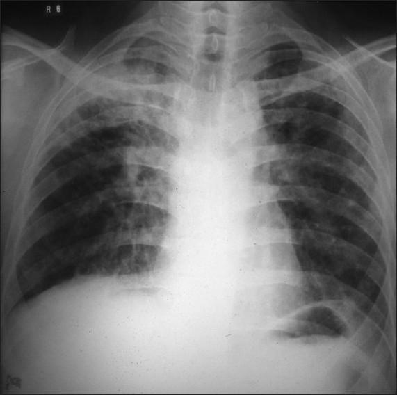
This diagnostic image is a posteroanterior (PA) chest X-ray demonstrating significant pulmonary pathology. The primary finding is located in the right upper lobe, where there are dense, heterogeneous exudative opacities consistent with lung consolidation. Within this region of increased density, a prominent, thick-walled radiolucent cavity is visible, indicating tissue necrosis or cavitation. The distribution of these findings is highly suggestive of secondary (reactivation) pulmonary tuberculosis. In contrast, the left lung field appears relatively clear, although subtle interstitial markings may be present. Key anatomical landmarks including the trachea, clavicles, and cardiac silhouette are visible, with the trachea appearing midline. This image serves as a classic educational example of cavitary lung disease and apical inflammatory processes, emphasizing the radiographic presentation of Mycobacterium tuberculosis infection in an adult patient.
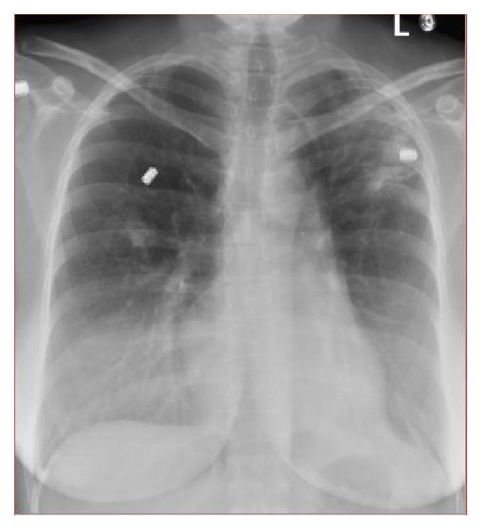
A posterior-anterior (PA) chest x-ray demonstrating bilateral pulmonary abnormalities. In the left upper lobe, there is a prominent hazy increased density or opacity, containing internal lucency suggestive of cavitary changes. The right lung shows a smaller, irregular density localized in the right middle lobe. These findings are clinically significant as they often represent infectious processes such as pulmonary tuberculosis or pyogenic pneumonia, or chronic fibrotic changes. The mediastinal contour and cardiothoracic ratio appear within normal limits. Radiopaque external markers are visible overlying the upper lung fields. This diagnostic image illustrates key radiological signs of pulmonary infiltration and cavitation, relevant for medical education regarding the diagnostic workup of mycobacterial infections and differential diagnosis of apical lung lesions.
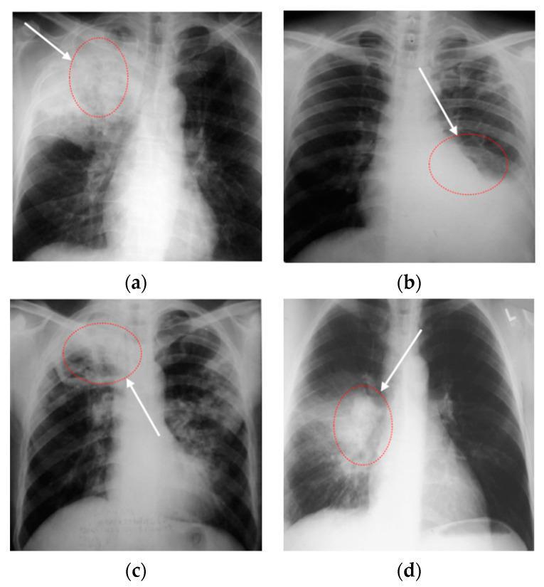
A set of four posterior-anterior (PA) chest X-ray radiographs (labeled a-d) demonstrating various manifestations of pulmonary tuberculosis. Image (a) shows dense consolidation in the right upper lobe containing radiolucent areas consistent with cavitary lesions. Image (b) illustrates tuberculous exudative pleurisy, characterized by a homogeneous basal opacity with a meniscus sign on the left side, indicating significant pleural effusion. Image (c) depicts secondary tuberculosis in the right upper lung, presenting as a large, heterogeneous, ill-defined opacity with associated pulmonary infiltrates. Image (d) demonstrates right hilar consolidation with prominent hilar lymphadenopathy, seen as rounded, increased densities in the mediastinal region. These diagnostic images highlight key radiological features of tuberculosis, including parenchymal consolidation, cavitation, pleural involvement, and hilar node enlargement, which are essential for clinical diagnosis and staging of the disease in infectious disease and radiology contexts.
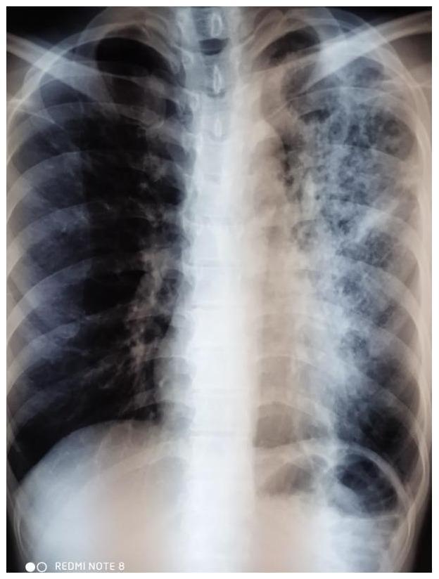
This diagnostic image is an anteroposterior (AP) chest X-ray demonstrating classic radiographic features of secondary pulmonary tuberculosis. The primary pathology is localized to the left lung field, which shows multiple thick-walled cavitating lesions in the upper lobe, indicative of tissue necrosis and cavitation. Associated patchy, ill-defined infiltrates and heterogeneous opacities are distributed throughout the left middle and lower zones, suggesting endobronchial spread or active consolidation. Global lung changes include signs of hyperinflation, evidenced by flattened diaphragms and increased radiolucency. There is notable blunting of the left costophrenic angle, consistent with a small pleural effusion or pleural thickening. The right lung field appears relatively clear but hyperexpanded. This image serves as an educational example of advanced cavitary tuberculosis and the typical distribution of post-primary tuberculous infection in an adult patient.
tuberculosis granuloma histology Langhans giant cells caseating necrosis
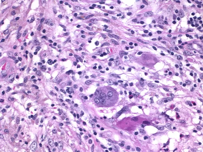
Histology of granulomatous mastitis on bright-field light microscopy. A dense inflammatory infiltrate forms granulomas centered on Langhans-type giant cells surrounded by epithelioid histiocytes, scattered lymphocytes, and occasional eosinophils. The image depicts a high-power view of the center of a granuloma within breast tissue biopsy; multinucleated giant cells with horseshoe-shaped nuclei are evident. The granulomatous architecture includes a peripheral rim of lymphocytes and histiocytes with minimal necrosis described. Staining by Hematoxylin and Eosin highlights basophilic nuclei and eosinophilic cytoplasm; pink collagenous stroma and adipose breast tissue may be visible in the background. Clinically, granulomatous mastitis can mimic infectious etiologies or neoplasia; the presence of Langhans giant cells strongly suggests a granulomatous process and warrants infectious workup for tuberculosis and fungal pathogens, as well as consideration of sarcoidosis or granulomatous reaction to foreign material. This image is diagnostic for granulomatous inflammation in breast tissue when correlated with clinical data and microbiologic studies; useful for education, differential diagnosis, and research on breast granulomatous disease. Additional educational value lies in distinguishing nonnecrotizing granulomas from caseating TB patterns, correlating histology with mammography and ultrasound, and guiding microbiologic testing and patient management in suspected granulomatous breast disease. The image supports teaching of granuloma biology and pathology.
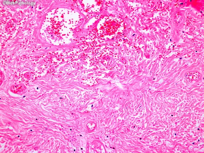
Light microscopy of an H&E-stained formalin-fixed tissue section at low to intermediate magnification reveals granulomatous inflammation characterized by well-formed nodular aggregates of epithelioid histiocytes and occasional Langhans-type multinucleated giant cells embedded in a fibrous stromal matrix. The granulomas appear in close association with accentuated lymphocytic cuffs and scattered plasma cells. Central areas show coagulative or caseating necrosis in some nodules, giving a pale eosinophilic center surrounded by a rim of epithelioid cells. The surrounding dermis/soft tissue shows delicate reticular fibers and vascular congestion with perivascular mononuclear infiltrates. There are no overt bacterial organisms evident with standard H&E, though organisms such as mycobacteria or fungi may require special stains (Ziehl-Neelsen, GMS) to confirm infection. The architecture suggests a granulomatous process rather than acute suppurative inflammation. Diagnostic significance: indicative of granulomatous disease; differential includes infectious etiologies (tuberculosis, fungal infections) and noninfectious conditions (sarcoidosis, foreign-body reaction). Clinical correlation recommended with exposure history, immunologic status, and targeted staining/cultures. Potential use cases: educational demonstration of granuloma morphology, pathology student review, differential diagnosis in infectious and granulomatous diseases, histology atlas reference. This description supports search for terms granuloma, epithelioid histiocytes, Langhans giant cells, caseation, TB, sarcoidosis, histopathology.
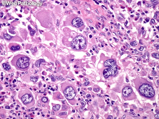
Histology image obtained with brightfield light microscopy on a hematoxylin and eosin stained tissue section. The specimen shows granulomatous inflammation characterized by aggregates of epithelioid macrophages (histiocytes) with elongated, pale to eosinophilic cytoplasm. Multinucleated giant cells, occasionally Langhans-type with peripheral, radiating nuclei, are interspersed among the epithelioid cells. Surrounding lymphocytes and a sparse fibrous stroma delineate nodular granulomas. In some fields there are crystalline or granular pink to eosinophilic deposits within the cytoplasm of histiocytes, and there may be mild necrosis at the center of larger granulomas, though necrosis is not uniformly present across the slide. The pattern is compatible with a granulomatous infectious or inflammatory process, requiring clinical correlation and microbiologic assessment. Differential diagnoses include mycobacterial infection (tuberculosis), fungal granulomas (histoplasmosis, blastomycosis), sarcoidosis, and foreign-body granulomatous reactions. The image emphasizes a macrophage-dominant infiltrate with discrete nodular architecture, preserving basic tissue planes while revealing cellular detail such as nuclear atypia is not prominent. This histologic phenotype is critical for guiding targeted tests (AFB stain, fungal stains, PCR) and therapeutic decisions, and it serves as an educational reference for recognizing granuloma formation in biopsy specimens. Correlation with clinical history and imaging is essential for diagnosis.
drug resistant tuberculosis treatment MDR-TB XDR-TB 2024
https://pakistanchestsociety.pk/wp-content/uploads/2020/08/P…
tuberculosis extrapulmonary types miliary TB spine Pott's disease lymphadenitis
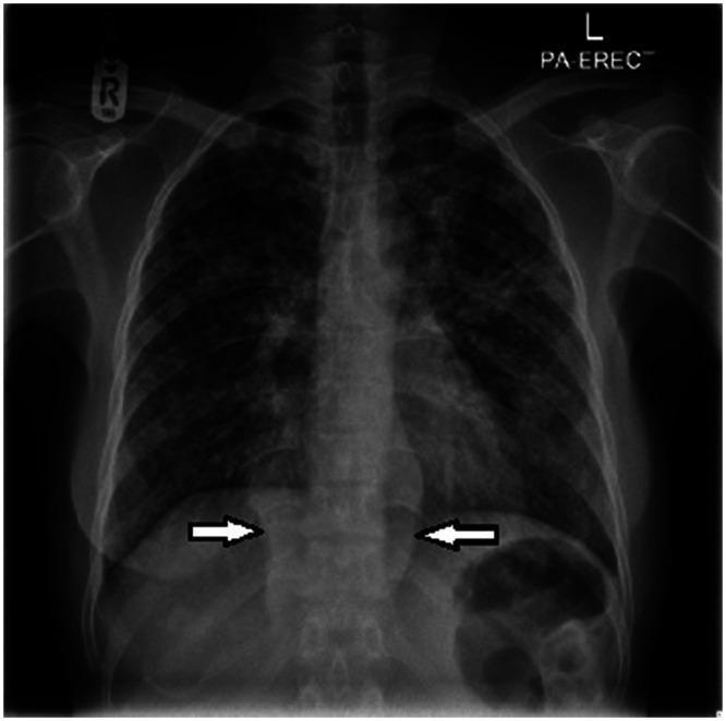
This posteroanterior (PA) erect chest radiograph demonstrates diffuse, bilateral pulmonary opacities and paraspinal soft tissue abnormalities. The lung fields exhibit extensive, small, nodular opacities ranging from discrete 'miliary' patterns to larger confluent areas, characteristic of hematogenous dissemination. In the lower thoracic region, bilateral homogeneous hyperdense shadows are visible adjacent to the spine (indicated by white arrows), suggesting paraspinal abscesses or collections. These findings are clinically significant for disseminated tuberculosis, presenting as miliary TB with associated Pott's disease (spondylodiscitis) and paraspinal involvement. The image illustrates key diagnostic markers for advanced mycobacterial infection, highlighting the relationship between parenchymal lung disease and extrapulmonary spinal manifestations.
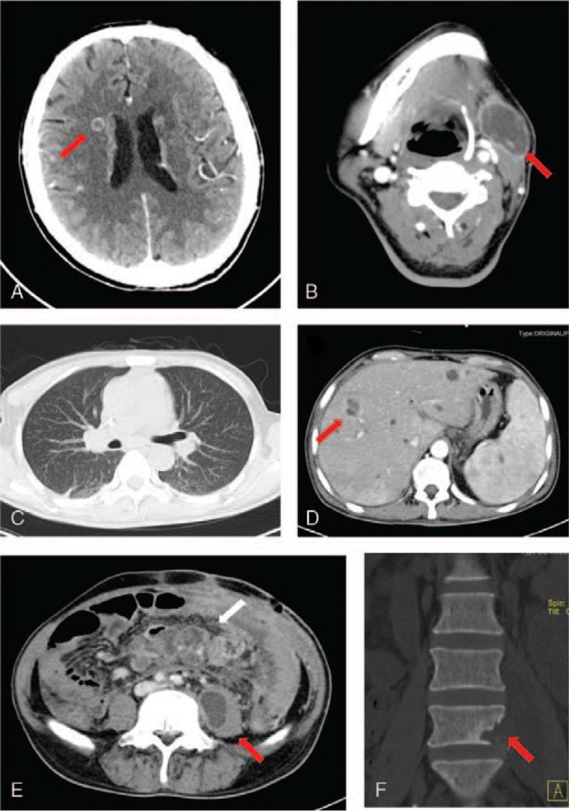
A composite of six computed tomography (CT) scans (A-F) demonstrating multi-organ manifestations of disseminated tuberculosis. (A) Axial brain CT showing a small, ring-enhancing nodule near the right lateral ventricle, consistent with a tuberculoma. (B) Axial neck CT revealing a large mass with peripheral annular enhancement under the left jaw, indicative of a cold abscess or tuberculous lymphadenitis. (C) Axial chest CT displaying diffuse, symmetric miliary nodules throughout both lungs. (D) Axial abdominal CT showing multiple hypoattenuating, ring-enhancing nodules within the liver parenchyma. (E) Axial pelvic CT demonstrating enlarged mesenteric lymph nodes with peripheral enhancement (white arrow) and a low-density fluid collection with annular enhancement in the left psoas major muscle (red arrow), characteristic of a psoas abscess. (F) Coronal CT of the lumbar spine showing focal bone destruction in the L5 vertebra, consistent with Pott disease. This series illustrates the systemic nature of disseminated TB, involving the central nervous system, respiratory system, lymphatic system, viscera, and musculoskeletal system.
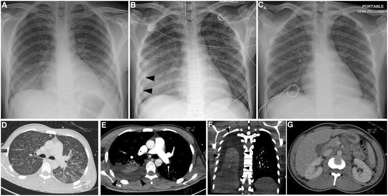
This composite figure displays a longitudinal imaging series of a 24-year-old male with miliary tuberculosis (TB). (A-C) Serial frontal chest radiographs show the progression and management of pleural complications. Initial radiograph (A) reveals bilateral, fine, micronodular interstitial opacities (miliary pattern). Follow-up (B) demonstrates the development of a right-sided pleural effusion, indicated by blunting of the costophrenic angle and peripheral opacification (arrowheads). Post-drainage film (C) shows resolution of the effusion with a pigtail catheter in situ. (D) Axial lung-window CT confirms diffuse, randomly distributed miliary nodules. (E-F) Axial and coronal mediastinal-window CTs demonstrate a moderate right pleural effusion (arrowheads) and associated compressive atelectasis in the right lower lobe. (G) Axial contrast-enhanced abdominal CT reveals extensive para-aortic and mesenteric lymphadenopathy, indicative of extrapulmonary TB involvement. The collection illustrates the classic radiographic and tomographic features of miliary TB, secondary pleural effusion, and associated abdominal lymphadenitis.
miliary tuberculosis chest X-ray millet seed pattern bilateral nodules
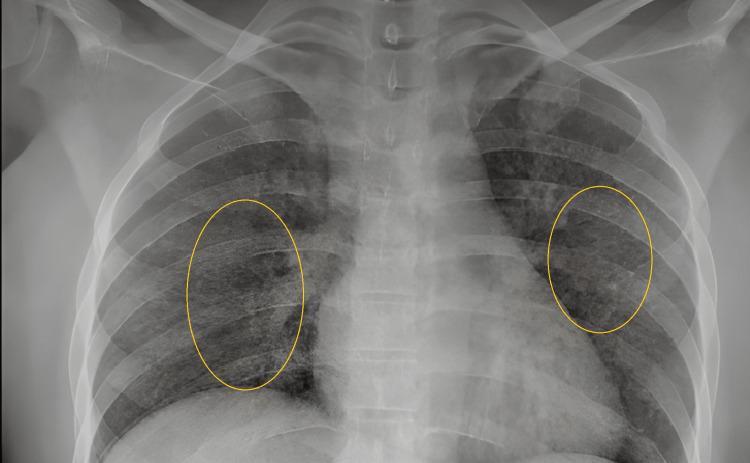
This diagnostic image is a posterior-anterior (PA) chest x-ray demonstrating classic features of miliary tuberculosis. The primary finding is a diffuse, bilateral, and symmetric distribution of small, discrete nodular opacities, approximately 1-3 mm in diameter, creating a 'miliary mottling' pattern throughout both lung fields. These millet-seed-sized nodules result in an overall granular appearance and increased density of the lung parenchyma. Yellow oval annotations highlight representative areas of the miliary pattern in the mid-lung zones. The cardiac silhouette, mediastinal contours, and diaphragmatic domes appear within normal limits without evidence of gross lymphadenopathy or pleural effusion. The visible skeletal structures, including the ribs and clavicles, show no obvious abnormalities. This radiological presentation is highly characteristic of hematogenous dissemination of Mycobacterium tuberculosis, making it a critical educational resource for understanding systemic mycobacterial disease manifestations in pulmonology and infectious disease.
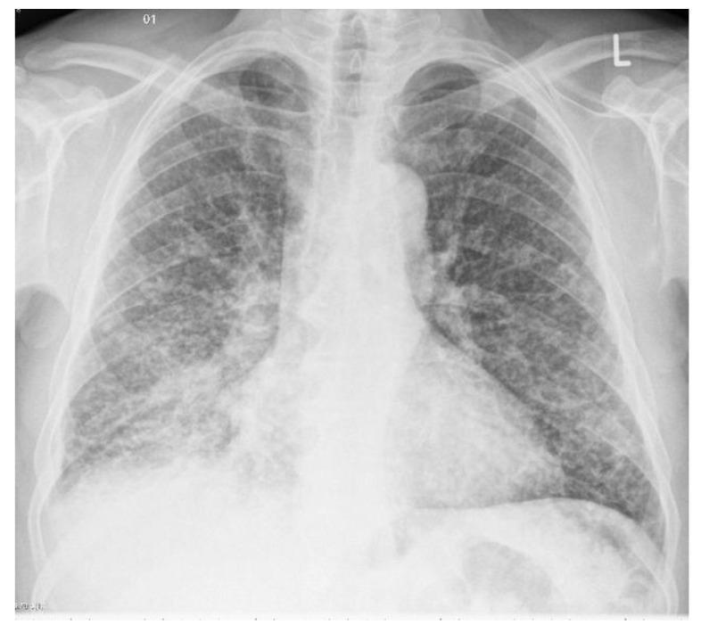
This diagnostic image is a posterior-anterior (PA) chest X-ray demonstrating miliary tuberculosis. The primary radiographic finding is a diffuse, bilateral, and uniform distribution of fine pulmonary nodules throughout both lung fields. These nodules are approximately 1–3 mm in diameter and are spread evenly from the apices to the lung bases, creating the characteristic 'miliary' (millet seed-like) pattern. The distribution involves all lobes without significant sparing, indicating hematogenous dissemination of Mycobacterium tuberculosis. The lung parenchyma shows a granular texture due to the density of these nodules. Cardiac silhouette and mediastinal contours appear within normal limits in this view, and no obvious pleural effusions are noted. This image serves as a classic educational example of miliary pulmonary involvement, emphasizing the importance of recognizing diffuse micronodular patterns in infectious disease diagnostics.
tuberculosis 2024 2025 new treatment WHO guidelines BPaL bedaquiline pretomanid
bedaquiline pretomanid linezolid tuberculosis treatment 2025
PMID: 40693952
tuberculin skin test Mantoux test reading induration
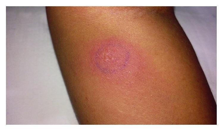
Clinical photograph of a forearm demonstrating a strongly positive Mantoux tuberculin skin test (TST). The visual finding consists of a significant area of induration and erythema. Centrally, the skin is visibly raised with a slightly irregular, 'orange-peel' (peau d'orange) texture, suggesting a potent delayed-type hypersensitivity reaction. The indurated area is circumscribed by a dark blue circular ink marking, used clinically to facilitate the measurement of the wheal. The surrounding skin is of a moderate tone with visible hair follicles and no evidence of generalized rash. This reaction measures approximately 25 mm in diameter, indicating exposure to Mycobacterium tuberculosis or related mycobacteria. The image serves as a teaching tool for recognizing high-degree induration associated with systemic tuberculosis infections, such as dural tuberculosis.
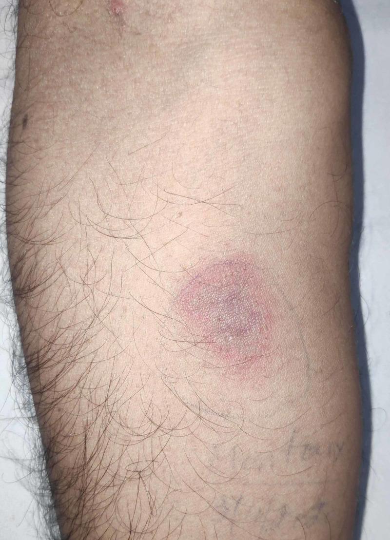
This clinical photograph displays a Mantoux tuberculin skin test site on the volar aspect of a human forearm, 72 hours post-intradermal injection of purified protein derivative (PPD). The visual findings demonstrate a positive reaction characterized by a central, well-defined zone of induration exhibiting a reddish-purple, dusky hue. Surrounding the induration is a peripheral ring of lighter pink erythema. The lesion is roughly circular in morphology, measuring approximately 24 mm in diameter. The surrounding skin appears otherwise normal with standard follicular distribution. This image is a classic educational representation used in infectious disease and pulmonology to illustrate delayed-type hypersensitivity (Type IV) reactions and screening for Mycobacterium tuberculosis infection. Key diagnostic features highlighted include the palpable firmness (induration) and the significant size threshold indicating a positive result in clinical diagnostic protocols.
Pakistan NTP TB categories treatment regimens 2024 MDR-TB XDR-TB protocols Cat 1 Cat 2
https://pakistanchestsociety.pk/wp-content/uploads/2020/08/P…
TUBERCULOSIS - Comprehensive Clinical Notes
Pakistan Context | International Guidelines | Clinical Practice
SECTION 1: EPIDEMIOLOGY & PAKISTAN BURDEN
Global Context
Pakistan's TB Burden
- Pakistan ranks 5th globally among high TB-burden countries
- Prevalence: 348 per 100,000, Incidence: 276 per 100,000, Mortality: 34 per 100,000
- Annual estimated incident cases: ~600,000; Case Detection Rate (CDR): ~64%
- Over 200,000 TB cases are missed annually - Pakistan contributes 7% of all globally missed cases
- TB was declared a national emergency in Pakistan in 2001
- Pakistan's National TB Control Program (NTP) operates under the National Institutes of Health (NIH), Islamabad
Risk Factors (Particularly Relevant in Pakistan)
- Poverty, overcrowding, malnutrition, poor housing
- Diabetes mellitus (very prevalent in Pakistan)
- HIV co-infection
- Silicosis, chronic lung disease
- Immunosuppression (steroids, biologics, post-transplant)
- Hodgkin lymphoma, hematological malignancies
- Prisoners, healthcare workers, refugees, displaced populations
SECTION 2: MICROBIOLOGY & PATHOGENESIS
Transmission
- Person-to-person via airborne droplet nuclei (1-5 microns; infectious dose = 1-10 bacilli)
- Droplets can remain suspended for hours in enclosed spaces
- High-risk settings: crowded homes, prisons, healthcare facilities, refugee camps (highly prevalent across Pakistan)
Pathogenesis Steps (Mnemonic: "I Feel My Granuloma Reactivating")
| Step | Process |
|---|---|
| Inhaled | Bacilli inhaled into alveoli |
| Fagocytosed | Macrophages engulf bacilli but cannot kill them |
| Macrophage activation | Delayed hypersensitivity (Type IV) develops over 2-8 weeks |
| Granuloma formation | Classic caseating granuloma with Langhans giant cells |
| Reactivation | Latent infection reactivates if immunity declines |
Primary vs. Secondary TB (Robbins/Kumar Pathology)
| Feature | Primary TB | Secondary (Reactivation) TB |
|---|---|---|
| Host | Previously unexposed | Previously sensitized |
| Location | Lower upper lobe or upper lower lobe, subpleural | Apex of upper lobe (bilateral) |
| Lesion | Ghon focus + lymph node = Ghon complex | Fibrocaseous cavitation |
| Calcification | Ranke complex (healed Ghon complex) | Satellite lesions, scarring |
| Lymphadenopathy | Prominent | Less prominent |
| Disease progression | 5% progress to significant disease | Reactivation in immunosuppressed |
| Transmission | Not infectious in most | Highly infectious |
Histology (Classic Teaching)
- Central caseous necrosis (cheese-like, amorphous, eosinophilic)
- Surrounding epithelioid macrophages (activated macrophages)
- Langhans giant cells (multinucleate, nuclei arranged in horseshoe/peripheral ring pattern)
- Outer lymphocytic cuff
- Eventually fibrosis and calcification
SECTION 3: CLASSIFICATION OF TUBERCULOSIS
By Site
A. Pulmonary TB (PTB) - ~80% of cases
- Primary PTB - Ghon complex, often self-limited
- Post-primary / Secondary PTB - Reactivation, apical cavitation, highly infectious
- Progressive Primary PTB - Immunocompromised patients (HIV, severe malnutrition)
- Miliary TB - Hematogenous dissemination; "millet seed" bilateral nodules
B. Extrapulmonary TB (EPTB) - ~20% of cases, higher in HIV+
| Site | Name | Key Features |
|---|---|---|
| Lymph nodes | Tuberculous lymphadenitis | Most common EPTB; painless cervical nodes; "cold abscess" |
| Spine | Pott's disease | L1-L2 most common; kyphosis, paraplegia risk |
| CNS | TB meningitis / Tuberculoma | Worst prognosis; 12-month treatment |
| Pleura | Tuberculous pleuritis | Exudative effusion, lymphocyte predominant |
| Peritoneum | Tuberculous peritonitis | Ascites, matted bowel loops |
| Genitourinary | Renal/GU TB | Sterile pyuria, "pipe-stem" ureter |
| Pericardium | Tuberculous pericarditis | Constrictive pericarditis risk |
| Bone/joints | Osteoarticular TB | Weight-bearing joints, hip/knee/spine |
| Skin | Lupus vulgaris, scrofuloderma | Scarring skin lesions |
| Abdominal | Intestinal TB | Ileocecal most common, strictures |
By Bacteriological Status
- Bacteriologically confirmed: positive smear, culture, or molecular test (GeneXpert)
- Clinically diagnosed: negative/unavailable tests but clinical/radiological diagnosis
By Drug Susceptibility (WHO Classification)
- DS-TB: Drug-susceptible TB (sensitive to Rifampicin and Isoniazid)
- RR-TB: Rifampicin-resistant TB (any resistance to rifampicin)
- MDR-TB: Multi-drug resistant (resistant to both Isoniazid + Rifampicin)
- Pre-XDR-TB: MDR/RR-TB + resistance to any fluoroquinolone
- XDR-TB: MDR/RR-TB + resistance to fluoroquinolone + bedaquiline or linezolid
By Treatment History (NTP Pakistan / WHO)
| Category | Definition |
|---|---|
| New case | Never treated or treated <1 month |
| Relapse | Previously declared cured/completed, now bacteriologically positive again |
| Treatment after failure | Smear/culture positive at 5th month or later during treatment |
| Treatment after lost to follow-up (TALF) | Interrupted treatment ≥2 consecutive months |
| Other previously treated | Known prior treatment, outcome unknown |
SECTION 4: HISTORY TAKING - PACT QUESTIONS
PACT Framework for TB History
- Duration and character of cough (>2 weeks = presumptive TB in Pakistan - "prolonged cough cardinal symptom")
- Hemoptysis (amount, frequency, streaks vs. massive)
- Dyspnea, chest pain, wheeze
- C - Cough (>2 weeks, productive)
- O - Orthopnea/dyspnea (pleural effusion or miliary)
- W - Weight loss (>10% body weight, gradual)
- S - Sweats at night (drenching, typical)
- F - Fever (low-grade, afternoon, remittent - "evening rise of temperature")
- A - Anorexia (loss of appetite, generalized weakness)
- D - Dyspnea (progressive breathlessness)
- E - Extra-pulmonary features (swollen nodes, back pain, hematuria, headache, ascites)
- Close household contact with a known TB patient (highest risk factor in Pakistan)
- Duration and intimacy of contact
- Occupational history (healthcare worker, prison guard, miners)
- Living conditions: overcrowding, ventilation, sunlight exposure
- Recent migration from endemic area
- History of prior TB treatment (crucial - if yes, get treatment details, compliance, outcome)
- BCG vaccination history (especially in children - scar on arm)
- HIV risk factors (sex history, injecting drug use, blood transfusion)
- Immunosuppressive drugs (steroids, DMARDs, biologics like TNF inhibitors)
- Diabetes mellitus (very common TB-DM comorbidity in Pakistan)
- Prior TB treatment: which drugs, for how long, compliance (DOT or self-administered)
- Outcome of prior treatment: cured, defaulted, failure?
- Current medications (especially corticosteroids, methotrexate, anti-TNF)
- Alcohol, smoking, drug use
- Socioeconomic status, food security, housing
- Family history of TB
- Travel history (Balochistan, KPK, rural Punjab - high burden areas)
SECTION 5: CLINICAL EXAMINATION FINDINGS
General Examination
- Wasting/cachexia - weight loss, loss of muscle bulk
- Pallor - chronic disease anemia
- Fever - low-grade (37.5-38.5°C), often evening rise
- Lymphadenopathy - especially cervical nodes (matted, firm, non-tender, rubbery); may have collar-stud abscess (pathognomonic)
- Clubbing (advanced/chronic PTB)
- BCG scar assessment in children
Respiratory Examination
| Finding | Significance |
|---|---|
| Tracheal deviation | Fibrosis/collapse pulling, effusion pushing |
| Reduced chest expansion | Affected side - consolidation, fibrosis |
| Dullness on percussion | Consolidation, effusion, collapse |
| Bronchial breathing | Consolidation (early active disease) |
| Amphoric breathing | Large cavity |
| Post-tussive crepitations | Classically over apices - highly suggestive of PTB |
| Absent/reduced breath sounds | Effusion, pneumothorax |
| Wheeze | Endobronchial TB, lymph node compression of airway |
Extrapulmonary Signs to Look For
- Neck: cervical lymphadenopathy, collar-stud abscess (discharge through fascia)
- Spine: kyphosis/gibbus (Pott's disease), point tenderness over vertebrae
- Abdomen: hepatomegaly, splenomegaly, ascites, tenderness, doughy feel
- CNS: neck stiffness, Kernig/Brudzinski sign, cranial nerve palsies (especially VI and VII), papilledema, altered consciousness
- Eye: choroidal tubercles (pathognomonic of miliary TB on fundoscopy)
- Skin: erythema nodosum, lupus vulgaris, scrofuloderma
SECTION 6: DIAGNOSIS OF TUBERCULOSIS
Diagnostic Algorithm
PRESUMPTIVE TB CASE
(Cough >2 weeks OR suggestive CXR OR close contact)
          |
          ↓
Sputum AFB smear x 2 (morning + spot)
+ GeneXpert MTB/RIF  ← First-line test (NTP Pakistan 2024)
          |
    ┌─────┴─────┐
  Positive    Negative
    |              |
Confirmed    High clinical suspicion?
DS-TB or         |
RR-TB          Yes → CXR + Sputum culture (LJ/MGIT)
               (if RR = refer for DST/MDR workup)
               No → Investigate other causes
A. Sputum Smear Microscopy (AFB Smear)
- Method: Ziehl-Neelsen (ZN) stain or fluorochrome (auramine-rhodamine) stain
- Specimens: 2 samples (early morning + spot) - Pakistan NTP protocol
- Grading: Scanty (1-9 AFB/100 fields), 1+ (10-99), 2+ (1-10/field), 3+ (>10/field)
- Sensitivity: 45-80% (poor in smear-negative disease, HIV+, children)
- Specificity: ~98% (but cannot distinguish M. tb from NTM)
- Advantage: Cheap, rapid (2 hours), widely available at NTP sites
B. GeneXpert MTB/RIF (Xpert MTB/RIF / Xpert Ultra)
- WHO-endorsed molecular test; NTP Pakistan first-line diagnostic test
- Detects M. tuberculosis DNA AND Rifampicin resistance simultaneously
- Time to result: ~2 hours
- Sensitivity: ~88% (smear-positive), ~68% (smear-negative pulmonary), ~80-90% (Ultra)
- Specificity: ~99%
- NTP Pakistan 2024 has rolled out Xpert MTB/RIF Ultra at key health centres
- Also used on CSF, pleural fluid, lymph node aspirates, gastric aspirates (children)
- Computer-Aided Detection (CAD) for CXR endorsed by NTP 2024 for adults/adolescents ≥15 years
C. Mycobacterial Culture
- Gold standard for diagnosis and drug susceptibility testing (DST)
- Löwenstein-Jensen (LJ) medium: Solid culture, 6-8 weeks (slow but cheap)
- MGIT (Mycobacterial Growth Indicator Tube): Liquid culture, 1-3 weeks (faster)
- Allows full DST including first and second-line drug sensitivities
- Required for all MDR/RR-TB cases
D. Tuberculin Skin Test (TST / Mantoux Test)
- Method: 0.1 mL PPD (purified protein derivative) injected intradermally into volar forearm
- Reading: 48-72 hours; measure induration (not erythema) in mm
| Induration | Positive In |
|---|---|
| ≥5 mm | HIV+, recent close contact, fibrotic CXR, immunosuppressed, transplant recipients |
| ≥10 mm | Recent immigrants from endemic countries, healthcare workers, prisoners, children <5y |
| ≥15 mm | No known risk factors |
- Limitations: False-negative in HIV, severe TB, malnutrition, steroids; False-positive with BCG vaccination and NTM (BCG given universally in Pakistan, so TST less specific)
- NTP Pakistan: TST less preferred over IGRA for LTBI diagnosis
E. IGRA (Interferon-Gamma Release Assay)
- Tests: QuantiFERON-TB Gold Plus (QFT-Plus), T-SPOT.TB
- Blood test measuring IFN-γ release by T cells stimulated with TB-specific antigens (ESAT-6, CFP-10)
- Advantages over TST: Not affected by BCG vaccination; more specific; no return visit needed
- Sensitivity: ~80-90%; Specificity: ~95-99%
- Preferred for LTBI in high-income settings and NTP Pakistan (where available)
- Limitations: expensive, requires laboratory processing; false-negative in immunosuppressed
F. Chest Radiography (CXR)
| TB Type | CXR Finding |
|---|---|
| Primary PTB | Unilateral consolidation (any lobe), hilar lymphadenopathy |
| Post-primary PTB | Upper lobe opacities, cavitation, streaky fibrosis, volume loss |
| Miliary TB | Bilateral, diffuse 1-3 mm nodules ("millet seeds") throughout both lungs |
| TB Pleuritis | Unilateral pleural effusion (homogeneous opacity, meniscus sign) |
| Advanced PTB | Bilateral cavities, destroyed lung, compensatory hyperinflation |
| Healed/Old TB | Calcified nodules (Ranke complex), pleural thickening, fibrous scars |
G. Additional Investigations by Site
- Pleural fluid: exudate (protein >3g/dL, LDH elevated), lymphocyte-predominant
- ADA (Adenosine Deaminase) >45 IU/L strongly suggests TB pleuritis (sensitivity 87%, specificity 93%)
- GeneXpert on pleural fluid (lower sensitivity ~16-20% but specific)
- Pleural biopsy: granulomas on histology (gold standard)
- CSF: clear/turbid, lymphocytic pleocytosis (100-500 cells/mm³), high protein (1-5 g/L), low glucose (<2.2 mmol/L or CSF:serum glucose ratio <0.5)
- AFB smear on CSF: ~37% sensitivity; culture: ~70%
- GeneXpert on CSF: ~80% sensitivity
- CT head: hydrocephalus, basal meningeal enhancement, infarcts, tuberculoma
- Ascitic fluid: exudate, lymphocyte-predominant; ADA >40 IU/L
- Ultrasound: matted bowel loops, mesenteric lymphadenopathy, ascites
- CT abdomen: ileocecal thickening, lymphadenopathy with central necrosis
- Colonoscopy + biopsy: granulomas (diagnosis gold standard)
- MRI spine: most sensitive (disc space narrowing, vertebral destruction, psoas abscess, cord compression)
- CT-guided biopsy of affected vertebra for culture/histology
SECTION 7: TREATMENT & MANAGEMENT
First-Line Anti-TB Drugs (Mnemonic: "RIPE")
| Drug | Code | Mechanism | Daily Dose (adult) | Key Side Effects |
|---|---|---|---|---|
| Rifampicin | R | Inhibits RNA polymerase (rpoB gene) | 10 mg/kg (max 600 mg) | Hepatotoxicity, red-orange urine/secretions, drug interactions (CYP450 inducer) |
| Isoniazid | H | Inhibits mycolic acid synthesis (katG, inhA) | 5 mg/kg (max 300 mg) | Peripheral neuropathy (give pyridoxine), hepatotoxicity, lupus-like syndrome |
| Pyrazinamide | Z | Disrupts membrane transport (active in acidic pH - kills intracellular organisms) | 25 mg/kg (max 2g) | Hyperuricemia/gout, hepatotoxicity |
| Ethambutol | E | Inhibits arabinogalactan synthesis (embB) | 15-20 mg/kg (max 1.6g) | Optic neuritis (check visual acuity monthly), red-green color blindness |
STANDARD TREATMENT REGIMENS
Pakistan NTP / WHO Standard Regimen for DS-TB (New Cases)
┌───────────────────────────────────────────────────────┐
│  INTENSIVE PHASE (2 months)  │  CONTINUATION PHASE (4 months)  │
│      2 H R Z E               │       4 H R E                   │
│ (Daily DOT)                  │  (Daily - NTP Pakistan)          │
└───────────────────────────────────────────────────────┘
Total Duration: 6 months
For Special Situations (US/WHO Guidelines - Harrison's 22E):
- Cavitary disease with positive 2-month cultures: Extend continuation to 7 months (total 9 months)
- TB meningitis: 2HRZE / 10HR (12 months total) - NTP Pakistan
- Bone/Joint TB: 6-9 months total (some guidelines 9-12 months)
- Pregnancy: Use 2HRZE/4HR; avoid Pyrazinamide (limited safety data in some guidelines, but WHO and NTP Pakistan permit it); Streptomycin is absolutely contraindicated
DRUG-RESISTANT TB TREATMENT
MDR-TB / RR-TB (NTP Pakistan / WHO 2024)
- Bedaquiline + Pretomanid + Linezolid ± Moxifloxacin
- Duration: 6 months (BPaLM) for eligible MDR/pre-XDR-TB
- This is the major recent update - ATS/CDC/ERS/IDSA 2025 Practice Guideline confirms all-oral shorter regimens are now recommended
- BPaL (without Mfx): For XDR-TB / pre-XDR-TB (6 months)
- Bdq-Lfx-Cfz-Z-Hhd-E-Pto (WHO approved shorter course)
- NTP Pakistan has protocols for this at PMDT (Programmatic Management of Drug-Resistant TB) sites
- Compose 5-drug regimen based on DST results using WHO Group A, B, C drugs
WHO Drug Classification for MDR-TB:
| Group | Drugs | Use |
|---|---|---|
| Group A (always use all 3 unless contraindicated) | Levofloxacin/Moxifloxacin, Bedaquiline, Linezolid | Backbone of MDR-TB treatment |
| Group B (add next) | Clofazimine, Cycloserine/Terizidone | Add to complete regimen |
| Group C (add when needed) | Ethambutol, Delamanid, Pyrazinamide, Imipenem-clavulanate, Meropenem-clavulanate, Amikacin, Ethionamide/Prothionamide, PAS | Adjunct |
DIRECTLY OBSERVED THERAPY (DOT)
- Core of DOTS strategy (5-point package) - both WHO and NTP Pakistan
- Requires healthcare worker or trained community DOT provider watching patient swallow every dose
- Reduces treatment failure, default, and drug resistance
- NTP Pakistan: Community DOT through Imams, schoolteachers, community leaders
TREATMENT MONITORING
Month 0 → Baseline: Sputum AFB x2, GeneXpert, CXR, LFTs, RFTs, visual acuity
Month 2 → Sputum smear/culture conversion: If positive → send for DST/culture
Month 5 → Sputum smear: If positive → sputum for culture + DST; consider MDR-TB
Month 6 → End of treatment: Sputum smear; Declare outcome
Treatment Outcomes (WHO/NTP Pakistan):
- Cured: Bacteriologically confirmed TB; negative smear at end of treatment AND at least one previous negative
- Treatment Completed: Completed treatment without bacteriological confirmation (smear-negative TB)
- Treatment Failure: Smear/culture positive at month 5 or later
- Died: For any reason during treatment
- Lost to Follow-Up (LTFU): Interrupted treatment ≥2 consecutive months
- Not Evaluated: No assigned outcome
SECTION 8: LATENT TB INFECTION (LTBI)
Definition
Who to Screen (PACT Questions for LTBI)
- Close contacts of bacteriologically-confirmed PTB cases
- HIV-positive individuals
- Patients starting immunosuppressants (anti-TNF, corticosteroids, chemotherapy, transplant)
- Healthcare workers in Pakistan (high-risk)
- Prisoners, homeless persons
- Immigrants from high-burden countries (Pakistan itself is high-burden)
Diagnosis of LTBI
Screen with IGRA (preferred) or TST
          |
    Positive?
     |        |
    Yes       No (if high-risk: immunosuppressed)
     |        |
Rule out     Consider empirical treatment
active TB    after ruling out active disease
(CXR, symptoms)
     |
If active TB excluded → Treat LTBI
LTBI Treatment Regimens (Harrison's 22E, Murray & Nadel's)
| Regimen | Duration | Notes |
|---|---|---|
| 3HP - Isoniazid + Rifapentine weekly | 3 months | PREFERRED (children >2y and adults including HIV+); DOT or self-administered |
| 4R - Rifampicin daily | 4 months | Good option, no return visit if self-admin; preferred over 9H |
| 3HR - Isoniazid + Rifampicin daily | 3 months | Alternative |
| 6H or 9H - Isoniazid daily | 6 or 9 months | Older regimen, less preferred due to lower completion rates |
- IGRA preferred over TST (due to universal BCG in Pakistan)
- Contact investigation of all household contacts of smear-positive PTB cases
- Children <5 years who are household contacts: give isoniazid preventive therapy (IPT) even if TST/IGRA negative (due to high risk)
- HIV-positive patients: 6H isoniazid or 3HP recommended
- Active TB must be excluded before initiating LTBI treatment
Pakistan-Specific Note on BCG
SECTION 9: TB EMERGENCIES
EMERGENCY 1: MASSIVE HEMOPTYSIS
- Clinical: bright red blood, large volumes, ± cardiovascular instability
- CXR: cavity, opacity (blood in lungs)
- CT chest: localizes bleeding site, identifies responsible vessel
- Bronchoscopy: identifies source, allows targeted intervention
MASSIVE HEMOPTYSIS
        |
ABC: Airway, Breathing, Circulation
IV access x2, CrossMatch, O2
Position: BLEEDING SIDE DOWN (prevents contralateral aspiration)
        |
        ↓
Resuscitation → IV fluids, blood transfusion if needed
        |
        ↓
Bronchoscopy (rigid preferred) → Balloon tamponade, clot removal
        |
        ↓
Bronchial Artery Embolization (BAE) - First-line intervention
        |
        ↓
Surgical resection (lobectomy/pneumonectomy) if BAE fails
EMERGENCY 2: TUBERCULOUS MENINGITIS (TBM)
- Grade I: GCS 15, no focal deficits, no altered consciousness
- Grade II: GCS 11-14 OR focal neurological deficit
- Grade III: GCS ≤10
- LP: clear/xanthochromic CSF, lymphocytic pleocytosis (100-500 cells), high protein, low glucose
- GeneXpert on CSF (~80% sensitivity - best available test)
- CT head: hydrocephalus, basal meningeal enhancement, infarcts
- MRI: superior to CT for early disease
TBM Treatment Regimen (NTP Pakistan 2024):
2HRZE / 10HR = 12 months total
(NOT 6 months like standard PTB)
- Grade I/II: Dexamethasone 0.4 mg/kg/day tapering over 6-8 weeks
- Grade III: Dexamethasone 0.4 mg/kg/day IV initially then oral, 8-week taper
- Reduces mortality and neurological sequelae
- Management of raised ICP: head elevation, mannitol, consider VP shunt for hydrocephalus
EMERGENCY 3: MILIARY TB / DISSEMINATED TB
- CXR: bilateral 1-3 mm millet-seed nodules (may be normal early)
- HRCT chest: random small nodules diffusely distributed
- Choroidal tubercles on fundoscopy: pathognomonic (present in 13-87%)
- Bone marrow biopsy: granulomas + AFB in severely immunocompromised
- Blood cultures (MGIT): mycobacteremia in immunosuppressed
- Hepatic biopsy: granulomas
- Standard 2HRZE/4HR (or 6-9 months if CNS involved → 12 months)
- Add corticosteroids if CNS or pericardial involvement
EMERGENCY 4: RESPIRATORY FAILURE FROM SEVERE TB
- Large bilateral cavitary TB with destroyed lung
- Miliary TB with hypoxic respiratory failure
- Massive pleural effusion
- Pneumothorax (spontaneous from rupture of subpleural focus/bulla)
- Supplemental O2, ICU admission, intubation/mechanical ventilation if needed
- Urgent anti-TB treatment initiation
- Thoracocentesis for large effusions
- Chest tube for pneumothorax
- Corticosteroids for miliary TB with severe hypoxia
EMERGENCY 5: PARADOXICAL REACTIONS (IRIS - Immune Reconstitution Inflammatory Syndrome)
- Do NOT stop TB treatment
- Do NOT stop ART
- NSAIDs (mild/moderate IRIS)
- Corticosteroids (severe IRIS) - Prednisolone 1.5 mg/kg/day for 2 weeks then taper
EMERGENCY 6: TENSION PNEUMOTHORAX FROM TB
- Rupture of peripheral TB cavity/bulla
- Clinical signs: respiratory distress, tracheal deviation away, absent breath sounds, hypotension
- Diagnosis: clinical (do not delay for CXR if unstable)
- Treatment: Immediate needle decompression (2nd ICS midclavicular line), then chest tube
EMERGENCY 7: HEMODYNAMIC COLLAPSE FROM PERICARDIAL TAMPONADE
- Serous/serosanguineous exudate, can progress to tamponade
- Beck's triad: hypotension + muffled heart sounds + raised JVP
- Diagnosis: echo (pericardial effusion, diastolic collapse)
- Treatment: Pericardiocentesis (urgent); anti-TB drugs + corticosteroids (reduces constriction risk)
SECTION 10: DIAGNOSIS OF INDIVIDUAL TB TYPES
Diagnostic Summary Table
| TB Type | Best Diagnostic Test | Supporting Investigations |
|---|---|---|
| Pulmonary TB (smear +ve) | GeneXpert + Sputum AFB smear | CXR, culture |
| Pulmonary TB (smear -ve) | GeneXpert on sputum | Sputum culture (MGIT), CXR, CT |
| TB Lymphadenitis | FNAC (granulomas + AFB) | GeneXpert on aspirate, culture, histology |
| TBM | GeneXpert on CSF | CSF cytology/chemistry, MRI brain |
| TB Pleuritis | ADA on pleural fluid | GeneXpert, pleural biopsy |
| TB Peritonitis | ADA on ascitic fluid + laparoscopic biopsy | GeneXpert on fluid, CT abdomen |
| Pott's Disease | MRI spine | CT-guided biopsy + culture/histology |
| Miliary TB | HRCT chest + fundoscopy | Bone marrow/liver biopsy, blood culture |
| Urogenital TB | Urine AFB x3 (early morning) | GeneXpert on urine, IVU, culture |
| Pericardial TB | Echo + ADA on pericardial fluid | GeneXpert, pericardial biopsy |
| Osteoarticular TB | MRI joint + biopsy | Culture, histology |
SECTION 11: PAKISTAN NTP vs. INTERNATIONAL GUIDELINES - COMPARISON
Key Differences Table
| Aspect | NTP Pakistan 2024 | WHO 2022/2024 | ATS/CDC/IDSA 2025 |
|---|---|---|---|
| DS-TB New Case | 2HRZE/4HRE | 2HRZE/4HR | 2HRZE/4HR or novel 4-month regimen |
| 4-month TB regimen | NOT for routine use (specialist only) | Recommended for adults with non-severe TB | Recommended for eligible patients |
| Children non-severe TB | 4-month regimen: specialist settings only | 4-month regimen recommended (3mo-16y) | Per WHO |
| TBM in children | 2HRZE/10HR (12 months) | 2HRZE/10HR OR 6HRZEto (alternative) | - |
| Pediatric diagnosis | PPA scoring card (2016) | Improved treatment decision algorithms (no mandatory lab result needed) | - |
| GeneXpert rollout | Endorsed; rolled out at key health centres | First-line test for all | First-line |
| CAD for CXR | Endorsed for adults/adolescents ≥15y | Endorsed | - |
| LTBI treatment | IPT (isoniazid) for contacts; IGRA preferred | 3HP preferred; multiple options | 3HP preferred |
| MDR-TB | Shorter STR + BPaL/BPaLM at PMDT sites | BPaLM 6 months preferred | BPaLM preferred |
| DOT | Mandatory (community DOT through trained providers) | Recommended | Recommended |
| Drug-sensitive continuation phase | 4HRE (with Ethambutol) | 4HR (without Ethambutol) | 4HR |
| MDR-TB Category II | No longer recommended (2HRZES/1HRZE/5HRE) | Abandoned | Abandoned |
| Delamanid in children | Endorsed for <6y with RR-TB | Endorsed | - |
Key Pakistan-Specific Protocols
- DOTS Programme: All TB patients must be registered and treated under NTP with DOT
- GeneXpert network: Expanded; all presumptive TB cases should get GeneXpert as first test
- PMDT Sites: Programmatic Management of Drug-Resistant TB - specialized centres for MDR/XDR-TB (Lahore, Karachi, Islamabad, Peshawar, Quetta)
- TB-HIV Co-infection: Increasing challenge; ART should start within 2 weeks of TB treatment (if CD4 <50) or within 2-8 weeks (if CD4 >50)
- TB-DM Comorbidity: Pakistan has very high DM burden; systematic review PMID 38346381 - DM worsens TB outcomes, requires extended monitoring
- Active Case Finding (ACF): NTP now actively screens prisons, refugees, brick kiln workers, healthcare workers
SECTION 12: RECENT UPDATES (2024-2026)
1. Novel 4-Month Regimen for DS-TB (TRUNCATE-TB / ZeNix / STOMP Trials)
- 2HRZE then 2HPaMZ (Rifapentine + Isoniazid + Pyrazinamide + Moxifloxacin): Non-inferior to 6-month regimen
- ATS/CDC/ERS/IDSA 2025 Clinical Practice Guideline (PMID: 40693952): Recommends 4-month regimen for eligible adults with pulmonary TB and children with non-severe TB
- NTP Pakistan: Has NOT adopted for routine use (specialist settings only as of 2024)
2. BPaLM / BPaL for MDR/XDR-TB (WHO 2022 update, confirmed 2024)
- BPaLM (Bedaquiline + Pretomanid + Linezolid + Moxifloxacin) for 6 months for MDR/pre-XDR-TB
- BPaL (without Mfx) for XDR-TB for 6-9 months
- Systematic review 2025 (PMID: 39813501): BPaLM shows high culture conversion rates and treatment success
- Meta-analysis 2025 (PMID: 40172415): Bedaquiline + Linezolid regimens significantly improve MDR-TB outcomes
- NTP Pakistan implementing at PMDT sites
3. LTBI Prevention - 1HP Monthly Regimen
- 1 month of daily Rifapentine + Isoniazid (1HP): non-inferior to 9H for HIV+ individuals (Harrison's 22E)
- Included in 2020 WHO LTBI treatment guidelines
4. Rifapentine-Isoniazid (3HP) for LTBI in HIV+
- Meta-analysis 2026 (PMID: 41715014): 3HP superior to 9H isoniazid for LTBI in PLHIV with higher completion rates
5. GeneXpert Ultra
- More sensitive than original GeneXpert (especially for paucibacillary disease, children)
- Detects trace positivity
- NTP Pakistan rolling out at key centres
6. Computer-Aided Detection (CAD) for CXR
- WHO and NTP Pakistan 2024 endorse CAD software for interpretation of digital CXR in adults/adolescents ≥15 years for TB screening and triage - reduces need for radiologist in resource-limited settings
7. TB-DM Comorbidity
- Pakistan has one of the highest DM burdens globally alongside high TB burden
- DM patients have 3x higher risk of active TB, worse treatment outcomes, higher mortality
- Screen all TB patients for DM (fasting glucose or HbA1c)
- Screen all DM patients for TB (especially if symptomatic)
SECTION 13: MEMORABLE MNEMONICS SUMMARY
1. Classic TB Symptoms - "COWS FADE"
2. First-Line Anti-TB Drugs - "RIPE"
3. Isoniazid Side Effects - "SHIPPER"
4. Rifampicin Side Effects - "HOBBIT"
5. TST Cut-offs - "5-10-15 Rule"
- 5 mm = highest risk (HIV, fibrotic CXR, immunosuppressed, recent contacts)
- 10 mm = moderate risk (immigrants, healthcare workers, children <5, prisoners)
- 15 mm = no risk factors
6. Pathogenesis - "I Feel My Granuloma Reactivating"
7. TB Emergencies - "HEMP-PIR"
8. Extrapulmonary TB sites - "SCAM-PBLG"
SECTION 14: DIAGNOSTIC & MANAGEMENT ALGORITHMS
Algorithm 1: Presumptive TB Case Workup (Pakistan NTP 2024)
PRESUMPTIVE TB (Cough ≥2 weeks or other TB symptoms)
                        |
             GeneXpert MTB/RIF (FIRST TEST)
            /                    \
  MTB detected               MTB not detected
      |                            |
Rifampicin resistance?      CXR + Clinical assessment
   /           \                   |
Sensitive     Resistant      High suspicion? → Sputum culture
   |              |          Low suspicion? → Other diagnoses
Treat DS-TB   Refer to
2HRZE/4HRE   PMDT site
             for DST &
           MDR regimen
Algorithm 2: Treatment Decision Tree - Pakistan NTP
NEW TB CASE DIAGNOSED
         |
Is it PULMONARY or EXTRAPULMONARY?
    /                 \
Pulmonary          Extrapulmonary
    |                    |
Smear/GeneXpert      Site-specific
    |                diagnosis
New case?            + Standard
    |                6-month tx
2HRZE/4HRE          (12 months if
(NTP Pakistan)       CNS/bone)
    |
Monitor at 2, 5, 6 months
(Sputum smear)
    |
Month 5 positive? → DST → MDR pathway
Month 5 negative? → Complete 6 months
Algorithm 3: MDR-TB Treatment (NTP Pakistan / WHO 2024)
RR-TB / MDR-TB CONFIRMED
           |
DST / LPA (Line Probe Assay)
           |
Fluoroquinolone sensitive?
    /               \
   Yes               No
    |                 |
BPaLM            BPaL (6-9m)
(6 months)       or individualized
at PMDT site     longer regimen
    |            based on DST
Regular ECG
monitoring
(QT prolongation -
Bedaquiline)
Algorithm 4: LTBI Management (WHO / NTP Pakistan)
RISK FACTOR FOR LTBI IDENTIFIED
(close contact, HIV+, immunosuppressed)
           |
Rule out ACTIVE TB
(CXR + symptoms + smear/GeneXpert if any symptoms)
           |
IGRA (preferred) or TST
           |
    Positive?
   /          \
  Yes          No (if high-risk: HIV, immunosuppressed)
   |                     |
Treat LTBI          Consider empirical
3HP (preferred)     treatment if very
or 4R or 6-9H       high risk
SOURCES & REFERENCES
- Harrison's Principles of Internal Medicine, 22nd Edition (2025) - Chapter 186, TB section, p. 1457-1469
- Robbins & Kumar Basic Pathology - Granulomatous inflammation, p. 1157-1230
- Robbins, Cotran & Kumar Pathologic Basis of Disease - p. 1779-1787
- Murray & Nadel's Textbook of Respiratory Medicine - TB chapters, blocks 9, 12, 23, 26
- Pakistan Chest Society (PCS) National Clinical Guidelines - Diagnosis & Management of Drug-Susceptible TB, 2020
- Pakistan NTP (National Tuberculosis Control Programme) - Revised National TB Management Guidelines, 2024
- ATS/CDC/ERS/IDSA 2025 Clinical Practice Guideline on TB Treatment Updates (PMID: 40693952)
- BPaLM Systematic Review 2025 (PMID: 39813501)
- Bedaquiline + Linezolid Meta-analysis 2025 (PMID: 40172415)
- Rifapentine-Isoniazid vs Isoniazid for LTBI in HIV (PMID: 41715014)
- WHO Consolidated Guidelines on Tuberculosis, Module 1-4 (2022/2024)
- MSF/TACTiC Country Advocacy Sheet Pakistan 2025
Key Pakistan-Specific Clinical Pearl: Pakistan uses 2HRZE/4HRE (with Ethambutol in continuation phase) unlike WHO's 2HRZE/4HR. The NTP has not adopted the WHO-recommended 4-month shorter regimen for routine use as of 2024 - it remains specialist-only. All TB cases in Pakistan must be registered under NTP and treated with DOT. For MDR/XDR-TB, referral to designated PMDT sites (Lahore, Karachi, Islamabad, Peshawar, Quetta) is mandatory for access to newer drugs like Bedaquiline, Pretomanid, and Linezolid.