
Hymen and its type
types of hymen anatomy diagram annular crescentic imperforate
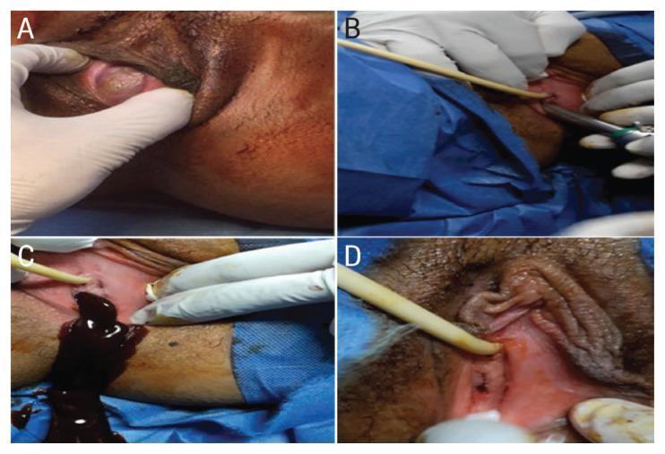
A series of four clinical photographs (A-D) documenting the surgical management of an imperforate hymen in a postpubertal patient. (A) Preoperative view showing a distended, bulging hymenal membrane with characteristic dark, bluish discoloration caused by hematocolpos (accumulated menstrual blood). (B) Intraoperative image demonstrating the controlled perforation of the hymenal membrane using a 10 mm laparoscopy trocar tip. (C) Clinical photograph capturing the drainage of retained menses through the trocar sleeve; the fluid is dark, thick, and viscous, consistent with long-term retention. (D) Postoperative follow-up image displaying the results of a reconstructive technique that preserved the integrity and patency of the annular hymenal ring while avoiding restenosis. These images illustrate the diagnostic presentation of primary amenorrhea due to outflow obstruction and a modified surgical approach for hymenotomy that aims to maintain anatomical structure.
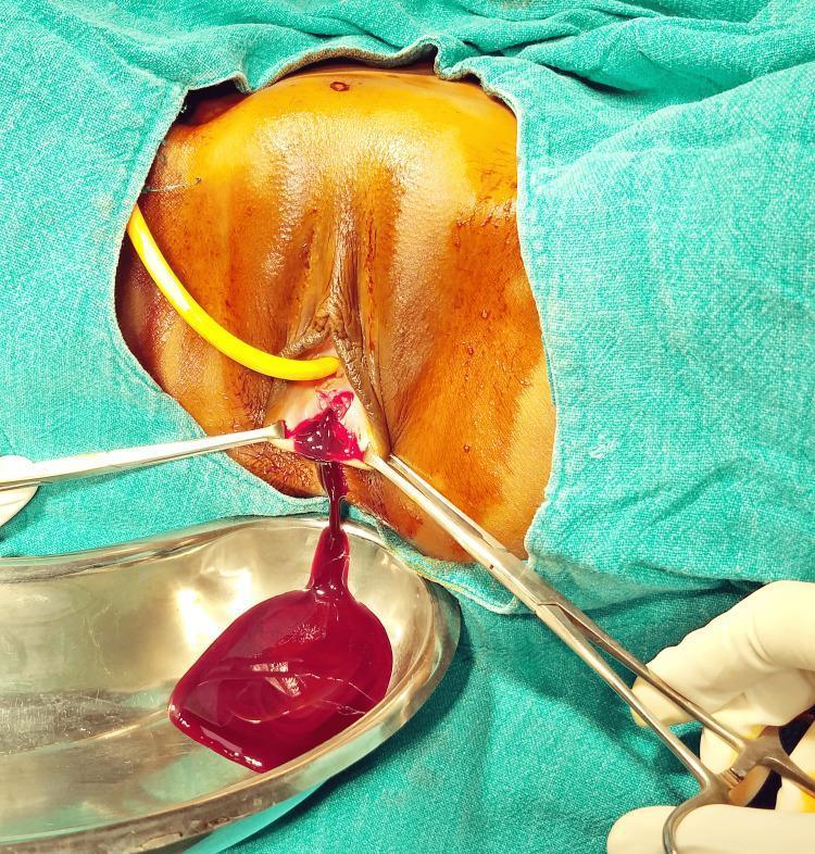
Clinical photograph of a surgical procedure illustrating the drainage of hematocolpos in a patient with an imperforate hymen. The patient is in the lithotomy position, with the perineal region prepped and draped using green surgical cloths. A cruciate incision has been made at the hymen, and two surgical forceps are seen retracting the edges to maintain patency. A large volume of dark red, viscous, ‘chocolate-like’ blood (retained menstrual fluid) is actively draining into a stainless steel kidney tray positioned below the introitus. A yellow Foley catheter is visible in the urethra to prevent urinary retention and clearly demarcate the urethral anatomy during the procedure. This visual demonstrates the surgical management of hematocolpometra and the typical appearance of stagnant menstrual blood. The image is relevant for gynecology and obstetrics, specifically in the context of adolescent health and congenital Müllerian anomalies.

Clinical photograph of the female external genitalia in two panels, demonstrating an imperforate hymen in a pediatric/adolescent patient. The top image shows a medical professional in gloves performing a physical examination. The central finding is a tense, bulging, and imperforate hymen with a characteristic bluish-purple discoloration, indicative of hematocolpos (trapped menstrual blood). The lower image provides a clearer view of the perineal region, showing normal pubic hair distribution for age and a vertical, slit-like introitus obstructed by the bulging membrane. Trace amounts of dark, altered blood/discharge are visible around the posterior fourchette and perineum. This visual is diagnostic for obstructive Mullerian anomalies presenting with primary amenorrhea and cyclical pelvic pain. It is used to teach clinical recognition of gynecological causes of acute urinary retention in adolescent females.
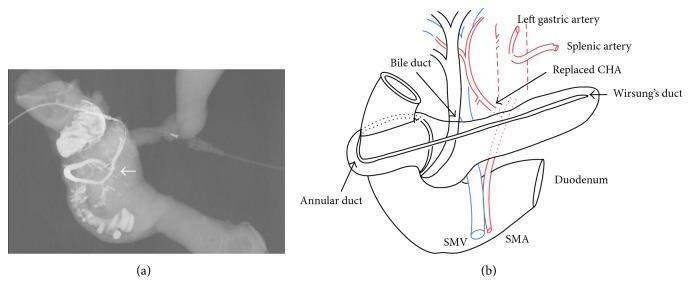
Educational image set illustrating annular pancreas anatomy. (a) A post-resection specimen cholangiopancreatogram using Urografin contrast. It demonstrates the annular pancreatic duct forming a complete circumferential loop around the second (descending) part of the duodenum, draining directly into the major duodenal papilla (indicated by an arrow). (b) A corresponding schematic diagram detailing the anatomical relationships between the pancreaticobiliary system and vascular anomalies. Key labeled structures include the annular duct, Wirsung’s duct, and the bile duct. Vascular variants are highlighted, specifically a replaced common hepatic artery (CHA) originating from the superior mesenteric artery (SMA) and passing posterior to the pancreatic head, alongside the superior mesenteric vein (SMV), splenic artery, and left gastric artery. This combination of fluoroscopic imaging and illustration demonstrates the developmental anomaly of the ventral pancreatic bud, emphasizing its clinical relevance in duodenal obstruction or associated biliary pathology.
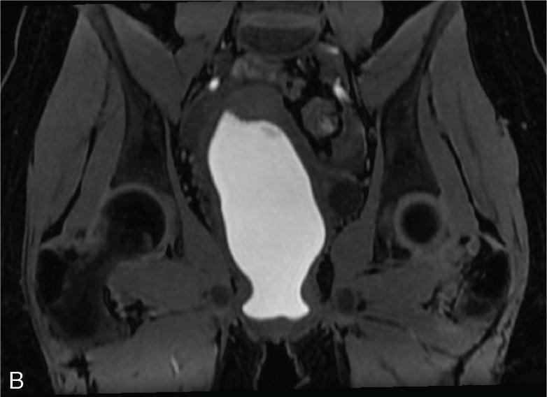
This diagnostic image is a coronal T1-weighted DIXON water MRI of the female pelvic region, illustrating significant pathology related to an imperforate hymen. The central feature is a large, hyperintense (bright), well-circumscribed structure occupying the vaginal canal and extending toward the uterine cavity. This high signal intensity on T1-weighting is characteristic of subacute hemorrhagic content, consistent with hematocolpos. The vaginal canal is markedly distended due to the accumulation of menstrual blood trapped by the distal obstruction. The surrounding pelvic anatomy, including the uterine walls and pelvic musculature, appears with lower signal intensity, providing high contrast against the centrally located fluid. This radiological presentation is classic for obstructive Müllerian anomalies in adolescent patients, where the inability to drain menstrual flow leads to progressive distention of the genital tract.
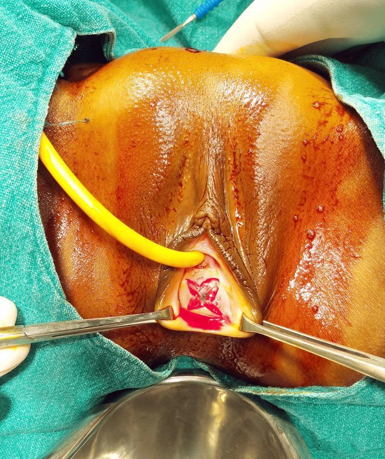
This clinical photograph captures a surgical procedure for an imperforate hymen. The image focus is on the perineal area, where a cruciate incision (X-shaped) has been made on the bulging hymen. Two surgical retractors are positioned to hold the vaginal introitus open, revealing the incision site marked by fresh blood and the underlying vaginal vault. A yellow urinary catheter is inserted into the urethra superior to the hymenal membrane to maintain patency and provide drainage during the procedure. The surrounding labial and perineal tissues appear edematous and erythematous, consistent with the obstructive symptoms of hematocolpometra. The patient is draped in a sterile green surgical field, and a metal basin is visible at the inferior aspect of the frame to catch drained fluid. This image illustrates the standard surgical approach (hymenectomy via cruciate incision) used to resolve hematocolpos and associated acute urinary retention in patients with imperforate hymen.
Hymen and Its Types
Definition
Types of Hymen
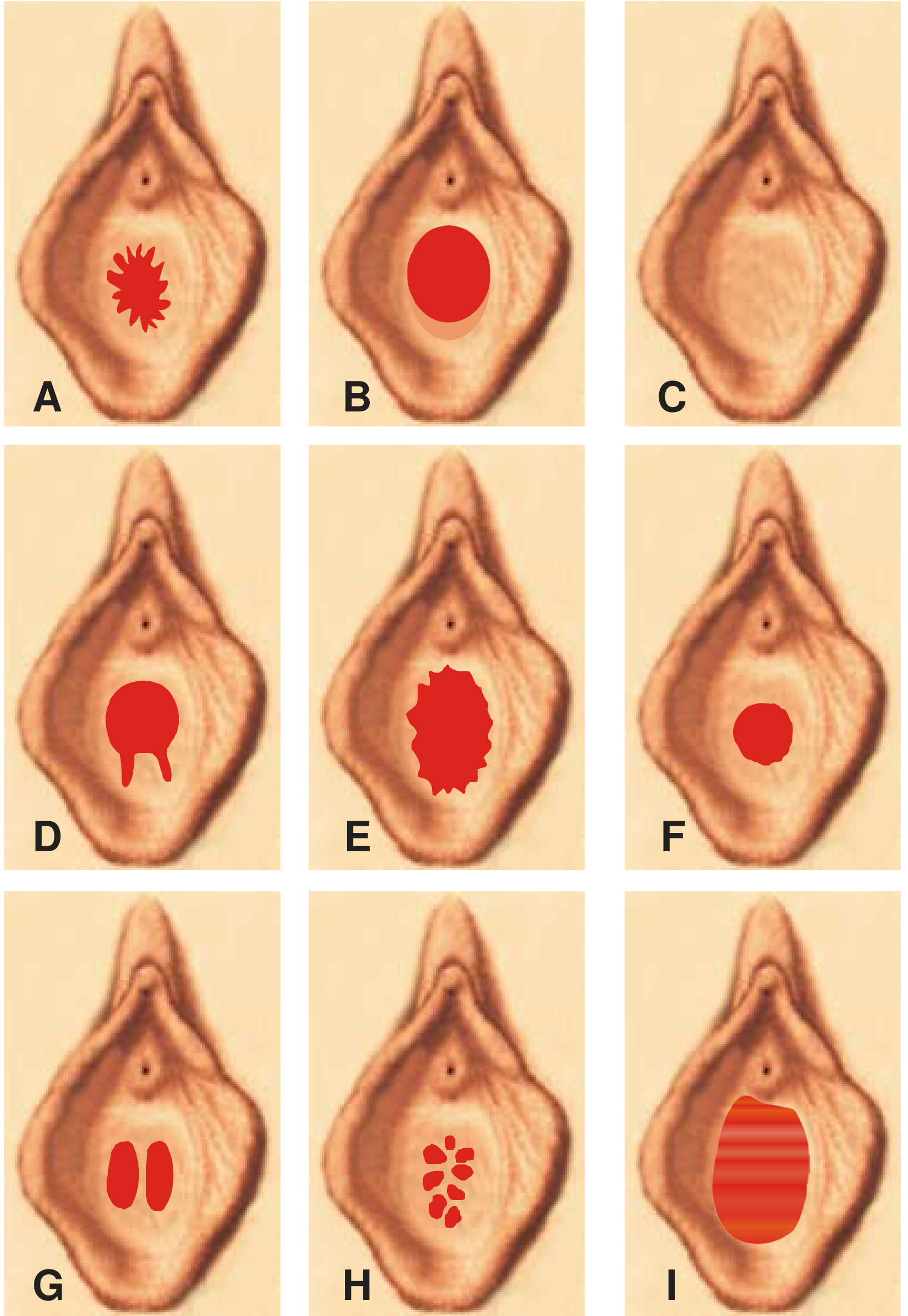
1. Semilunar (Crescentic)
- The hymenal tissue is present at the lateral and posterior margins of the vaginal opening.
- The opening faces anteriorly (toward the urethra).
- This is one of the most commonly encountered types in nulliparous females.
2. Annular
- The hymenal opening is oval and centrally placed, encircling the vaginal opening.
- The hymenal membrane forms a complete ring around the orifice.
3. Infantile
- There is a small linear opening in the midline.
- Seen in infants and young girls; the tissue is thicker and less elastic.
4. Cribriform (Sieve-like)
- Multiple small hymenal openings are present.
- Resembles a sieve or colander.
- Clinically: menstrual blood may drain slowly and cause dysmenorrhea.
5. Vertical
- A single vertical hymenal opening is present.
6. Septate
- There are two lateral openings side by side, separated by a band (septum) of tissue running across the middle.
- The band can sometimes cause difficulty during intercourse and may need to be divided surgically.
7. Imperforate
- No hymenal opening is present at all.
- This is a clinically significant abnormality.
- At puberty it causes hematocolpos (accumulation of menstrual blood in the vagina) presenting as:
- Primary amenorrhea
- Cyclical pelvic pain
- A bluish, bulging membrane at the introitus
- Urinary retention in severe cases
- Treatment: Surgical hymenotomy (cruciate incision).
8. Fimbriated (Notched / Frilly-edged)
- The free margin of the hymen has natural notches, giving it a frilly or fimbriated appearance.
- Must be differentiated from a ruptured hymen (important in forensic medicine):
| Feature | Fimbriated Hymen | Ruptured Hymen |
|---|---|---|
| Site of notches | Anteriorly; do not reach the base | Posteriorly (6 o'clock position); extend to attachment |
| Shape | Bilaterally symmetrical | Asymmetrical |
| Mucous membrane | Intact over notches | Not intact |
| Cause | Natural anatomical variant | Sexual intercourse or foreign body |
Microperforate Hymen (Additional Type)
Post-Coital / Post-Delivery States
- Hymenal tears (lacerations): After sexual intercourse, the hymen typically tears posteriorly (at 5, 6, and 7 o'clock positions). The tears heal as rounded tags.
- Carunculae myrtiformes: Irregular fleshy tags (remnants of the hymen) seen after delivery. Their presence confirms a parous state.
- Parous introitus: Seen after childbirth - the vaginal opening is widely gaping with only remnant tags.
Forensic Significance
- An intact hymen does not automatically prove virginity - sexual intercourse can occur without hymenal rupture (elastic hymen), and rupture can occur without intercourse (trauma, instrumentation, tampons, surgery).
- The most common site of rupture from sexual intercourse is the posterior commissure (6 o'clock / posterolateral positions).
- Causes of non-coital rupture include: accidental falls, masturbation, foreign bodies, ulceration (diphtheria, fungal), scratching, surgical procedures, and sanitary tampons.
P C Dikshit - Textbook of Forensic Medicine and Toxicology, pp. 346-347