
Spaces of the ischoorectal fossa
Reading File
The ischioanal (ischiorectal) fossa is a wedge-shaped fat-filled space on either side of the anal canal, important in the spread of perianal infections and abscesses. It communicates with several key spaces.
Searching sources...
Finding Sources
Finding Sources
Reading File
Reading File
Finding Sources
Finding Sources
Reading File
Reading File
Reading File
Loading Image
Reading File
I now have comprehensive information from multiple surgical textbooks. Here is a full, well-structured answer:
Spaces of the Ischiorectal (Ischioanal) Fossa
Nomenclature note: The term ischiorectal fossa is still widely used clinically but the modern anatomical name is ischioanal fossa, since the space does not directly adjoin the rectum but lies lateral to the anal canal.
The Ischioanal Fossa Itself
The ischioanal fossa is a pyramid-shaped, fat-filled space on either side of the anal canal. Its tip (apex) points anteriorly toward the symphysis pubis, and its base faces posteriorly/inferiorly toward the perineal skin.
Boundaries:
| Wall | Structure |
|---|---|
| Superomedial | Levator ani + inferior fascia of the pelvic diaphragm |
| Lateral | Ischial tuberosity + obturator fascia (obturator internus) |
| Inferior (base) | Perineal skin |
| Posterior | Gluteus maximus + sacrotuberous ligament |
| Anterior | Posterior border of the urogenital diaphragm (deep transverse perineal) |
The fat pad within the fossa acts as a mobile cushion that slides during defecation and childbirth. Running through it are branches of the internal pudendal vessels and pudendal nerve, whose main trunks travel in the pudendal (Alcock's) canal - a fascial tunnel in the lateral wall formed by the obturator fascia.
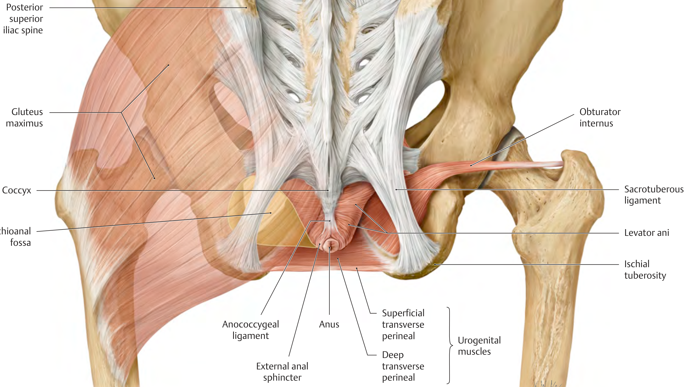
Spaces of (and around) the Ischiorectal/Ischioanal Fossa
The ischiorectal/ischioanal fossa is not a single isolated space - it communicates with several named spaces that are surgically important for understanding abscess spread.
1. Perianal Space
- Surrounds the anal canal superficially (subcutaneous)
- Contains the external hemorrhoidal plexus
- Laterally becomes continuous with the buttock fat
- Communicates with the ischioanal space; together they form the ischioanal fossa
2. Ischioanal (Ischiorectal) Space
- The main bulk of the fossa - extends laterally and superiorly from the perianal skin up to the levator ani
- Medial wall: external sphincter + levator ani
- Lateral wall: obturator fascia
- Contains the pudendal neurovascular bundle in Alcock's canal
- The two ischioanal spaces communicate posteriorly via the deep postanal space (see below)
3. Intersphincteric Space
- Located between the internal and external anal sphincters
- Continuous with the perianal space distally
- Extends cephalad into the rectal wall
- Origin of most cryptoglandular infections (infected anal glands open into anal crypts at the dentate line, and abscesses form here first before spreading)
- Abscess here causes severe pain with no external signs but exquisite tenderness on digital rectal exam
4. Superficial Postanal Space
- Located posteriorly, below the anococcygeal ligament
- Connects the right and left perianal spaces posteriorly
5. Deep Postanal Space (Space of Courtney)
- Located posteriorly, above the anococcygeal ligament, between the levator ani (above) and external sphincter (below)
- Connects the two ischioanal spaces posteriorly, forming the classic horseshoe abscess pathway
- The anococcygeal ligament divides the postanal space into superficial and deep; if this ligament is not adequately divided during drainage, the deep component is missed
6. Supralevator Space
- Lies above the levator ani on either side of the rectum
- Extends superiorly to the peritoneum at the rectosacral fascia
- Right and left sides communicate posteriorly
- Abscesses here arise from upward extension of cryptoglandular sepsis OR downward extension of intra-abdominal pathology (e.g., perforated diverticulitis)
7. Submucosal Space
- Between the internal anal sphincter and the anal mucosa, distal to the dentate line
- Contains the internal hemorrhoidal plexus
- Proximally becomes the submucosa of the rectum
8. Retrorectal Space
- Above the rectosacral fascia (Waldeyer's fascia)
- Between the upper two-thirds of the rectum and the sacrum
- Distinct from the supralevator space
Clinical Significance: Spread of Anorectal Sepsis
Understanding these spaces explains the pattern of abscess spread from the cryptoglandular origin:
Anal gland infection (intersphincteric space)
         |
    Spreads to:
    ├── Downward → Perianal abscess (most common, ~40-45%)
    ├── Laterally through external sphincter → Ischiorectal abscess
    ├── Upward (intersphincteric) → Supralevator abscess
    └── Posteriorly → Deep postanal space → Horseshoe abscess
                      (bilateral ischioanal involvement)
Horseshoe abscess: Infection spreads from the deep postanal space (Space of Courtney) laterally into both ischioanal fossae, creating a U-shaped or horseshoe-shaped collection that encircles the anal canal posteriorly. Adequate drainage requires opening the deep postanal space by dividing the anococcygeal ligament.
Summary Table
| Space | Location | Key Clinical Point |
|---|---|---|
| Perianal | Subcutaneous around anus | Most common abscess site |
| Ischioanal | Lateral to anal canal | Large, may not be visible externally |
| Intersphincteric | Between sphincters | Pain without external signs |
| Superficial postanal | Below anococcygeal lig., posterior | Connects perianal spaces |
| Deep postanal (Courtney) | Above anococcygeal lig., posterior | Key to horseshoe abscess |
| Supralevator | Above levators | Intra-abdominal or upward spread |
| Submucosal | Between IAS and mucosa | Contains internal hemorrhoidal plexus |
| Retrorectal | Rectum-sacrum (above rectosacral fascia) | Retrorectal tumors/cysts |
- Mulholland and Greenfield's Surgery, 7th ed., p. 3183
- Schwartz's Principles of Surgery, 11th ed., p. 1341
- Yamada's Textbook of Gastroenterology, 7th ed., p. 179
- Current Surgical Therapy, 14th ed.
- THIEME Atlas of General Anatomy and Musculoskeletal System, p. 581
This is a shared conversation. Sign in to Orris to start your own chat.