I now have everything I need to write a complete, clear answer. Here it is:
Respiratory Tract - Organs and Lung Anatomy
Organs of the Respiratory Tract
Upper Respiratory Tract
These are the structures above the larynx that conduct air into the body:
| Organ | Function |
|---|
| Nose (external + nasal cavity) | Filters, warms, and humidifies air |
| Paranasal sinuses | Lighten skull, resonate voice |
| Pharynx (nasopharynx, oropharynx, laryngopharynx) | Common passage for air and food |
| Larynx | Voice production, protects airway |
Lower Respiratory Tract
These are the structures from the trachea downward involved in gas exchange:
| Organ | Function |
|---|
| Trachea | Conducts air to bronchi |
| Primary (main) bronchi (right & left) | Air passage into each lung |
| Secondary (lobar) bronchi | Supplies each lobe |
| Tertiary (segmental) bronchi | Supplies each bronchopulmonary segment |
| Bronchioles | Smaller airways, no cartilage |
| Alveolar ducts and alveoli | Site of actual gas exchange |
| Lungs (right & left) | Main organs of respiration |
| Pleura | Serous covering of lungs |
THE LUNGS - Detailed Description
a) Size and Shape
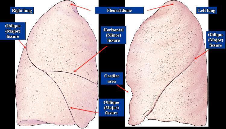
The lungs are the paired, cone-shaped organs of respiration sitting in the thoracic cavity on either side of the mediastinum. Each lung has:
- Shape: Like a half-cone, with a rounded apex (top) and a concave base (bottom)
- Apex: Projects about 2.5 cm above the clavicle into the root of the neck
- Base: Concave, sitting on the diaphragm
- Three surfaces: Costal (facing ribs), Mediastinal (facing the heart), and Diaphragmatic (base)
- Two borders: Anterior border and inferior border
Right lung:
- Heavier (~625 g), shorter and wider
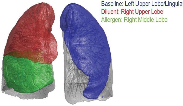
- Divided into 3 lobes by two fissures: oblique fissure + horizontal (minor) fissure
- Lobes: Superior, Middle, Inferior
Left lung:
- Lighter (~565 g), longer and narrower
- Has only an oblique fissure, dividing it into 2 lobes: Superior and Inferior
- Has a cardiac notch on its anterior border (where the heart bulges in)
- Has a lingula - a tongue-like projection of the upper lobe that extends over the heart
(- Color Atlas of Human Anatomy Vol. 2, Internal Organs)
b) Surface Anatomy
The key surface markings of the lungs:
Apex:
- Projects 2.5 cm above the medial third of the clavicle
Anterior border:
- Right lung: runs nearly vertical from the apex to the 6th costal cartilage
- Left lung: similar but deviated left at the 4th costal cartilage due to the cardiac notch
Lower border (inferior margin):
- Crosses rib 6 at the midclavicular line, rib 8 at the midaxillary line, and rib 10 at the back
- The pleura extends about 2 ribs lower than the lung itself
Fissures on surface:
- Oblique fissure (both lungs): begins at T3/T4 spine posteriorly, runs forward and downward to the 6th rib anteriorly
- Horizontal fissure (right lung only): runs from the oblique fissure at the midaxillary line horizontally to the 4th costal cartilage anteriorly
c) Blood Supply
The lungs have two separate blood supplies - functional and nutritive:
1. Functional blood supply (for gas exchange) - Pulmonary circulation:
- Pulmonary arteries carry deoxygenated blood from the right ventricle to the lungs
- The pulmonary trunk divides into right and left pulmonary arteries at the level of T4/T5
- These arteries branch alongside the bronchi, reaching every bronchopulmonary segment
- After gas exchange, oxygenated blood returns via pulmonary veins to the left atrium
- There are two pulmonary veins on each side (4 total), entering the left atrium
2. Nutritive blood supply (for lung tissue itself) - Bronchial circulation:
- Bronchial arteries arise from the thoracic aorta (and sometimes upper intercostal arteries)
- They supply the walls of bronchi, bronchioles, and pleura
- They run along the esophagus and enter the lung at the hilum
- They do NOT reach the alveolar walls (which get O2 directly from air)
(- Fishman's Pulmonary Diseases and Disorders)
d) Venous and Lymphatic Drainage
Venous drainage:
- Pulmonary veins (2 on each side, 4 total) carry oxygenated blood back to the left atrium
- Unlike pulmonary arteries, pulmonary veins are valveless
- They run in an intersegmental course (between segments) unlike arteries which run centrally within segments
- Bronchial veins drain the larger bronchi and drain into the azygos vein (right side) or hemiazygos vein (left side)
- Smaller bronchial veins drain into pulmonary veins
Lymphatic drainage:
The lymphatic system of the lung has two networks:
| Network | Location | Drains to |
|---|
| Deep (peribronchial) network | Runs along bronchi and vessels deep within lung | Bronchopulmonary (hilar) nodes at bronchial divisions |
| Superficial (subpleural) network | Begins under the pleura, in septa between segments | Follows pulmonary veins → tracheobronchial nodes |
Chain of lymph nodes:
Bronchopulmonary nodes → Tracheobronchial nodes (at carina) → Paratracheal nodes → Mediastinal nodes
Clinical note: Hilar and paratracheal lymph nodes are major filtering stations in tuberculosis and lung cancer - enlargement of these nodes is an important diagnostic sign.
(- Color Atlas of Human Anatomy Vol. 2, Internal Organs)
e) Nerve Supply
The lungs are supplied by the autonomic nervous system. The nerve fibers form the pulmonary plexus at the hilum of each lung.
Parasympathetic fibers:
- From the vagus nerve (CN X)
- Effect: bronchoconstriction (narrows airways) + increases secretions of glands
- Sensory fibers from vagus carry the cough reflex and Hering-Breuer reflex signals
Sympathetic fibers:
- From upper thoracic (T2-T5) and cervical sympathetic ganglia
- Effect: bronchodilation (widens airways) + vasoconstriction
Pulmonary plexus:
- Vagal and sympathetic fibers meet at the hilum to form the pulmonary plexus
- From there, nerve bundles follow the bronchi and blood vessels deep into the lung
- Nerves supply smooth muscle of bronchi, blood vessels, and glands
- Some fibers reach the visceral pleura
(- Fishman's Pulmonary Diseases and Disorders; Color Atlas of Human Anatomy Vol. 2)
f) Relations
The lungs are closely related to many important structures. These relations differ between the two sides.
RIGHT LUNG:
| Surface | Important Relations |
|---|
| Mediastinal surface | Heart (right atrium, superior vena cava) |
| Apex | Right subclavian artery and vein, brachiocephalic trunk, trachea |
| Hilum | Azygos vein arches over it from behind |
| Posterior | Esophagus, right vagus nerve |
| Other | Right phrenic nerve runs between lung and pericardium |
LEFT LUNG:
| Surface | Important Relations |
|---|
| Mediastinal surface | Heart (left ventricle mainly), aortic arch, thoracic aorta |
| Apex | Left subclavian artery and vein, aortic arch |
| Posterior | Esophagus, thoracic aorta |
| Mediastinal surface | Cardiac notch and lingula related to the heart's projection |
Both lungs are also related to:
- The parietal pleura externally
- The ribs and intercostal muscles via the costal surface
- The diaphragm via the diaphragmatic surface
(- Gray's Anatomy for Students)
g) Bronchopulmonary Segments
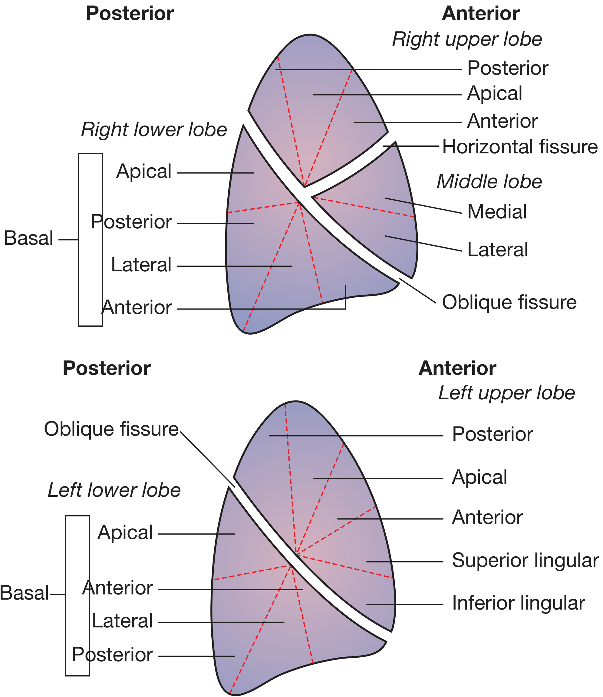
A bronchopulmonary segment is the area of lung supplied by one segmental (tertiary) bronchus along with its accompanying branch of the pulmonary artery.
Key features:
- Each segment is shaped like an irregular cone with the apex toward the hilum and base toward the lung surface
- Each segment is functionally independent - it can be surgically removed without affecting adjacent segments
- The pulmonary artery runs centrally within the segment (intrasegmental)
- The pulmonary veins run along the edges between segments (intersegmental)
- There are 10 segments in the right lung and 8-10 in the left (some fuse on the left side)
(- Gray's Anatomy for Students; Color Atlas of Human Anatomy Vol. 2)
RIGHT LUNG - 10 segments:
| Lobe | Segment Number | Segment Name |
|---|
| Upper (Superior) | 1 | Apical |
| 2 | Posterior |
| 3 | Anterior |
| Middle | 4 | Lateral |
| 5 | Medial |
| Lower (Inferior) | 6 | Apical (Superior) |
| 7 | Medial basal |
| 8 | Anterior basal |
| 9 | Lateral basal |
| 10 | Posterior basal |
LEFT LUNG - 8-10 segments (some fuse):
| Lobe | Segment Number | Segment Name |
|---|
| Upper (Superior) | 1+2 | Apicoposterior (fused) |
| 3 | Anterior |
| 4 | Superior lingular |
| 5 | Inferior lingular |
| Lower (Inferior) | 6 | Apical (Superior) |
| 7+8 | Anteromedial basal (fused) |
| 9 | Lateral basal |
| 10 | Posterior basal |
Diagram of bronchopulmonary segments:
Clinical importance: Knowledge of bronchopulmonary segments is essential for:
- Bronchoscopy - to locate a lesion
- Postural drainage - positioning the patient to drain a specific segment
- Segmentectomy - surgical removal of a single segment (e.g., for small lung tumours)
- Lung cancer staging - identifying which segment a tumour occupies
(Sources: Gray's Anatomy for Students; Bailey and Love's Short Practice of Surgery 28th Ed.; Color Atlas of Human Anatomy Vol. 2; Fishman's Pulmonary Diseases and Disorders)