
In detail about neuropathic joint or charchot arthropathy for Orthopaedic postgraduate exam in simple words cover all aspects properly
Reading File
Now let me search the medical library for Charcot arthropathy comprehensively.
Finding Sources
Finding Sources
Excellent - multiple excellent sources found. Let me read all key sections in parallel.
Reading File
Reading File
Reading File
Excellent data. Now let me read the Miller's staging table + treatment section, Campbell's full treatment section, and the Rheumatology section:
Reading File
Reading File
Reading File
Now let me get the clinical features, differential diagnosis, and treatment sections plus fetch key radiograph images:
Reading File
Reading File
Loading Image
Loading Image
Now let me get the rest of the treatment section from Harrison's and the Campbell's pathogenesis/neurovascular theory + fetch the midfoot X-ray image:
Reading File
Reading File
Loading Image
Loading Image
I now have comprehensive data from multiple authoritative sources. Let me compile the complete, detailed orthopaedic PG exam answer.
Neuropathic Joint / Charcot Arthropathy
A Complete Guide for the Orthopaedic Postgraduate Exam
1. DEFINITION
Charcot arthropathy (neuropathic joint) is a chronic, progressive, destructive arthritis affecting bone architecture and joint alignment in patients who have lost protective sensation (pain, proprioception, or both). Without these protective mechanisms, joints undergo repeated microtrauma leading to progressive cartilage and bone destruction.
- Harrison's Principles of Internal Medicine 22E, p. 3012
- Miller's Review of Orthopaedics 9th Ed, p. 602
2. HISTORY
- First described by Jean-Martin Charcot in 1868 in patients with tabes dorsalis (neurosyphilis)
- Today, diabetes mellitus is the most common cause
- The term "Charcot joint" honors him; "neuropathic arthropathy" is the more accurate modern term
3. CAUSES (Etiology) - HIGH-YIELD
Remember: "DeLiSy A-SPICS"
| Cause | Joint most affected |
|---|---|
| Diabetes mellitus (most common today) | Midtarsal, tarsometatarsal, MTP |
| Leprosy | Tarsal, tarsometatarsal |
| Syringomyelia | Shoulder, elbow, wrist |
| Amyloidosis | Knee, ankle |
| Spina bifida / Myelomeningocele | Lower limb joints |
| Syphilis (Tabes dorsalis) | Knee, hip, ankle |
| Peripheral neuropathy (alcohol, avitaminosis) | Various |
| Intraarticular steroid injections (repeated) | Any injected joint |
| Congenital insensitivity to pain | Knee, ankle, intertarsal |
| Spinal cord injury | Below-injury joints |
Key memory tip: In tabes dorsalis - knee is #1; in syringomyelia - upper limb joints (shoulder, elbow, wrist); in diabetes - foot/ankle joints.
- Rheumatology 2-Volume Set 2022, p. 2088
- Campbell's Operative Orthopaedics 15th Ed, Box 7.7
4. PATHOPHYSIOLOGY - TWO THEORIES
Two competing theories exist; the true pathogenesis is a combination of both.
Theory 1: Neurovascular Theory (French theory - Charcot's own)
- Neurologic damage → autonomic dysfunction → arteriovenous shunting → increased local blood flow
- Increased blood flow → bone resorption (osteoclastic > osteoblastic activity)
- TNF-alpha and IL-1B → increased NF-kB → increased osteoclast production → bone loss
- This is the "vascular" mechanism - bone melts away
Theory 2: Neurotraumatic Theory (German theory - Virchow & Volkmann)
- Loss of deep pain and proprioception → protective neuromuscular reflexes gone
- Joint subjected to repeated unperceived microtrauma
- Ligament tears, microfractures accumulate without healing response
- This is the "mechanical" mechanism - joint is beaten to destruction
Bottom line: Bone resorption + repeated unprotected trauma = progressive joint disorganization.
- Campbell's Operative Orthopaedics 15th Ed (foot & ankle chapter)
5. JOINT DISTRIBUTION BY DISEASE
| Disease | Joints Affected |
|---|---|
| Diabetes mellitus | Midtarsal, tarsometatarsal (TMT), MTP, talotibial |
| Tabes dorsalis | Knee (most common), hip, ankle |
| Syringomyelia | Shoulder, elbow, wrist (upper limb) |
| Amyloidosis | Knee, ankle |
| Congenital sensory neuropathy | Knee, ankle, intertarsal, MTP |
| Leprosy | Tarsal, tarsometatarsal |
Exam tip: Syringomyelia → upper limb Charcot. All others → lower limb predominantly.
6. INCIDENCE
- Occurs in approximately 1-1.5% of diabetic patients
- Up to 7.5% of diabetics with neuropathy
- Usually occurs at age ≥50 years in someone with diabetes for several years
- Up to 35% present with bilateral disease
- Pain is present in up to 50% of cases (not always painless!)
7. CLINICAL FEATURES
Symptoms:
- Swelling, warmth, redness, and deformity of the joint
- Disproportionately little pain for the degree of radiographic destruction (classic)
- Pain may occur in up to 50% from intraarticular fractures of osteophytes
- History of trivial or minor trauma triggering acute presentation
- Swelling and redness often improve in the morning (worse with activity)
Signs:
- Progressive joint enlargement (bony overgrowth + effusion)
- Joint instability, hypermobility
- Deformity - "rocker-bottom foot" in diabetic Charcot foot (downward collapse of tarsal bones → convexity of sole)
- Large bony prominences palpable (osteophytes protruding from top of foot)
- Calluses over metatarsal heads → pressure ulcers → osteomyelitis
- Crepitus, loose bodies
Classic Diabetic Charcot Foot:
-
Warm, swollen, erythematous foot
-
Arch collapse - rocker-bottom deformity
-
Often confused with cellulitis or osteomyelitis
-
No remembered significant trauma (or only trivial trauma)
-
Harrison's 22E, p. 3012
8. RADIOLOGICAL FEATURES
Radiographs are the key investigation. The classic X-ray features can be remembered as the "6 Ds":
- Distension of joint (effusion)
- Density increased (sclerosis/eburnation)
- Detritis (loose bodies, intraarticular fragments)
- Dislocation / subluxation
- Degenerative changes (joint space loss, osteophytes)
- Destruction / Disorganization (fragmentation of bone)
Additional features:
- Periarticular bone fragmentation
- Subchondral bone sclerosis (eburnation)
- Large osteophytes at joint margins
- Marked joint destruction out of proportion to clinical pain
- Resorption and tapering of distal metatarsal bones (in diabetic foot)
- "Lisfranc fracture-dislocation" pattern at tarsometatarsal joints
Joint margins in neuropathic joint = DISTINCT (vs. osteomyelitis where they are blurred)
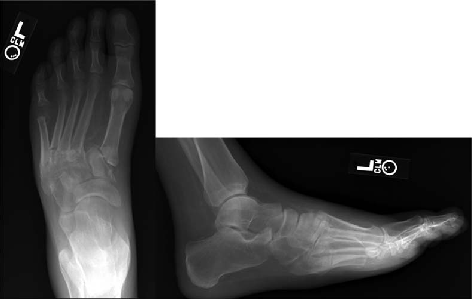
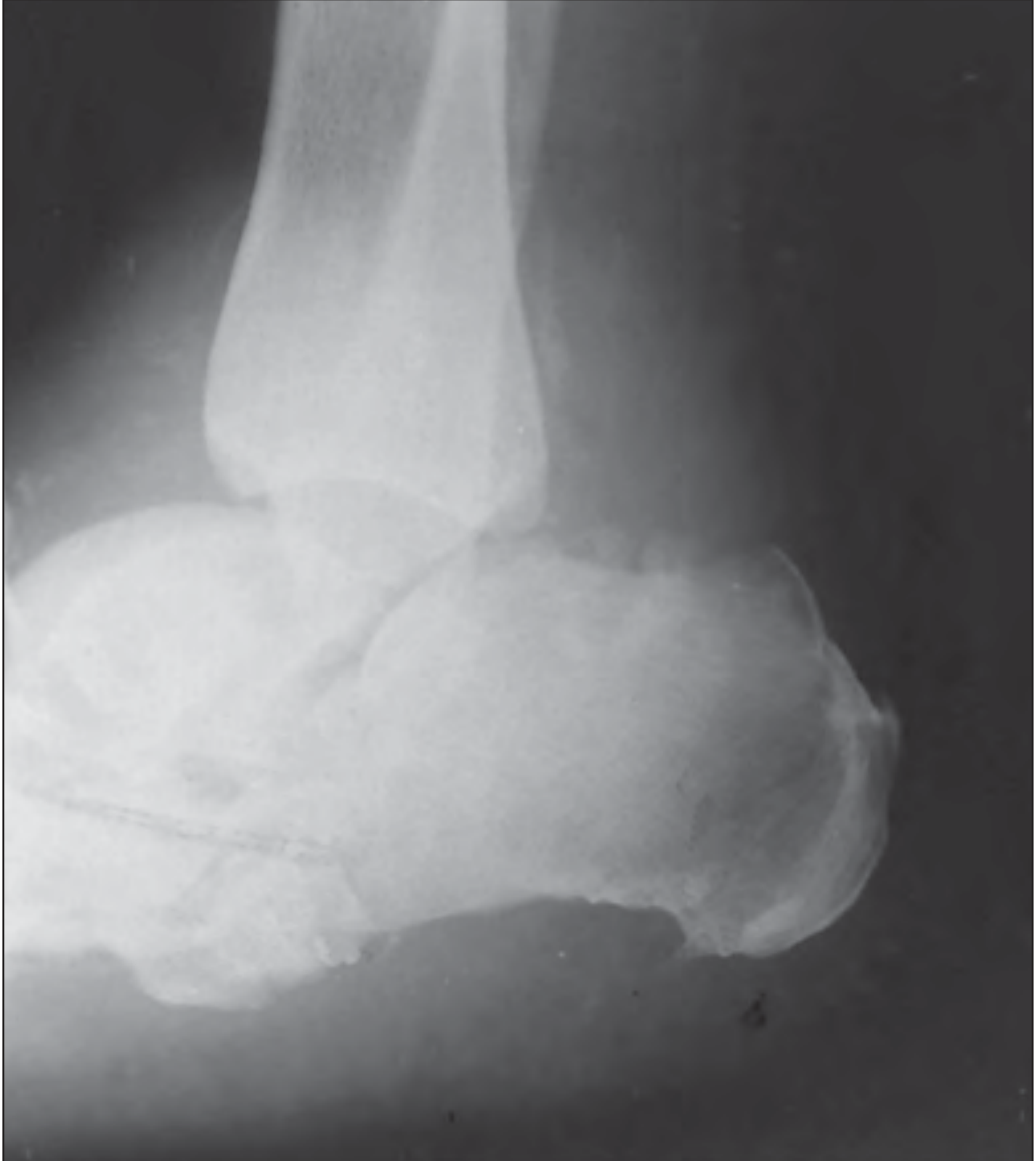
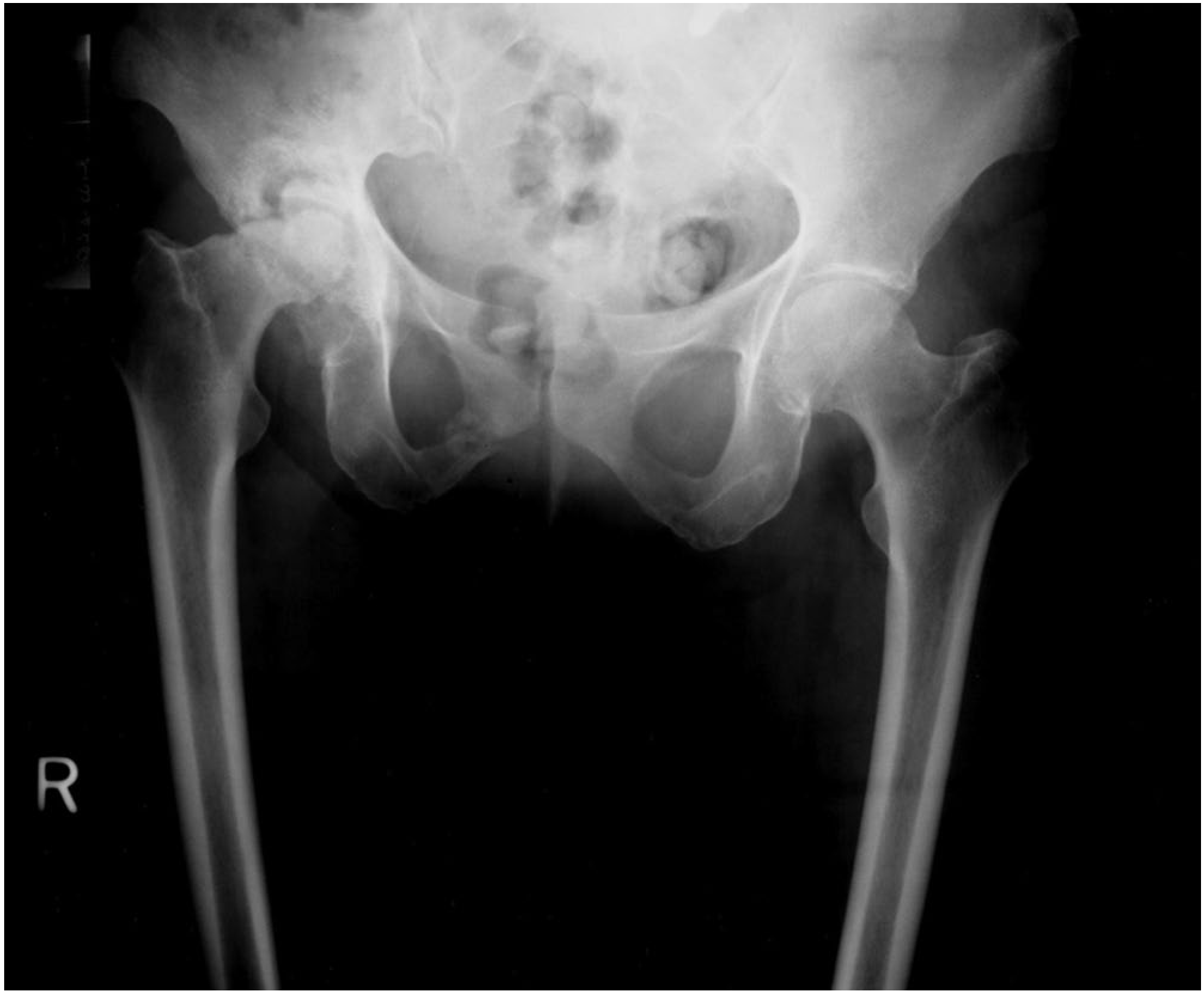
9. HISTOLOGY
On synovial biopsy:
- Osteochondral fragments embedded within the synovium - this is diagnostic of neuropathic arthropathy
- Similar to severe OA: cartilage fragmentation, subchondral eburnation, osteophytes, loose bodies
- More advanced: erosions, devitalized bone, microscopic bone/cartilage fragments
- Synovial fluid: noninflammatory, may be xanthochromic or bloody, contains fragments of synovium, cartilage, and bone
10. STAGING - EICHENHOLTZ CLASSIFICATION (Most Important)
This is the most exam-tested classification. It is based on clinical signs, symptoms, and radiographic changes over time.
| Stage | Name | Clinical Features | Radiograph Findings |
|---|---|---|---|
| 0 | Clinical (Pre-fragmentation) | Acute inflammation; confused with infection; earliest stage | Regional bone demineralization only |
| 1 | Dissolution (Fragmentation) | Acute inflammation, swelling, erythema, warmth | Bone demineralization + periarticular fragmentation, joint dislocation |
| 2 | Coalescence | Less inflammation, decreasing swelling and erythema | Absorption of bone debris; early bone healing, periosteal new bone |
| 3 | Resolution | Resolved erythema, swelling, warmth; consolidation | Smoothed bone edges, bony/fibrous ankylosis |
Continuum from bone resorption and fragmentation to bone formation and consolidation takes 6 to 18 months.
Treatment goal: Reach Stage 3 (resolution) while maintaining alignment and ambulatory status, minimizing soft tissue breakdown.
11. BRODSKY CLASSIFICATION (Anatomic - Exam Favourite)
Classifies Charcot foot by location:
| Type | Location | Frequency |
|---|---|---|
| 1 | Midfoot (tarsometatarsal joints) - most common | ~60% |
| 2 | Hindfoot (subtalar, talonavicular, calcaneocuboid joints) | ~10% |
| 3A | Tibiotalar (ankle) joint | ~20% |
| 3B | Calcaneal tuberosity fracture | <10% |
| 4 | Combination of areas | <10% |
| 5 | Forefoot | <10% |
Type 1 (midfoot) → hindfoot valgus caused by forefoot abduction with contracted gastrocsoleus complex
Type 2 → rocker-bottom deformity; varus or valgus hindfoot
Type 3A → ulcerations over prominent malleoli; generally requires surgery
Type 3B → pathologic calcaneal fracture
12. INVESTIGATIONS
Imaging:
- Plain X-ray - first line; shows the "6 Ds" as above
- MRI - early detection; differentiates from osteomyelitis; shows bone marrow edema, joint effusion
- Bone scan (Technetium-99m) - increased uptake in BOTH neuropathic joint AND osteomyelitis - cannot differentiate
- Indium-111 labeled WBC scan - increased uptake in osteomyelitis but NOT in neuropathic joint - useful for differentiation
- CT scan - better characterizes bony fragmentation and deformity
Lab:
- ESR, CRP - may be elevated in acute phase
- Blood glucose, HbA1c - to diagnose/control diabetes
- Serologic tests for syphilis (VDRL, TPHA) if suspected
- Synovial fluid analysis: noninflammatory; fragmented cartilage/bone; absence of crystals and low neutrophils (high neutrophils suggest osteomyelitis)
13. DIFFERENTIAL DIAGNOSIS
The key differentials are:
| Condition | Differentiating Features |
|---|---|
| Osteomyelitis | Most important DD in diabetic foot; blurred joint margins on X-ray; Indium-111 WBC scan positive; cultures positive |
| Avascular Necrosis | History, location, MRI pattern different |
| Advanced OA | No neuropathy; slower progression; less fragmentation |
| Stress fracture | Location, mechanism different |
| CPPD (Calcium pyrophosphate deposition) | Crystals on joint fluid analysis |
| Septic arthritis | Acute fever, high WBC count, positive cultures |
| Cellulitis | No bony changes on X-ray |
Key differentiator from osteomyelitis:
- Neuropathic joint: distinct joint margins, Indium-111 WBC scan NEGATIVE
- Osteomyelitis: blurred joint margins, Indium-111 WBC scan POSITIVE
- Technetium bone scan: positive in BOTH - not helpful for differentiation
14. TREATMENT
Goals:
- Achieve Eichenholtz Stage 3 (resolution)
- Maintain alignment and plantigrade foot
- Preserve ambulatory status
- Minimize soft tissue breakdown and ulceration
A. NON-OPERATIVE (Conservative) - First line
Step 1: Immobilization + Non-weight-bearing
- Total Contact Cast (TCC) - gold standard for acute Charcot foot
- Non-weight-bearing for at least 8 weeks (Harrison's) to 4-6 weeks (acute phase)
- Distributes pressure evenly, reduces edema
- Continued until warmth, swelling subside
- Once swelling/warmth resolve → transition to custom brace
- AFO (Ankle-Foot Orthosis)
- CROW boot (Charcot Restraint Orthotic Walker)
Step 2: Medical management
- Bisphosphonates (e.g., pamidronate, alendronate) - reduce osteoclast activity; useful in acute phase
- Neuroleptic medications (for neuropathic pain)
- Antidepressants (amitriptyline, duloxetine) - neuropathic pain
- Topical anesthetics
- Treatment of underlying cause (glucose control in diabetes)
- Protective inserts and orthotics + regular foot examination - extremely important for prevention
Success rate: Non-operative treatment successful in up to 75% of cases
B. OPERATIVE TREATMENT
Indications:
- Stable deformity with recurrent ulcers secondary to bony prominence
- Unstable/unbraceable deformity
- Ankle and hindfoot Charcot (high failure rate of conservative management)
- Failed conservative treatment
Procedures:
| Indication | Surgery |
|---|---|
| Bony prominence causing recurrent ulcers | Exostectomy (removal of the prominence) |
| Unstable midfoot deformity | Arthrodesis (fusion) |
| Hindfoot/ankle Charcot | Tibiotalocalcaneal (TTC) arthrodesis |
| Midfoot deformity (rocker-bottom) | Plantar closed-wedge osteotomy + arthrodesis |
| Achilles contracture | TAL (Tendo-Achilles Lengthening) - almost universally required |
Fixation for ankle/hindfoot:
- Intramedullary (IM) rod - provides internal stability; can be left indefinitely
- Multiplanar external fixator - alternative option
- Even with radiographic nonunion, many patients can be pain-free and functional
Arthroplasty (TKA/THA) in Charcot joint:
- Generally a relative contraindication
- TKA: fair results reported; requires attention to alignment, ligamentous balancing, bone grafting
- THA: high rates of recurrent dislocation and loosening; surgery only for severe disability
15. COMPLICATIONS
- Pressure ulcers - over bony prominences/metatarsal heads
- Osteomyelitis - from infected ulcers; major cause of morbidity
- Recurrent deformity - especially with high BMI
- Amputation - in severe, uncontrolled cases with osteomyelitis
- Deep vein thrombosis - with prolonged immobilization
- Nonunion after surgical fusion
- Dislocation/loosening after arthroplasty
- Bilateral disease in up to 35%
16. PROGNOSIS / NATURAL HISTORY
- Natural history: acute inflammation → bone resorption → consolidation → stable deformity
- Takes 6-18 months to progress through stages
- Development of Charcot in diabetics is not related to body weight
- Recurrence linked to higher BMI
- Medial column of foot fails before lateral (pull of posterior tibial tendon)
- Sagittal plane deformities more likely to cause ulceration than transverse plane abnormalities
- Early immobilization prevents progression to advanced stages
- Diabetic patients with Charcot have lower quality of life than diabetics without it
17. KEY EXAM POINTS SUMMARY
| Fact | Answer |
|---|---|
| First described by | Jean-Martin Charcot, 1868, in tabes dorsalis |
| Most common cause today | Diabetes mellitus |
| Most common joint in diabetes | Midfoot (tarsometatarsal) |
| Most common joint in tabes dorsalis | Knee |
| Most common joint in syringomyelia | Shoulder |
| Most important classification | Eichenholtz (stages 0-3) |
| Most common Brodsky type | Type 1 (midfoot) ~60% |
| Gold standard treatment | Total Contact Cast + Non-weight bearing |
| Best imaging to differentiate from osteomyelitis | Indium-111 WBC scan |
| Technetium scan | Positive in BOTH - not useful for differentiation |
| Gold standard for ankle/hindfoot Charcot | TTC arthrodesis |
| Histological diagnosis | Osteochondral fragments embedded in synovium |
| Joint fluid character | Noninflammatory, xanthochromic/bloody, fragmented debris |
| Bilateral disease incidence | Up to 35% |
| Pain incidence | Up to 50% have some pain (not always "painless") |
| Non-operative success rate | 75% |
Sources:
- Campbell's Operative Orthopaedics 15th Edition 2026
- Miller's Review of Orthopaedics 9th Edition
- Harrison's Principles of Internal Medicine 22E (2025)
- Rheumatology 2-Volume Set (2022, Elsevier)
In detail about complete foot drop and its evaluation for Orthopaedic postgraduate exam in simple words
Finding Sources
Finding Sources
Finding Sources
Finding Sources
Reading File
Reading File
Reading File
Reading File
Reading File
Reading File
Reading File
Finding Sources
Loading Image
Reading File
Finding Sources
Finding Sources
Reading File
Reading File
Reading File
I now have comprehensive data. Let me compile the complete, detailed answer for the orthopaedic PG exam.
Foot Drop - Complete Guide for Orthopaedic Postgraduate Exam
1. DEFINITION
Foot drop (drop foot) is the inability to dorsiflex the foot and toes, resulting from weakness or paralysis of the ankle dorsiflexors. The foot hangs in a plantar-flexed (and often inverted) position at rest and cannot be lifted upward during the swing phase of walking.
It is a symptom/sign, not a diagnosis - always find the cause.
2. ANATOMY - THE FOUNDATION (Know This Inside Out)
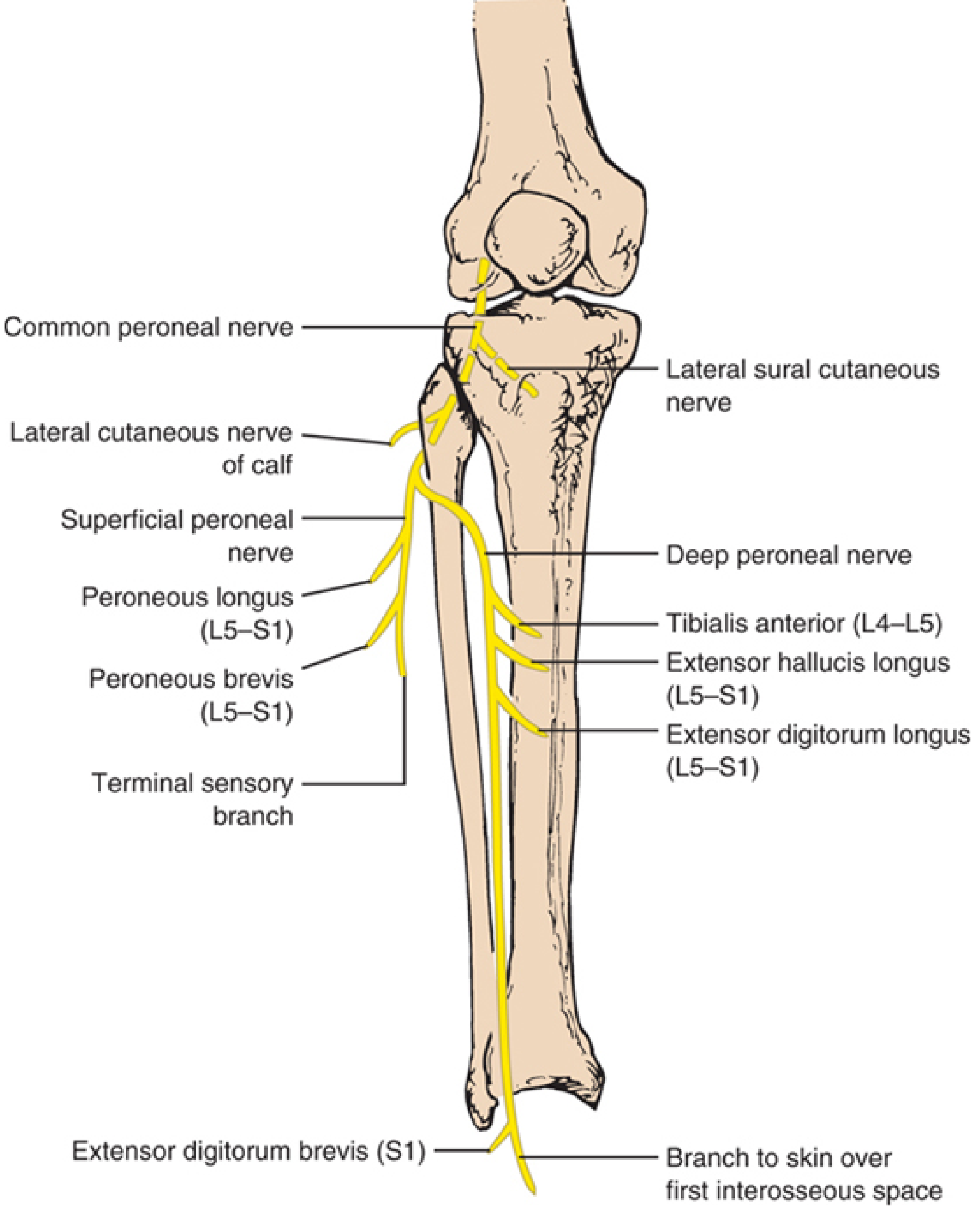
The Common Peroneal (Fibular) Nerve
- Origin: Terminal branch of sciatic nerve (L4, L5, S1, S2)
- Course: Descends in popliteal fossa → winds around the neck/head of fibula (most vulnerable point) → enters peroneus longus muscle → divides into two terminal branches
Two Terminal Branches:
A. Deep Peroneal Nerve (Anterior Tibial Nerve)
Supplies the dorsiflexors - the muscles lost in foot drop:
| Muscle | Root | Function | Test |
|---|---|---|---|
| Tibialis Anterior | L4-L5 | Dorsiflexion + inversion | Heel walking; resist dorsiflexion |
| Extensor Hallucis Longus (EHL) | L5-S1 | Extension of big toe + dorsiflexion | Dorsiflex big toe against resistance |
| Extensor Digitorum Longus (EDL) | L5-S1 | Extension of 4 toes + dorsiflexion | Dorsiflex toes against resistance |
| Extensor Digitorum Brevis (EDB) | L5-S1 | Extension of toes | Dorsiflex proximal phalanges |
| Peroneus Tertius | L5-S1 | Dorsiflexion + eversion | Part of EDL |
Sensory: Only the first web space (skin between first and second toes)
B. Superficial Peroneal Nerve
Supplies the evertors:
| Muscle | Root | Function | Test |
|---|---|---|---|
| Peroneus Longus | L5-S1 | Plantarflexion + eversion | Evert foot against resistance |
| Peroneus Brevis | L5-S1 | Plantarflexion + eversion | Evert foot against resistance |
Sensory: Lateral two-thirds of leg + dorsum of foot (EXCEPT first web space)
Key Point: Complete common peroneal palsy = foot drop + loss of eversion + sensory loss over lateral leg and dorsum of foot (except sole which is tibial nerve territory).
3. GAIT IN FOOT DROP - STEPPAGE GAIT
In foot drop, the patient cannot lift the foot during the swing phase. The foot would drag on the floor. To compensate:
Steppage Gait (High-Stepping Gait):
- Exaggerated hip and knee flexion during swing phase
- Raises the leg higher than normal to prevent the foot dragging
- Foot "slaps" down on heel strike
- Characteristic "slap" sound with each step
- May also circumduct the leg to clear the foot
This is tested clinically by asking the patient to walk on their heels - they cannot do so on the affected side.
4. CAUSES - LEVEL-BY-LEVEL APPROACH (HIGH YIELD)
Organize causes by anatomical level. This is critical for exams.
Level 1: Cortical / Upper Motor Neuron
- Cerebral stroke (parasagittal cortical lesion - leg area)
- Brain tumor
- Cerebral palsy (spastic type)
- Multiple sclerosis
(Note: UMN foot drop has spasticity + brisk reflexes + extensor plantar - different from LMN)
Level 2: Spinal Cord
- Spinal cord injury / trauma
- Spinal cord compression (disc, tumor, abscess)
- Motor neuron disease (ALS)
- Transverse myelitis
Level 3: Nerve Root (L4-L5 Radiculopathy) ← VERY COMMON
- L4-L5 disc prolapse (most common spinal cause)
- L5 root compression by disc or osteophyte
- Cauda equina syndrome
- Key: L5 radiculopathy - also has back pain + dermatomal sensory loss over dorsum of foot
Level 4: Lumbosacral Plexus
- Lumbar plexopathy (diabetes, malignancy)
- Retroperitoneal hemorrhage
- Post-delivery (obstetric palsy - baby's head pressing on lumbosacral trunk)
Level 5: Sciatic Nerve
- Sciatic nerve palsy (deep injection in buttock - commonest iatrogenic cause in India)
- Hip dislocation / acetabular fractures
- Posterior surgical approach to hip
Level 6: Common Peroneal (Fibular) Nerve ← MOST COMMON PERIPHERAL CAUSE
This is the most common compressive neuropathy of the lower limb.
Causes at the fibular head (most common site):
- Compression (most frequent cause):
- Prolonged squatting - "strawberry picker's palsy", yoga foot drop
- Leg crossing - habitual leg crossers, especially after weight loss
- Cast or tight bandage at fibula head
- Bedridden patients - pressure on outer knee
- Intraoperative positioning (lateral decubitus, lithotomy position)
- Post-arthroplasty (TKA/THA) - "post-arthroplasty foot drop"
- Trauma:
- Proximal fibular fracture
- Knee dislocation (peroneal nerve injured in 25-45% of knee dislocations!)
- Tibial plateau fracture
- Laceration/penetrating injury
- Masses:
- Intraneural ganglion (from superior tibiofibular joint capsule) - underdiagnosed but treatable
- Baker cyst
- Ganglion cyst
- Osteochondroma
- Schwannoma
- Leprosy (common in India)
- Stretch injury - forced foot inversion + plantarflexion ("punter's palsy" in footballers)
- Weight loss - nerve becomes vulnerable without subcutaneous fat padding
Specific iatrogenic causes:
- TKA - retractor tip compressing nerve against proximal fibula (most common TKA nerve injury)
- THA - especially with valgus-flexion deformity correction
- Tibial osteotomy - EHL nerve supply at risk
Level 7: Anterior Compartment
- Anterior compartment syndrome - pressure necrosis of tibialis anterior and deep peroneal nerve
Level 8: Ankle (Anterior Tarsal Tunnel Syndrome)
- Compression of deep peroneal nerve under the inferior extensor retinaculum
- Causes: fractures, tight shoes, ankle sprain
- Only EDB affected; sensory loss only in first web space
Level 9: Muscle / Tendon
- Tibialis anterior tendon rupture
- Myopathies (rare)
- Scapuloperoneal syndromes
Level 10: Peripheral Neuropathies (Bilateral foot drop)
- Diabetes mellitus
- Charcot-Marie-Tooth disease (hereditary motor-sensory neuropathy)
- Alcohol neuropathy
- Lead poisoning
- Guillain-Barré syndrome
- Leprosy
5. CLINICAL FEATURES OF COMMON PERONEAL NERVE PALSY (Most Common Cause)
History:
- Inability to lift the foot/toes
- Foot drag while walking
- Frequent tripping/falling
- Numbness/tingling over dorsum of foot and lateral leg
- History of recent weight loss, leg crossing, prolonged squatting, trauma, surgery
Examination:
Motor (the key findings):
- Loss of dorsiflexion (ankle) - tibialis anterior
- Loss of toe extension (EHL, EDL)
- Loss of foot eversion (if whole common peroneal involved)
- Plantar flexion and inversion PRESERVED (tibial nerve intact)
- Wasting of anterior and lateral compartment muscles in chronic cases
Sensory:
- Loss over lateral leg (lower 2/3) and dorsum of foot
- First web space involved if deep peroneal involved
- Sole of foot NORMAL (tibial nerve territory) - this distinguishes it from sciatic nerve palsy
Reflexes:
- Ankle jerk (S1) PRESERVED (gastrocnemius-soleus is tibial nerve)
- Knee jerk normal
- Plantar response: normal (flexor)
Other findings:
- Foot rests in plantarflexion and inversion at rest (equinovarus posture)
- No inversion of foot on walking (tibialis posterior - tibial nerve - still works)
- Tinel's sign may be positive at fibular head
6. DISTINGUISHING FOOT DROP CAUSES - KEY DIFFERENTIAL POINTS
| Feature | Common Peroneal Palsy | L5 Radiculopathy | Sciatic Nerve Palsy |
|---|---|---|---|
| Inversion (Tibialis Posterior) | NORMAL | WEAK (L5 supplies TP too) | WEAK |
| Ankle jerk | Normal | Normal (L5-S1 root) | ABSENT (S1 tibial division) |
| Plantar flexion | Normal | Normal | WEAK |
| Sole sensation | Normal | Normal | LOST |
| Hamstrings | Normal | May be weak | WEAK |
| Back pain | Absent | Present (radicular) | Absent |
| EMG - Short head biceps femoris | Normal | Normal | Abnormal (this is key) |
| EMG - Tibialis posterior | Normal | Abnormal | Abnormal |
| Tinel at fibular head | Often positive | Negative | Negative |
Critical point for exams:
- Tibialis posterior (inversion) is spared in peroneal palsy but weak in L5 radiculopathy - this is the key distinguishing muscle
- Short head of biceps femoris is supplied by the common peroneal division of sciatic nerve in the thigh - if abnormal on EMG, lesion is ABOVE the fibular head (sciatic nerve), not at fibular head
7. EVALUATION / INVESTIGATIONS
Step 1: Clinical Localization
Examine to determine: UMN vs LMN, and what level of LMN.
Bedside tests:
- Walk on heels (tests dorsiflexion - will fail)
- Observe gait - steppage gait
- Test all muscles systematically (grade 0-5 MRC scale)
- Test sensation precisely
- Check reflexes
- Tinel sign at fibular head
- Spine examination (SLR, dermatomes)
Step 2: Electrodiagnostic Studies (Most Important Investigation)
Nerve Conduction Study (NCS):
- Tests deep and superficial peroneal motor and sensory responses
- Can demonstrate focal conduction block across fibular head
- Compare both sides
- Fibular motor amplitude recording tibialis anterior = best prognostic indicator
- Low/absent amplitude = axonal loss = poor prognosis
- Conduction block across fibula head = demyelination = good prognosis
Electromyography (EMG):
- Denervation (fibrillations, positive sharp waves) in peroneal-innervated muscles
- Key muscles to test:
- Tibialis anterior
- EHL, EDL, EDB
- Peroneus longus/brevis
- Short head of biceps femoris - if abnormal, lesion is in thigh/sciatic nerve
- Tibialis posterior - if abnormal, suggests L5 radiculopathy, NOT peroneal palsy
- Paraspinal muscles (L4-L5 level) - if abnormal, suggests radiculopathy
EMG timing matters:
- Fibrillations appear 2-3 weeks after nerve injury
- Early EMG (within days) only shows reduced/absent motor unit action potentials
- Repeat at 4-6 weeks for full picture
Step 3: Imaging
X-ray:
- Proximal fibula (fracture, tumor)
- Knee joint (dislocation, degenerative changes, osteochondroma at fibular head)
- Lumbar spine (disc space narrowing, osteophytes, spondylolisthesis)
MRI:
- MRI lumbar spine - for radiculopathy (disc herniation, spinal stenosis, tumor)
- MRI knee - for intraneural ganglion, Baker cyst, tumors, soft tissue masses at fibular head
- Cross-sectional area of nerve on MRI
- Better than ultrasound for spinal pathology
Ultrasound:
- Preferred imaging for fibular head lesions - slightly more accurate than MRI for compressive fibular neuropathies
- Shows thickened nerve (cross-sectional area >8 mm² is abnormal)
- Identifies intraneural ganglia, tumors, ganglia, Baker cysts
- Can identify involvement of anterior fascicles (deep peroneal fibers)
- Dynamic assessment possible
CT scan:
- For bony lesions (tumors, osteochondroma, fractures)
- CT myelogram if MRI contraindicated
Step 4: Blood Tests
- Blood glucose / HbA1c - diabetes
- ESR, CRP - inflammatory/infective causes
- Serum B12, folate - nutritional neuropathy
- Heavy metals (lead) - if occupational exposure
- Nerve biopsy - if hereditary neuropathy suspected (CMT)
8. COMPLETE vs INCOMPLETE FOOT DROP
| Feature | Incomplete | Complete |
|---|---|---|
| EMG/NCS | Partial denervation; some MUAPs present | Complete denervation |
| Partial motor recovery | Common (87% full recovery) | Less common (38% full recovery) |
| Management | Conservative first | Surgical exploration if no recovery |
(Campbell's Operative Orthopaedics data on knee dislocation peroneal nerve injuries)
9. TREATMENT
A. Non-Surgical (Conservative)
Immediate:
- Remove cause - remove tight cast, unpad the knee, change position of bed-bound patient
- For post-TKA peroneal palsy: Remove compressive dressings and flex the knee (reduces tension on nerve) - first treatment
- Protect foot from pressure sores
Orthotic support:
- AFO (Ankle-Foot Orthosis) - custom-made plastic AFO
- Holds foot in 90° or slight dorsiflexion
- Prevents equinus contracture
- Allows functional gait
- Used while awaiting nerve recovery
- Functional electrical stimulation (FES) - emerging option
Physiotherapy:
- Passive range-of-motion exercises (prevent contractures)
- Stretching of Achilles tendon
- Strengthening of partially active muscles
- Gait training with AFO
Medical:
- Treat underlying cause (control diabetes, treat leprosy, supplement B12)
- Nerve regeneration: no proven pharmacological agents
- Pain management for neuropathic pain
Wait for recovery:
- If nerve not cut: most palsies show return within 3 months (for neuropraxia)
- Axonotmesis: slower recovery, 1 mm/day nerve regeneration
- Watch for: Tinel sign advancing distally = progressing regeneration
B. Surgical
Indications:
- No recovery at 3 months + EMG showing no evidence of reinnervation
- Complete palsy with known structural cause (ganglion, tumor, entrapment)
- Persistent foot drop >1 year with no recovery
- Known transection (neurotmesis) - early repair
Surgical options:
1. Nerve decompression/exploration:
- For intraneural ganglia, fibular tunnel syndrome, entrapment
- Results: good for demyelinating lesions
2. Nerve repair / grafting:
- Direct end-to-end repair if gap small
- Sural nerve graft if large gap
- Best results within first 6 months
3. Neurolysis:
- Release of scar tissue around nerve
- For traction injuries
4. Tendon Transfer (Most important for established foot drop):
The goal is to restore active dorsiflexion when nerve recovery is not expected.
Most common transfer: Tibialis Posterior Tendon Transfer (TPTT)
- Tibialis posterior (tibial nerve, L4-L5) rerouted to dorsum of foot
- Transferred through interosseous membrane ("transmembranous route") to dorsum of foot
- Attached to mid-dorsum (third cuneiform / base of second metatarsal)
- Prerequisites:
- Tibialis posterior must be grade 4-5 strength
- Foot must be passively correctable
- Joint must be mobile
- TAL (Tendo-Achilles Lengthening) if equinus present
- Indication: Persistent foot drop >1 year with no neurological recovery
Other tendon transfers:
- Peroneus longus to dorsum
- EHL to dorsum (if hindfoot stable)
5. Arthrodesis (Ankle/Triple fusion):
- For rigid deformity with irreversible foot drop
- Provides stable plantigrade foot
- Triple arthrodesis (subtalar + talonavicular + calcaneocuboid) - for fixed equinovarus
- Lambrinudi's arthrodesis - special triple arthrodesis with dorsal wedge at talonavicular to correct foot drop passively
6. Functional electrical stimulation (FES)/neuroprosthesis:
- Implantable devices for irreversible cases
10. PROGNOSIS - KEY NUMBERS
| Situation | Prognosis |
|---|---|
| Neuropraxia (compressive) | Very good; recovery in days-weeks (3 months) |
| Incomplete common peroneal palsy (knee dislocation) | 87% full motor recovery |
| Complete common peroneal palsy (knee dislocation) | Only 38% full motor recovery |
| Axonotmesis | Recovery at rate of 1 mm/day (approx 1 inch/month) |
| Neurotmesis (transection) | Poor without surgical repair |
| Distal fibular motor amplitude (NCS) | Best prognostic indicator |
Rule: Acute demyelinating lesions - uniformly good prognosis. Axonal loss and stretch injuries - delayed and incomplete recovery.
11. POST-TKA / POST-THA FOOT DROP - SPECIAL SCENARIO
After TKA:
- Most common nerve injury after TKA
- Usually due to retractor tip compressing nerve against proximal fibula
- Also with valgus + flexion contracture correction
- Immediate management: Remove compressive dressings + FLEX THE KNEE
- Most recover within 3 months if nerve not cut
- If no recovery at 3 months: EMG/NCS → explore and decompress
After THA:
- Peroneal division of sciatic nerve most at risk (because it has less movement capacity)
- Risk factors: cementless prosthesis, revision surgery, leg lengthening >2.7 cm
- Immediate management: flex hip, flex knee (reduce tension on sciatic nerve)
12. KEY EXAM SUMMARY TABLE
| Fact | Answer |
|---|---|
| Most common cause of foot drop | L4-L5 disc prolapse (spinal) / Common peroneal nerve palsy (peripheral) |
| Most common compressive neuropathy of lower limb | Common peroneal nerve at fibular head |
| Gait in foot drop | Steppage gait (high-stepping gait) |
| Muscle that distinguishes peroneal palsy from L5 root | Tibialis posterior (weak in L5 root, normal in peroneal palsy) |
| Sole sensation in peroneal palsy | Normal (tibial nerve) |
| Ankle jerk in peroneal palsy | Normal (S1, tibial nerve) |
| Best EMG prognostic indicator | Fibular motor amplitude recording tibialis anterior |
| Immediate treatment post-TKA peroneal palsy | Remove compressive dressings + flex the knee |
| Gold standard AFO for foot drop | Custom-made plastic ankle-foot orthosis |
| Best tendon transfer for established foot drop | Tibialis posterior tendon transfer (transmembranous route) |
| Prerequisite for TPTT | TP muscle strength grade 4-5; passive correctability |
| Recovery rate of complete peroneal palsy post knee dislocation | Only 38% full recovery |
| EMG timing for denervation changes | 2-3 weeks after injury |
| Nerve crossing at fibular head - surgical landmark | 10-12 cm below knee joint; wraps around fibular neck in peroneus longus origin |
| "Strawberry picker's palsy" | Peroneal palsy from prolonged squatting |
| "Punter's palsy" | Peroneal palsy from forced foot inversion in football |
| Intraneural ganglion source | Superior tibiofibular joint capsule disruption |
Sources:
- Localization in Clinical Neurology 8th Edition - comprehensive nerve anatomy and lesion localization
- Bradley and Daroff's Neurology in Clinical Practice - differential diagnosis and electrodiagnostics
- Campbell's Operative Orthopaedics 15th Ed 2026 - surgical management and TKA peroneal palsy
- Miller's Review of Orthopaedics 9th Ed - gait analysis and classification
- Gray's Anatomy for Students - anatomy of common fibular nerve
In detail about management of claw hand for Orthopaedic postgraduate exam
Finding Sources
Finding Sources
Finding Sources
Finding Sources
Reading File
Reading File
Reading File
Reading File
Finding Sources
Finding Sources
Loading Image
Reading File
Reading File
Reading File
Loading Image
Loading Image
I now have comprehensive data. Let me compile the complete orthopaedic PG exam answer on claw hand management.
Management of Claw Hand - Complete Orthopaedic PG Exam Guide
1. DEFINITION
Claw hand (intrinsic-minus hand) is a deformity characterized by:
- Hyperextension at the metacarpophalangeal (MCP) joints
- Flexion at the proximal interphalangeal (PIP) and distal interphalangeal (DIP) joints
It results from paralysis of the intrinsic muscles of the hand (lumbricals and interossei), with the long flexors and extensors acting unopposed.
2. ANATOMY - WHY THE CLAW FORMS (The Key Concept)
To understand claw hand, you must understand the balanced action of intrinsics vs extrinsics.
Normal Intrinsic Function:
The lumbricals and interossei do two things:
- Flex the MCP joint (via their insertion into the volar plate/flexor aspect)
- Extend the PIP and DIP joints (via their insertion into the lateral bands of the extensor hood)
What happens when intrinsics are paralyzed:
| Muscle group | Action lost | Result |
|---|---|---|
| Interossei + Lumbricals lost | MCP flexion lost | Long extensors (EDC) hyperextend MCP |
| Interossei + Lumbricals lost | PIP/DIP extension lost | Long flexors (FDP, FDS) flex PIP and DIP |
Result: MCP hyperextension + PIP/DIP flexion = CLAW posture
The "Paradox of Ulnar Clawing" - High vs Low Lesion:
Low ulnar nerve palsy → MORE clawing (ring + little fingers)
High ulnar nerve palsy → LESS clawing (ring + little fingers)
Why? In high ulnar palsy, FDP (ring + little) is also paralyzed → cannot flex the PIP/DIP → less claw. This is the "ulnar paradox".
3. CAUSES OF CLAW HAND
A. Ulnar nerve palsy (Most common cause)
- Ring + little fingers only (ulnar 2 fingers)
- Why? Lumbricals to ring and little fingers = ulnar nerve. Lumbricals to index and middle = median nerve
- Degree of clawing: Ring > little (lumbrical to index/middle fingers partially corrects by median nerve)
B. Combined median + ulnar nerve palsy
- All four fingers involved
- Called "complete intrinsic minus hand" or "ape hand" + claw combined
C. Leprosy (Hansen's disease)
- Most common cause in India
- Ulnar nerve affected at elbow → ulnar claw
- Combined median + ulnar involvement = all finger clawing
- May have fixed contractures
D. Volkmann's ischemic contracture (severe form)
- Post-compartment syndrome
- Intrinsic-minus or claw hand deformity
- Fibrotic muscles, contractures
E. Rheumatoid arthritis
- Intrinsic muscle tightening → intrinsic-plus deformity (opposite) OR
- Intrinsic atrophy from nerve involvement → claw pattern
F. Cerebral palsy / Spastic conditions
- UMN type - different from true claw (has spasticity)
G. Charcot-Marie-Tooth disease
- Hereditary motor-sensory neuropathy
- Progressive clawing of all fingers
4. CLINICAL ASSESSMENT - THE EXAMINATION SEQUENCE
Inspection:
- Which fingers are clawed? (Ulnar 2 only? All 4?)
- Degree of clawing (mild to severe)
- Wasting of interosseous spaces (hollowing of dorsal web spaces)
- Hypothenar wasting (ulnar palsy)
- Thenar wasting (median palsy)
- Skin changes - anaesthetic skin, ulcers, scars (leprosy)
Assessment of deformity:
Is the deformity FLEXIBLE or FIXED?
- Flexible: can passively correct MCP hyperextension → surgical candidate
- Fixed: contractures present → needs preliminary treatment before tendon transfer
Key Clinical Tests:
1. Bouvier's Test (Most Important for Surgical Planning)
Purpose: Determines if the extensor mechanism (lateral bands) is competent.
How to do: Manually block (prevent) hyperextension of the MCP joint and ask patient to actively extend the PIP joint.
Interpretation:
| Result | Meaning | Treatment implication |
|---|---|---|
| Bouvier POSITIVE = PIP extends when MCP blocked | Extensor mechanism is competent | Static procedure alone sufficient (e.g., Zancolli capsulodesis) |
| Bouvier NEGATIVE = PIP cannot extend even when MCP blocked | Extensor mechanism is incompetent | Dynamic procedure needed (active tendon transfer) |
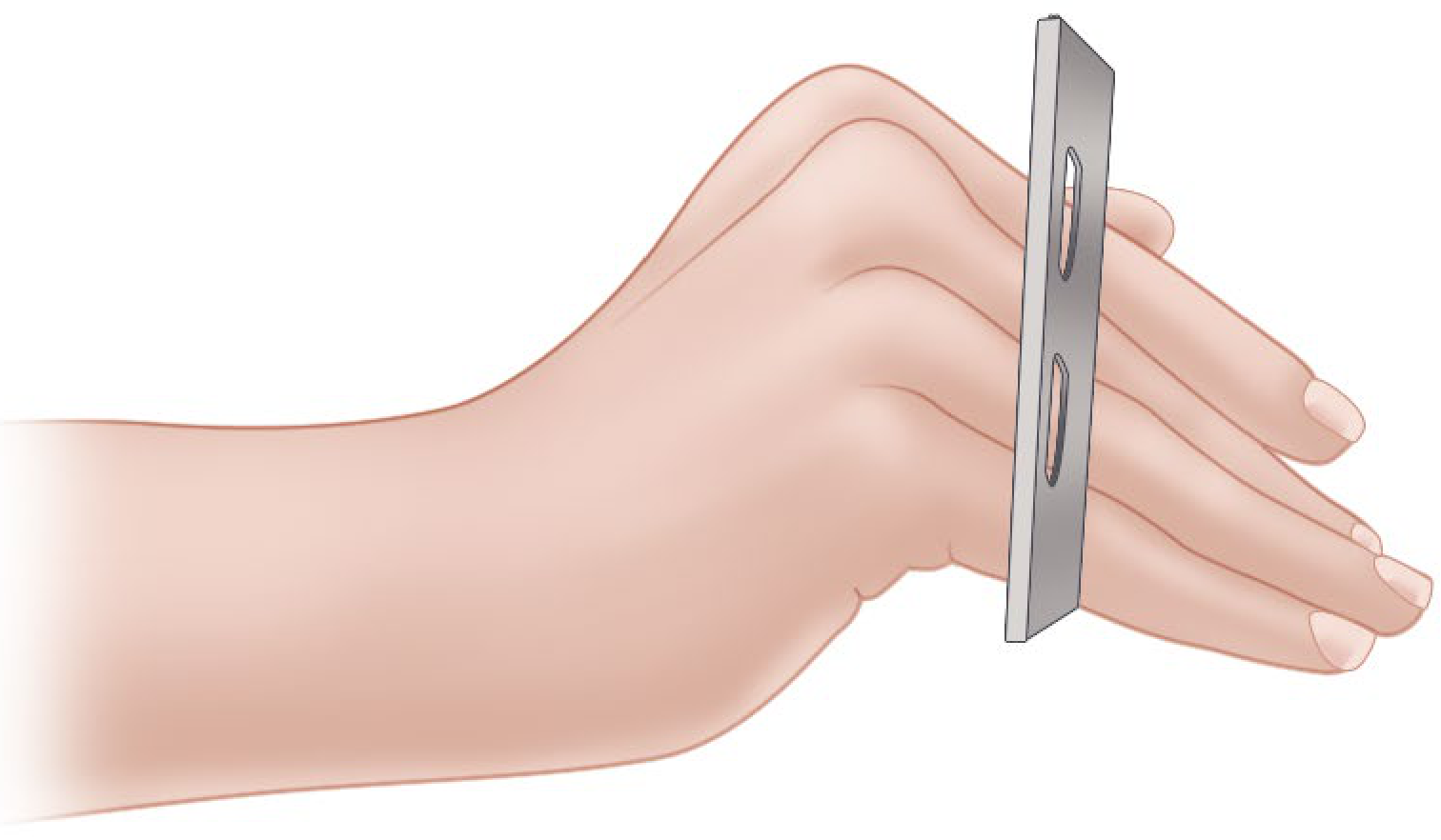
2. Froment's Sign (for adductor pollicis weakness in ulnar palsy)
- Ask patient to hold a flat object between thumb and index finger
- In ulnar palsy: adductor pollicis paralyzed → compensates by flexing thumb IP joint using FPL (median nerve)
- Positive: IP joint flexes during pinch → FPL compensating
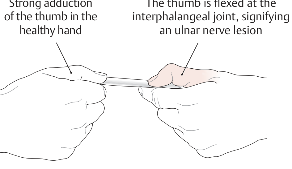
3. Assessment of passive correctability:
- Can you passively flex the MCP and extend the PIP? → Prerequisite for tendon transfer
- If NOT passively correctable → splinting first, or PIP arthrodesis
4. Muscle testing (MRC grading 0-5):
- Test all intrinsic muscles: interossei (abduction/adduction), lumbricals
- Test extrinsic muscles: FDP, FDS, FPL, EPL, EDC
5. Sensory assessment:
- Two-point discrimination, monofilament testing
- Maps distribution (ulnar half of ring + entire little finger = ulnar nerve)
5. PRINCIPLES OF MANAGEMENT
The overall management strategy follows three steps:
Step 1: Treat the cause (if possible)
- Nerve repair/decompression if nerve injury is recent and accessible
- Leprosy treatment (MDT - multidrug therapy)
- Release of entrapment (cubital tunnel release for ulnar nerve)
- If nerve can recover → wait for recovery (up to 18-24 months)
Step 2: Prevent/correct contractures (Splinting)
- Lively/dynamic splints - hold MCP in slight flexion to prevent fixed hyperextension contracture
- Preserve passive range of motion
- Prerequisites before any tendon transfer:
- Passive MCP flexion must be possible
- Passive PIP extension must be possible
- Skin must be healthy, no ulcers
Step 3: Surgical correction (Tendon transfers or static procedures)
6. SURGICAL MANAGEMENT - DETAILED
The Goal of Surgery:
To restore MCP flexion (and PIP/DIP extension) while preserving grip and pinch function.
Surgery is planned based on:
- Bouvier's test result → static vs dynamic
- Available muscles (not paralyzed, grade 4+, expendable)
- Pattern of nerve palsy (ulnar only vs combined)
- Passive correctability of joints
A. STATIC PROCEDURES
(Used when Bouvier's test is POSITIVE - extensor mechanism intact)
1. Zancolli Capsulodesis (Most Important Static Procedure)
Principle: Tighten the volar plate of the MCP joint to create a 10-30° flexion contracture at the MCP → prevents hyperextension → allows long extensors to work on IP joints.
Technique:
- Transverse incision in palm at distal palmar crease
- Expose flexor tendon sheaths at each MCP joint
- Retract flexor tendons → expose MCP volar plate
- Resect an elliptical segment of the volar fibrocartilaginous plate (or advance it proximally)
- Close volar plate with non-absorbable sutures → creates MCP flexion contracture of 10-30°
- K-wires to maintain position if needed
- Dorsal splint with MCP in flexion + wrist in extension
Post-op: Cast for 3 weeks → then MCP exercises
Advantage: Simpler; no donor muscle sacrifice
Limitation: Does NOT restore grip strength; only corrects clawing
2. Fowler Tenodesis
Principle: A tendon graft looped through extensor retinaculum acts as a passive tenodesis - when wrist flexes, tension on graft passively extends IP joints.
- Activated by wrist flexion (passive, not active)
- Not useful in patients with wrist flexion weakness
3. Riordan Tenodesis
Principle: ECRB and ECU tendons cut in half at midforearm (distal insertion intact), split ends passed through lumbrical canals to radial finger.
- NOT activated by wrist motion (unlike Fowler)
4. Bone Block (Mikhail)
- Dorsal bone block at MCP joint prevents hyperextension
- Rarely used
5. MCP Joint Arthrodesis
- For severe, irreversible cases
- Sacrifices motion
B. DYNAMIC PROCEDURES
(Used when Bouvier's test is NEGATIVE - extensor mechanism incompetent)
(Also when grip strength restoration is needed)
These are tendon transfers - redirect an expendable functioning muscle to restore intrinsic function.
The Route of Transfer
All transfers pass through the lumbrical canal (volar to the deep transverse metacarpal ligament) and attach to the radial side of the lateral band of the extensor aponeurosis.
Why radial side? Attaching to the radial lateral band corrects both clawing AND restores MCP flexion with IP extension in the correct direction.
1. Modified Stiles-Bunnell Procedure
Donor: FDS (flexor digitorum superficialis) of ring or middle finger
Route: Through lumbrical canal → radial lateral band of finger
Key: One FDS can be split into four tails for all four fingers
Problem with original Bunnell (all FDS tendons): Too strong → PIP hyperextension = intrinsic-plus deformity (opposite of claw)
Modification: Use only ONE FDS split into 4 tails → reduces this risk
Insertion sites (in order of preference - decreasing intrinsic-plus risk):
- Into bone at base of proximal phalanx (safest)
- Into flexor sheath
- Into lateral band (highest risk of intrinsic-plus)
2. Zancolli Lasso Procedure
Donor: FDS (of the clawed finger itself)
How it works:
- FDS tendon is divided distally
- Passed through a slit in the flexor pulley sheath just distal to the MCP joint
- Looped back and sutured to itself
- Creates a dynamic block - when FDS contracts, it flexes the MCP
- Technically a dynamic transfer because FDS is still a functioning muscle-tendon unit
Advantage: Simple, elegant, preserves the original FDS in its original position
Indication: Very commonly used in low ulnar palsy when FDS is intact
3. Brand Transfer (Paul Brand - Leprosy surgeon)
Donor: ECRB (extensor carpi radialis brevis) extended by plantaris tendon graft
Route: Through interosseous spaces → lumbrical canals → radial lateral band
Two techniques:
- Palmar route (through carpal tunnel): Passes under palmar ligaments
- Dorsal route (through interosseous spaces): More common, safer for median nerve
Advantage: Powerful transfer; restores grip strength well
Disadvantage: Reeducation difficult; may compress median nerve (palmar route)
Useful in leprosy/combined nerve palsies where FDS is unavailable
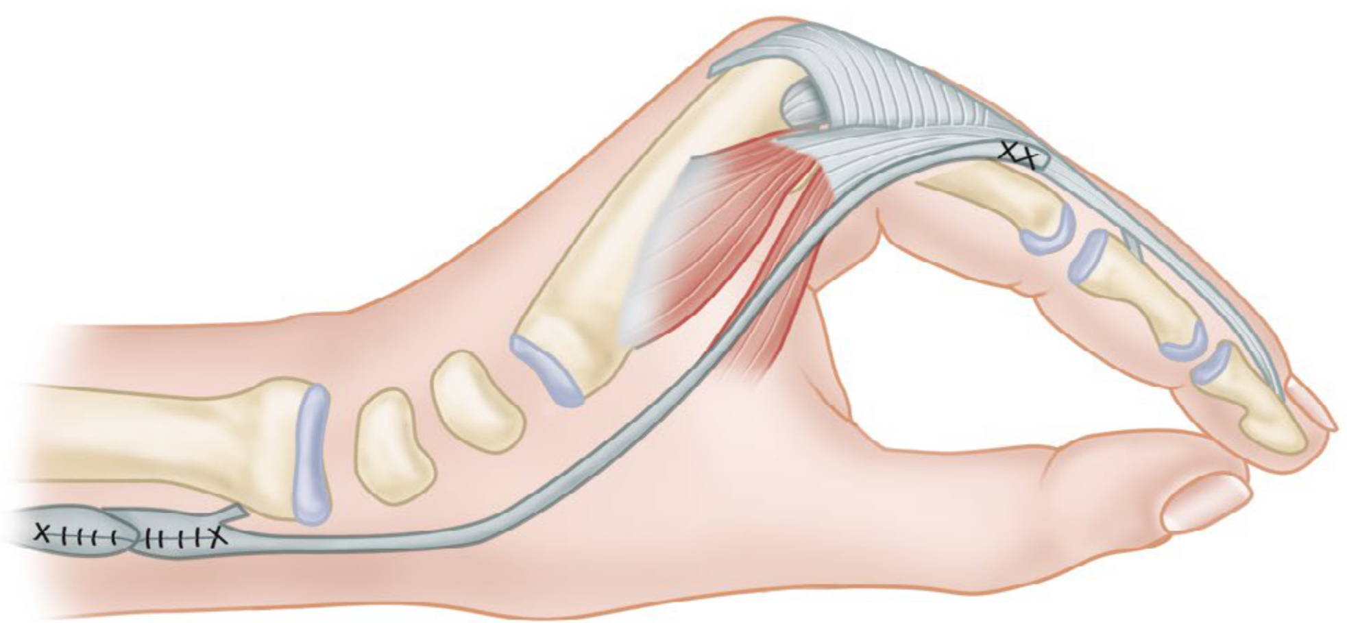
4. Riordan Transfer (FCR-based)
Donor: FCR (flexor carpi radialis) passed radially + extended with tendon graft (plantaris)
Route: Through lumbrical canals → radial lateral band
Note: Useful if patient has strong wrist flexion (which weakens Bunnell/intrinsic transfers)
5. Fowler Transfer (Riordan modification - most commonly tested)
Donor: EIP (extensor indicis proprius) + EDQ (extensor digiti quinti)
Riordan modification: EIP to ring/little fingers; Palmaris longus (extended with graft) to index/middle
Passed: Volar to deep transverse metacarpal ligament → radial lateral band
Advantage: Available even in combined nerve palsies (radial nerve preserved)
C. COMBINED NERVE PALSIES
Low Median + Low Ulnar Palsy (at wrist)
- All four fingers clawed + thumb opposition + adduction lost
- Clawing: Brand transfer (ECRB with graft) - best option
- Opposition: Riordan transfer (FCR-based)
- Thumb adduction: EIP through third intermetacarpal space to adductor insertion
Prerequisites: Must first correct any thumb web contracture and joint contractures.
High Median + High Ulnar Palsy (above elbow)
- FDS, FDP (all fingers), all intrinsics, thenar, hypothenar all paralyzed
- Only radial nerve muscles available for transfer
- Treatment:
- Arthrodesis of thumb MCP joint
- Zancolli capsulodesis of all MCP joints (for clawing)
- ECRL transfer for finger flexion
- ECRB for thumb and finger extension
7. SPECIAL CONSIDERATIONS
Leprosy Claw Hand Management
- Most important message: Prevent the claw from becoming FIXED
- Use dynamic splints early during active nerve inflammation
- Prednisolone for acute leprous neuritis (reduces nerve damage)
- Once deformity is fixed → surgical correction
Surgery in leprosy:
- If disease is inactive and joints are mobile:
- Brand transfer or Riordan transfer
- If skin is anaesthetic: Extra care to avoid pressure ulcers post-surgery
- Thumb: Opponensplasty for opposition loss (median nerve)
Post-Volkmann's Ischemic Contracture
- Claw hand from intrinsic fibrosis
- Treatment:
- Muscle slide (origin release of intrinsic muscles) for early/mild cases
- Tendon lengthening
- Limited goals; contracture releases improve function partially
8. PREREQUISITES FOR TENDON TRANSFER
Before any transfer surgery, all must be present:
- Passive correctability - MCP must flex passively, PIP must extend passively
- Donor muscle grade 4-5 MRC strength (will lose 1 grade after transfer)
- Stable skeletal framework - no untreated fractures
- Good soft tissue cover - no active infection, healing scars
- Patient motivation + compliance for rehabilitation
- Joint contractures corrected first (by splinting or surgery)
- If nerve can still recover - wait at least 18-24 months before tendon transfer
9. SUMMARY TABLE - PROCEDURES AT A GLANCE
| Procedure | Type | Donor | Best for |
|---|---|---|---|
| Zancolli Capsulodesis | Static | Volar plate | Bouvier +ve; simple claw |
| Zancolli Lasso | Dynamic | FDS (same finger) | Low ulnar; FDS intact |
| Modified Stiles-Bunnell | Dynamic | FDS (ring/middle) | All 4 fingers |
| Brand Transfer | Dynamic | ECRB + plantaris graft | Leprosy; combined palsy |
| Riordan Transfer | Dynamic | FCR + graft | Strong wrist flexors |
| Fowler/Riordan modification | Dynamic | EIP + EDQ | Radial nerve only available |
| Fowler Tenodesis | Static | Tendon graft from retinaculum | Passive correction |
10. KEY EXAM POINTS SUMMARY
| Fact | Answer |
|---|---|
| Most common cause of claw hand | Ulnar nerve palsy |
| Which fingers in ulnar palsy | Ring and little (ulnar 2) |
| Why index and middle less clawed | Lumbricals 1 and 2 supplied by median nerve |
| Ulnar paradox | High ulnar palsy = LESS claw (FDP also paralyzed) |
| Most important pre-op test | Bouvier's test |
| Bouvier positive → | Static procedure sufficient (Zancolli capsulodesis) |
| Bouvier negative → | Dynamic procedure needed (tendon transfer) |
| Route of all intrinsic replacements | Volar to deep transverse metacarpal ligament → radial lateral band |
| Simplest static procedure | Zancolli capsulodesis |
| Best transfer when only radial nerve intact | Fowler transfer (EIP + EDQ) |
| Best transfer in leprosy/combined palsy | Brand transfer (ECRB + plantaris graft) |
| Complication of Bunnell transfer | Intrinsic-plus deformity (PIP hyperextension) |
| Sign of adductor pollicis weakness | Froment's sign |
| Dose FDS gives 1 grade loss | Yes - always plan for grade 4+ donor |
| India most common cause of claw | Leprosy (Hansen's disease) affecting ulnar nerve at elbow |
Sources:
- Campbell's Operative Orthopaedics 15th Edition 2026 - Chapter 76 (Tendon transfers, intrinsic reconstruction)
- THIEME Atlas of Anatomy - General Anatomy and Musculoskeletal System - Ulnar nerve lesions
- Miller's Review of Orthopaedics 9th Edition - Hand surgery
In detail about rickets , vitamin D dependent rickets and vitamin D resistant rickets for Orthopaedic postgraduate exam in simple words
Finding Sources
Finding Sources
Reading File
Reading File
Reading File
Reading File
Reading File
Finding Sources
Finding Sources
Finding Sources
Finding Sources
Reading File
Reading File
Reading File
Reading File
Reading File
Finding Sources
Finding Sources
Reading File
Reading File
Finding Sources
I now have comprehensive data from multiple authoritative textbooks. Let me compile the complete orthopaedic PG exam answer.
Rickets, Vitamin D-Dependent Rickets, and Vitamin D-Resistant Rickets
Complete Orthopaedic Postgraduate Exam Guide
SECTION 1: RICKETS - THE FOUNDATION
1.1 Definition
Rickets is a disease of growing children characterized by failure of mineralization of newly formed osteoid (uncalcified bone matrix) at the growth plate and at cortical/cancellous bone. The same process in adults (after epiphyseal fusion) is called osteomalacia.
The core problem: Calcium and/or phosphate are insufficient in the extracellular fluid → osteoid cannot mineralize → soft, weak bones + disrupted growth plate.
1.2 Vitamin D Metabolism - Know This First
The pathway is essential to understand all three types:
SKIN (sunlight on 7-dehydrocholesterol)
↓
Vitamin D3 (cholecalciferol) - inactive
↓ [Liver 25-hydroxylase (CYP2R1)]
25(OH)D - calcidiol - still inactive (measured to assess stores)
↓ [Kidney 1α-hydroxylase (CYP27B1)] ← STIMULATED BY PTH
1,25(OH)2D3 - calcitriol - ACTIVE form (acts on gut, bone, kidney)
↓
Actions:
• ↑ intestinal absorption of Ca and PO4
• ↑ renal reabsorption of Ca and PO4
• ↑ bone mineralization
• ↑ bone resorption (with PTH)
Key points:
- Two hydroxylation steps: Liver (25th position) then Kidney (1st position)
- Active form = 1,25(OH)2D3 = calcitriol = acts on Vitamin D Receptor (VDR)
- FGF23 (Fibroblast Growth Factor 23) - secreted by bone - INHIBITS the 1α-hydroxylase → reduces active vitamin D + promotes phosphate excretion
1.3 Classification of Rickets
| Type | Mechanism |
|---|---|
| 1. Nutritional (Calciopenic) | Vitamin D deficiency / Calcium deficiency / Phosphate deficiency |
| 2. Vitamin D-Dependent Rickets (VDDR) | Defect in vitamin D activation or receptor |
| 3. Vitamin D-Resistant Rickets (VDRR) / Hypophosphatemic | Renal phosphate wasting - unresponsive to normal doses of Vit D |
| 4. Renal Osteodystrophy | Chronic renal failure → poor 1α-hydroxylation |
| 5. Drug-induced | Phenytoin, rifampicin → increase catabolism of Vit D |
SECTION 2: NUTRITIONAL RICKETS (Classic Vitamin D Deficiency)
2.1 Causes
- Inadequate sunlight exposure (most common cause worldwide)
- Dietary deficiency of vitamin D or calcium
- Exclusive breastfeeding beyond 3-6 months (breast milk low in Vit D)
- Prematurity - insufficient stores
- Malabsorption (celiac sprue, Crohn's, fat malabsorption) - Vit D is fat-soluble
- Dark skin in temperate climates - less UV penetration
- Maternal Vit D deficiency (congenital rickets - rare, as Vit D preferentially given to fetus)
- Phytate/oxalate-rich diet (binds calcium in gut) - common cause in India, Africa, Bangladesh
2.2 Pathophysiology - Step by Step
- Low Vit D → ↓ intestinal absorption of Ca and PO4
- ↓ serum Ca → PTH rises (secondary hyperparathyroidism)
- PTH does two things:
- Stimulates osteoclasts → bone resorption to maintain serum Ca
- Stimulates kidney → phosphate excretion increases (phosphaturia)
- Result: serum Ca maintained near normal, but serum PO4 falls sharply
- Both Ca × PO4 product insufficient → osteoid cannot mineralize
- In the growth plate: zone of provisional calcification fails → widened, disorganized physis
- Osteoblasts keep laying down unmineralized osteoid → soft, expandable bone
Key teaching point: In rickets, Ca is near normal (PTH maintains it) but PO4 is low (PTH wastes it). Calcium falls only in late/severe rickets → then tetany occurs.
Tetany in rickets: Ca falls below 7 mg/dL → tetanic spasm → can be life-threatening (laryngospasm)
2.3 Clinical Features - Grouped by System
Skull:
- Craniotabes - softening of skull bones; ping-pong ball consistency on pressing occipital/parietal bones (earliest sign - from 3-6 months)
- Frontal bossing - excess osteoid deposition → prominent forehead
- Delayed closure of fontanelles (anterior fontanelle stays open beyond 18 months)
- Squared head (caput quadratum)
Chest:
- Rachitic rosary (Rickety rosary) - beaded nodularity at costochondral junctions (enlarged ends of ribs)
- Harrison's groove (Harrison's sulcus) - horizontal groove along lower chest margin where diaphragm pulls weakened ribs inward during inspiration
- Pigeon chest (pectus carinatum) - sternum pushed forward
- Barrel chest
Spine:
- Kyphosis or Kyphoscoliosis (especially lumbar region)
- Exaggerated lumbar lordosis in ambulatory children
Long Bones / Limbs:
- Widening of wrists and ankles (expanded metaphyses)
- Genu varum (bow legs) - from medial bowing of tibia/femur - most common in toddlers beginning to walk
- Genu valgum (knock knees) - in older children
- Coxa vara - femoral neck deformity
- Bowing of forearm bones - radius and ulna
- Pathologic fractures (greenstick fractures)
Other:
- Short stature - height below 5th percentile
- Dental abnormalities - delayed teething, enamel defects, dental caries
- Hypotonia and muscle weakness - characteristic; causes "frog posture" in infants
- Waddling gait - from coxa vara + proximal muscle weakness
- Delayed motor milestones (walking, sitting)
- Tetany - carpopedal spasm, Chvostek sign, Trousseau sign (in hypocalcemic form)
- Anemia
2.4 Radiological Features (HIGH YIELD)
Changes are best seen at:
- Wrist (distal radius/ulna) - most useful
- Knee (distal femur / proximal tibia)
- Costochondral junctions
Key radiograph findings:
- Widening of the growth plate (physis) - hallmark sign
- Cupping of the metaphysis - concavity of the metaphyseal margin
- Fraying of the metaphysis - irregular, frayed, "paintbrush" appearance
- Loss of zone of provisional calcification - normally a bright dense line at physis; absent in rickets
- Metaphyseal haziness - blurred, poorly defined metaphysis
- General osteopenia - reduced bone density throughout
- Bowing of long bones - genu varum (medial bowing of tibia/fibula), coxa vara
- Codfishing of vertebrae - biconcave vertebral bodies
- Looser zones (Milkman's fractures / pseudofractures) - more in osteomalacia (adults) - transverse radiolucent bands on compression side of bones; common in femoral neck, pubic rami, scapula
- Delayed bone age - ossification centres appear late
In adults (osteomalacia):
- Decreased cortical thickness
- Relative radiolucency
- Looser zones / pseudofractures - pathognomonic
2.5 Laboratory Findings
| Parameter | Nutritional Rickets |
|---|---|
| Serum Calcium | Normal or slightly ↓ |
| Serum Phosphate | ↓↓ (markedly low) |
| Alkaline Phosphatase (ALP) | ↑↑ (markedly elevated) - most consistent |
| 25(OH)D | ↓↓ |
| 1,25(OH)2D | Normal or ↑ (PTH drives it up) |
| PTH | ↑↑ (secondary hyperparathyroidism) |
| Urine Calcium | ↓ |
| Urine Phosphate | ↑ (PTH effect) |
Key: ALP is the most reliable screening test. 25(OH)D level is the best test to confirm deficiency.
2.6 Treatment of Nutritional Rickets
Medical (First line):
1. Vitamin D supplementation:
- Recommended daily intake: 600 IU (age 1-70 years) / 800 IU (>70 years)
- Deficiency treatment: 50,000 IU weekly for 3-12 weeks → then maintenance 800 IU/day
- In children: 1,000-6,000 IU daily based on weight
Stoss therapy (single high dose):
- Single oral/IM dose of 600,000 IU (or 100,000-300,000 IU)
- Used in non-compliant patients or where daily therapy not feasible
- Results in healing within weeks
2. Calcium supplementation:
- 1.5-2 g/day of elemental calcium
- Essential alongside Vit D (without calcium, Vit D alone gives incomplete response)
Monitoring:
- Normocalcemia within 1 week of therapy
- PTH and ALP normalize in 3-6 months
- Radiographic healing: cupping/fraying begins resolving in 3-6 months
- Most deformities (bowing) resolve spontaneously with medical treatment if treated early
Vitamin D toxicity: Only at doses >40,000 IU/day chronically (safe margin is large)
Surgical (For residual/progressive deformities):
Indications for corrective osteotomy:
- Deformity persists or worsens despite adequate medical treatment
- Active rickets must first be treated medically - never operate on active/uncontrolled rickets (bones too soft, poor healing)
- Significant angular deformity causing functional disability
- Age: generally after 3-4 years old when bone is stronger
Types of osteotomy:
- Valgus osteotomy of distal femur and/or proximal tibia - for genu varum
- Stapling / guided growth (hemiepiphysiodesis) - for ongoing deformity correction in growing children
- Dome osteotomy / closed wedge osteotomy - for severe cases
- Fixation: K-wires in plaster, or internal fixation depending on age/severity
SECTION 3: VITAMIN D-DEPENDENT RICKETS (VDDR)
These are genetic disorders where vitamin D intake and serum levels may be normal, but there is a defect in either activation of vitamin D or in the receptor.
Think of it this way: Plenty of Vit D is available but the factory can't process it (Type I) or the target organ doesn't respond (Type II).
3.1 VDDR Type I (Pseudovitamin D-Deficiency Rickets)
Also called: 1α-hydroxylase deficiency / Pseudo-Vit D resistant rickets
Gene: CYP27B1 (encodes renal 25(OH)D 1α-hydroxylase)
Inheritance: Autosomal recessive (AR) - gene on chromosome 12q14
Defect: Renal 1α-hydroxylase enzyme is absent/deficient → cannot convert 25(OH)D to active 1,25(OH)2D
Biochemistry:
| Parameter | VDDR Type I |
|---|---|
| Calcium | ↓ |
| Phosphate | ↓ |
| 25(OH)D | Normal |
| 1,25(OH)2D | ↓↓ (very low) - KEY |
| PTH | ↑ |
| ALP | ↑ |
Key distinguishing feature: 25(OH)D is NORMAL (diet/sunlight fine) but 1,25(OH)2D is LOW (can't activate it)
Clinical Features:
- Presents in children under 2 years (earlier/more severe than nutritional)
- Severe bony changes of rickets
- Hypocalcemic tetany and seizures (characteristic)
- Muscle hypotonia
- Radiological features identical to nutritional rickets
Treatment:
- Calcitriol (1,25(OH)2D = the active hormone) - bypasses the deficient enzyme
- Dose: 0.25-1 μg/day (physiologic doses sufficient - this is what distinguishes it from Type II)
- OR Alfacalcidol (1α-hydroxyvitamin D) - also bypasses the kidney step
- Calcium supplementation (1.5-2 g/day)
- Responds well to physiologic doses of calcitriol - excellent prognosis with treatment
3.2 VDDR Type II (Hereditary Vitamin D-Resistant Rickets)
Also called: Vitamin D Receptor (VDR) mutation / True VDDR
Gene: VDR gene (encodes the Vitamin D Receptor protein)
Inheritance: Autosomal recessive (AR)
Defect: VDR is non-functional → even abundant 1,25(OH)2D cannot act on target organs (gut, bone, kidney) → complete end-organ resistance
Biochemistry:
| Parameter | VDDR Type II |
|---|---|
| Calcium | ↓ |
| Phosphate | ↓ |
| 25(OH)D | Normal |
| 1,25(OH)2D | ↑↑↑ (very HIGH) - KEY |
| PTH | ↑ |
| ALP | ↑ |
Key distinguishing feature: 1,25(OH)2D is massively ELEVATED (body produces lots of active vitamin D but organs can't respond to it → feedback accumulates)
Clinical Features:
- Presents in infants under 1 year (earlier and more severe than Type I)
- Similar bony changes to nutritional rickets
- Alopecia (partial or total) - pathognomonic feature! Hair follicles have VDR; without functioning VDR → alopecia
- Poor dentition
- Hypocalcemic tetany and seizures
- Does NOT respond to normal or even high doses of vitamin D
Treatment:
- Very difficult to treat due to end-organ resistance
- Long-term nocturnal calcium infusions (IV calcium drip overnight) - normalizes PTH → reduces urinary phosphate → improves rickets and growth
- High-dose calcitriol (supraphysiologic) - may partially help mild cases
- Hair growth does NOT restore with treatment (alopecia is permanent)
- Outcome: improved skeletal disease but not alopecia
3.3 VDDR Type III (Rare)
- Mutation in CYP3A4 gene → accelerated catabolism of Vit D metabolites
- Both 25(OH)D and 1,25(OH)2D are low
- Similar to phenytoin-induced Vit D deficiency
SECTION 4: VITAMIN D-RESISTANT RICKETS (VDRR) / HYPOPHOSPHATEMIC RICKETS
This is a completely different mechanism from VDDR. Here, the problem is renal phosphate wasting, not Vitamin D pathway.
Think of it as "phosphate diabetes" - the kidney spills phosphate into urine like the diabetic kidney spills glucose.
4.1 Pathophysiology - The FGF23 Story
FGF23 (Fibroblast Growth Factor 23):
- Produced by osteocytes and osteoblasts in bone
- Functions:
- Inhibits renal tubular phosphate reabsorption → phosphaturia → hypophosphatemia
- Inhibits renal 1α-hydroxylase → reduces 1,25(OH)2D production
In X-linked hypophosphatemic rickets:
- PHEX gene mutation (phosphate-regulating endopeptidase on X chromosome) → PHEX normally degrades FGF23 → without PHEX, FGF23 accumulates → extreme phosphaturia + suppressed Vit D activation
The cascade:
PHEX mutation → ↑ FGF23 → ↑ renal PO4 excretion → ↓ serum PO4
→ ↓ 1α-hydroxylase → ↓ 1,25(OH)2D → ↓ gut PO4 absorption
Result: SEVERE hypophosphatemia + INAPPROPRIATELY LOW 1,25(OH)2D (should rise to compensate, but doesn't)
4.2 Types of Hypophosphatemic Rickets
| Type | Genetics | Gene | Mechanism |
|---|---|---|---|
| X-linked hypophosphatemic rickets (XLH) - Most common | X-linked dominant | PHEX | Loss of PHEX → ↑ FGF23 |
| Autosomal dominant (ADHR) | AD | FGF23 (mutation renders FGF23 resistant to cleavage) | FGF23 accumulates |
| Autosomal recessive (ARHR) | AR | DMP1 (dentine matrix protein) | ↑ FGF23 |
| Hereditary hypophosphatemic with hypercalciuria | AR | NaPi2c renal transporter | No FGF23 elevation |
| Oncogenic osteomalacia (acquired) | - | Tumor secreting FGF23 | ↑ FGF23 from tumor |
| Fanconi syndrome | Various | Multiple tubular defects | PO4 + glucose + amino acid + bicarb loss |
4.3 X-linked Hypophosphatemic Rickets (XLH) - The Classic Form
Inheritance: X-linked dominant (both males and females affected; males more severely)
Gene: PHEX on X chromosome
Most common hereditary rickets and most common heritable form overall
Biochemistry:
| Parameter | XLH / VDRR |
|---|---|
| Calcium | Normal |
| Phosphate | ↓↓ (very low) |
| 25(OH)D | Normal |
| 1,25(OH)2D | ↓ or low-normal (inappropriately low) |
| PTH | Normal |
| ALP | ↑ |
| Urine Phosphate | ↑↑ (phosphaturia) |
Critical distinguishing features vs nutritional rickets:
- Ca is NORMAL (not low) - no secondary hyperparathyroidism
- PTH is NORMAL (no hypocalcemia to stimulate it)
- 25(OH)D is NORMAL (sunlight exposure/diet fine)
- Phosphaturia - key finding
Clinical Features:
- Usually presents after child begins to walk (1-2 years)
- Severe bowing deformities - more pronounced than nutritional rickets
- Short stature - disproportionate, lower limbs especially shortened
- Dental abnormalities - abscess, enamel defects, pulp anomalies
- Generally no tetany (Ca is normal)
- Muscle weakness less prominent than nutritional rickets
- May have craniosynostosis
- Rachitic bone changes on X-ray
- Does NOT respond to normal doses of vitamin D - hence "resistant"
Treatment:
Traditional treatment (Second-line now):
- Oral phosphate supplementation (elemental phosphate 1-2 g/day in divided doses, 4-5 times/day)
- Multiple daily doses required (phosphate poorly retained)
- Side effects: diarrhea, secondary hyperparathyroidism
- Calcitriol (0.5-1 μg/day for adults; 0.025-0.05 μg/kg/day in children)
- To counteract the FGF23-induced suppression of 1,25(OH)2D
- Monitor for hypercalcemia and nephrocalcinosis
New treatment - First line (Gold standard now):
- Burosumab (Crysvita) - anti-FGF23 monoclonal antibody (IgG1)
- Binds and neutralizes FGF23 → restores phosphate reabsorption + normalizes 1,25(OH)2D
- FDA approved for children (≥1 year) and adults with XLH
- Dose in children: 0.8-2 mg/kg every 2 weeks subcutaneously
- Dramatically superior to phosphate + calcitriol for growth, rickets healing, bowing
- Monitor: serum phosphate, alkaline phosphatase, 1,25(OH)2D
Surgical treatment:
- Corrective osteotomy for residual bowing after medical control
- Only after disease is biochemically controlled
- Valgus osteotomy of distal femur + proximal tibia
- External fixator or internal fixation
- High recurrence rate if medical treatment not maintained
SECTION 5: COMPARISON TABLE - THE KEY EXAM DIFFERENTIATOR
| Feature | Nutritional Rickets | VDDR Type I | VDDR Type II | VDRR (XLH) |
|---|---|---|---|---|
| Defect | Vit D deficiency | 1α-hydroxylase absent | VDR mutation | PHEX mutation → ↑ FGF23 |
| Inheritance | None (dietary) | AR | AR | X-linked dominant |
| Calcium | ↓ or normal | ↓ | ↓ | Normal |
| Phosphate | ↓ | ↓ | ↓ | ↓↓ (very low) |
| 25(OH)D | ↓↓ | Normal | Normal | Normal |
| 1,25(OH)2D | Normal or ↑ | ↓↓ | ↑↑↑ | ↓ or low-normal |
| PTH | ↑ | ↑ | ↑ | Normal |
| ALP | ↑ | ↑ | ↑ | ↑ |
| Urine PO4 | ↑ (PTH effect) | ↑ | ↑ | ↑↑ (primary phosphaturia) |
| Tetany | Yes (late) | Yes (early, severe) | Yes (severe) | No |
| Alopecia | No | No | Yes - pathognomonic | No |
| Response to Vit D | Excellent | Good (calcitriol) | Poor/none | None (resistant) |
| Treatment | Vit D + Ca | Calcitriol (physiologic dose) | IV Ca infusions | Burosumab / PO4 + calcitriol |
| Key lab clue | Low 25(OH)D | Low 1,25(OH)2D with normal 25(OH)D | Very HIGH 1,25(OH)2D | Normal Ca, low PO4, normal PTH |
SECTION 6: OTHER RELATED CONDITIONS
Renal Osteodystrophy
- Chronic renal failure → impaired 1α-hydroxylation → ↓ 1,25(OH)2D
- PLUS: phosphate retention → hypocalcemia → secondary hyperparathyroidism
- Combined features of rickets/osteomalacia + osteitis fibrosa cystica (from PTH)
- Radiograph: rickets features + subperiosteal resorption + "salt and pepper skull"
- Treatment: calcitriol + phosphate binders + calcium
Oncogenic Osteomalacia (Tumor-Induced)
- Rare mesenchymal tumors secrete FGF23 → identical to XLH
- Phosphatonin-mediated
- Treatment: find and remove the tumor → cure; if not found: phosphate + calcitriol or burosumab
Drug-Induced Rickets
- Phenytoin, rifampicin → induce CYP enzymes → accelerate catabolism of Vit D metabolites → functional deficiency
- Treatment: higher doses of Vit D supplementation
SECTION 7: ORTHOPAEDIC SURGICAL MANAGEMENT OF DEFORMITIES
When to Operate
- Deformity persists after 1-2 years of adequate medical treatment
- Deformity is progressive or severe causing disability
- Active rickets MUST be controlled first - biochemically quiescent disease
- Child old enough for surgery (typically ≥4 years)
Types of Deformity Correction
| Deformity | Surgical Option |
|---|---|
| Genu varum (bow legs) | Medial tibial/femoral osteotomy (valgus osteotomy) |
| Genu valgum (knock knees) | Lateral femoral/tibial osteotomy or hemiepiphysiodesis |
| Coxa vara | Subtrochanteric valgus osteotomy |
| Tibial torsion | Tibial derotation osteotomy |
Post-operative Care
- Immobilization in plaster/cast
- Continue medical treatment throughout (phosphate + calcitriol for VDRR)
- Monitor healing carefully - may be delayed if disease reactivates
- Recurrence is high in VDRR if medical treatment not maintained
SECTION 8: EXAM SUMMARY - QUICK REVISION TABLE
| Fact | Answer |
|---|---|
| Rickets is a disease of | Growing children (failure of mineralization at growth plate) |
| Same process in adults | Osteomalacia |
| First hydroxylation of Vit D | Liver (25-position) → 25(OH)D |
| Second hydroxylation | Kidney (1α-position) → 1,25(OH)2D = calcitriol (active) |
| Why PO4 falls more than Ca in rickets | PTH maintains Ca by bone resorption + increases urinary PO4 loss |
| Earliest sign of rickets | Craniotabes |
| Rachitic rosary | Enlarged costochondral junctions |
| Harrison's groove | Horizontal groove along lower chest from diaphragm pull |
| Best screening lab for Vit D status | Serum 25(OH)D level |
| Most consistent lab abnormality | Elevated alkaline phosphatase |
| Key radiograph finding | Cupping + fraying of metaphysis + widened physis |
| Looser's zones = | Pseudofractures in osteomalacia (compression side of bones) |
| VDDR Type I defect | 1α-hydroxylase deficiency (CYP27B1 gene mutation) |
| VDDR Type II defect | VDR mutation (end-organ resistance) |
| VDDR Type II pathognomonic feature | Alopecia |
| VDDR Type II 1,25(OH)2D level | ↑↑↑ (very high) |
| VDRR most common form | X-linked hypophosphatemic (XLH) |
| VDRR gene | PHEX (X chromosome) |
| VDRR mechanism | ↑ FGF23 → phosphaturia + ↓ 1,25(OH)2D |
| Calcium in VDRR | NORMAL (key differentiator) |
| PTH in VDRR | Normal |
| New drug for XLH | Burosumab (anti-FGF23 antibody) |
| Traditional treatment XLH | Oral phosphate 4-5x/day + calcitriol |
| Vitamin D deficiency treatment | 50,000 IU weekly × 3-12 weeks → 800 IU/day maintenance |
| Never operate on | Active rickets - must control medically first |
| Recurrence of deformity after surgery in VDRR | High - if medical treatment stopped |
Sources:
- Harrison's Principles of Internal Medicine 22E (2025, McGraw Hill) - Vit D metabolism, VDDR types
- Grainger & Allison's Diagnostic Radiology - X-ray features
- Guyton & Hall Textbook of Medical Physiology - Pathophysiology
- Miller's Review of Orthopaedics 9th Edition - Classification and orthopaedic management
- Robbins & Kumar Basic Pathology - Morphology and histology
- Rheumatology 2-Volume Set (Elsevier, 2022) - Biochemistry comparison table
- Rockwood & Green's Fractures in Adults 10th Ed (2025) - Causes and mechanisms
In detail about soft tissue flaps for Orthopaedic postgraduate exam in simple words don't miss any points
Finding Sources
Finding Sources
Reading File
Reading File
Reading File
Reading File
Reading File
Finding Sources
Finding Sources
Reading File
Reading File
Loading Image
Reading File
Finding Sources
Reading File
Finding Sources
Loading Image
Reading File
Reading File
I now have comprehensive data. Let me compile the complete detailed answer.
Soft Tissue Flaps - Complete Orthopaedic Postgraduate Exam Guide
1. DEFINITION
A flap is a block of tissue that is transferred from one location (donor site) to another (recipient site) while maintaining its own blood supply. This is the fundamental difference from a graft, which has no intrinsic blood supply and relies entirely on the recipient bed for nutrition.
Graft vs Flap:
| Feature | Graft | Flap |
|---|---|---|
| Blood supply | None - depends on recipient bed | Brings its own blood supply |
| Indication | Healthy vascular recipient bed | Exposed bone, tendon, hardware, poor vascularity |
| Complexity | Simpler | More complex |
| Coverage ability | Limited | Extensive |
2. THE RECONSTRUCTIVE LADDER
The reconstructive ladder is the surgeon's decision-making framework - always consider simpler options first, escalate to more complex when needed.
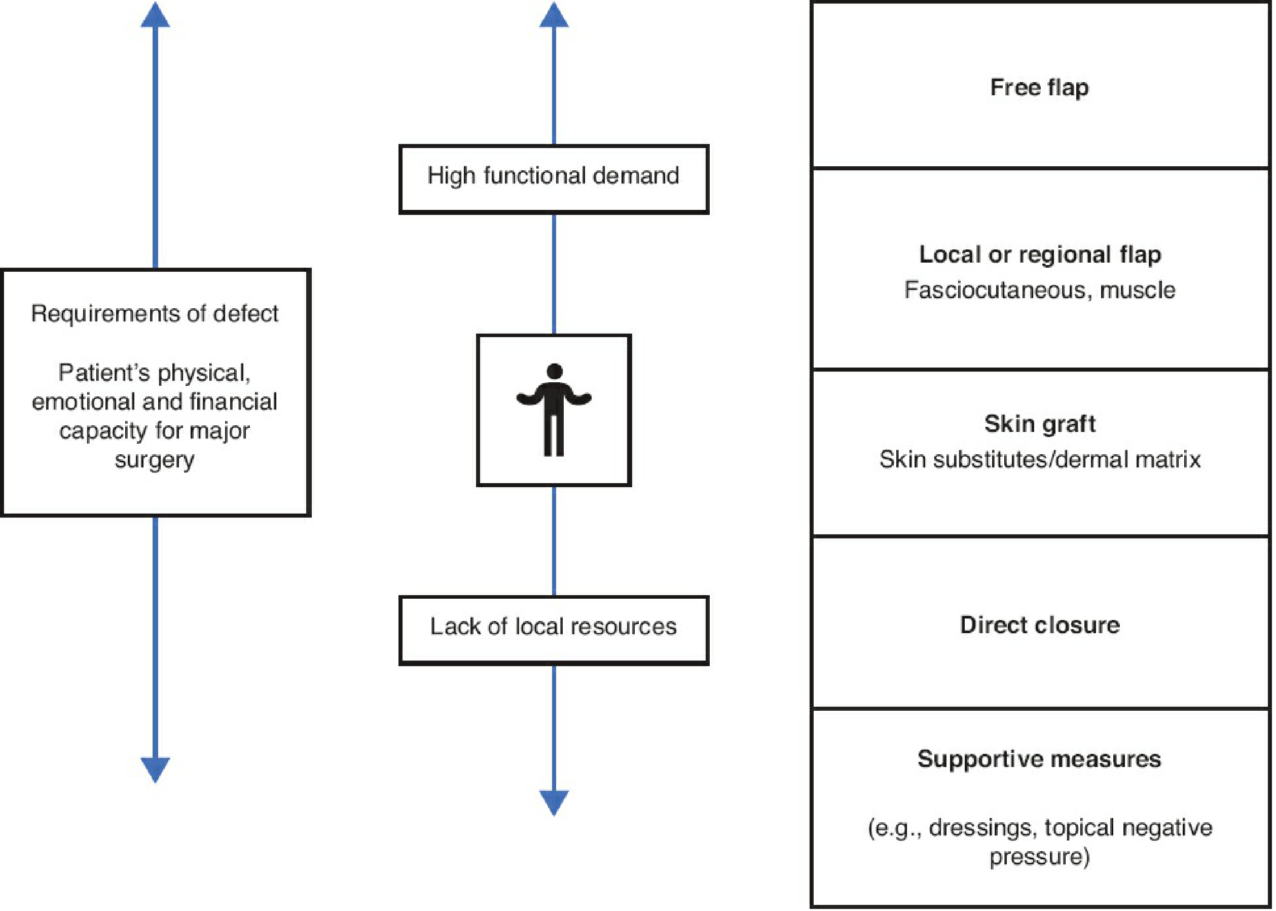
Rungs (bottom to top - simple to complex):
- Supportive measures - dressings, negative pressure wound therapy (NPWT/VAC)
- Direct/Primary closure - simplest; tension-free approximation
- Secondary intention healing - granulation, small wounds
- Skin graft - STSG or FTSG; needs vascular recipient bed
- Local or regional flap - fasciocutaneous, muscle (brings blood supply)
- Free flap - microsurgical transfer for complex/large defects
Key principle: Choose based on defect requirements + patient's physical, emotional, and financial capacity. The best option is not always the simplest - sometimes a free flap is more appropriate than multiple local procedures.
3. INDICATIONS FOR FLAP COVERAGE
Flap coverage is required when a skin graft is NOT suitable - i.e., the recipient bed cannot support graft take:
- Exposed bone stripped of periosteum
- Exposed tendon stripped of paratenon
- Exposed joint/articular cartilage
- Exposed orthopaedic implant (nail, plate, external fixator pin tract)
- Exposed nerve or vessel
- Irradiated tissue (poor vascularity)
- Infected wounds where a vascularized flap adds defense
- Large defects requiring bulk/dead space filling
- Sensate coverage needed (e.g., weight-bearing areas of sole)
4. CLASSIFICATION OF FLAPS
Flaps can be classified in four ways:
A. By Blood Supply (Most Important Classification)
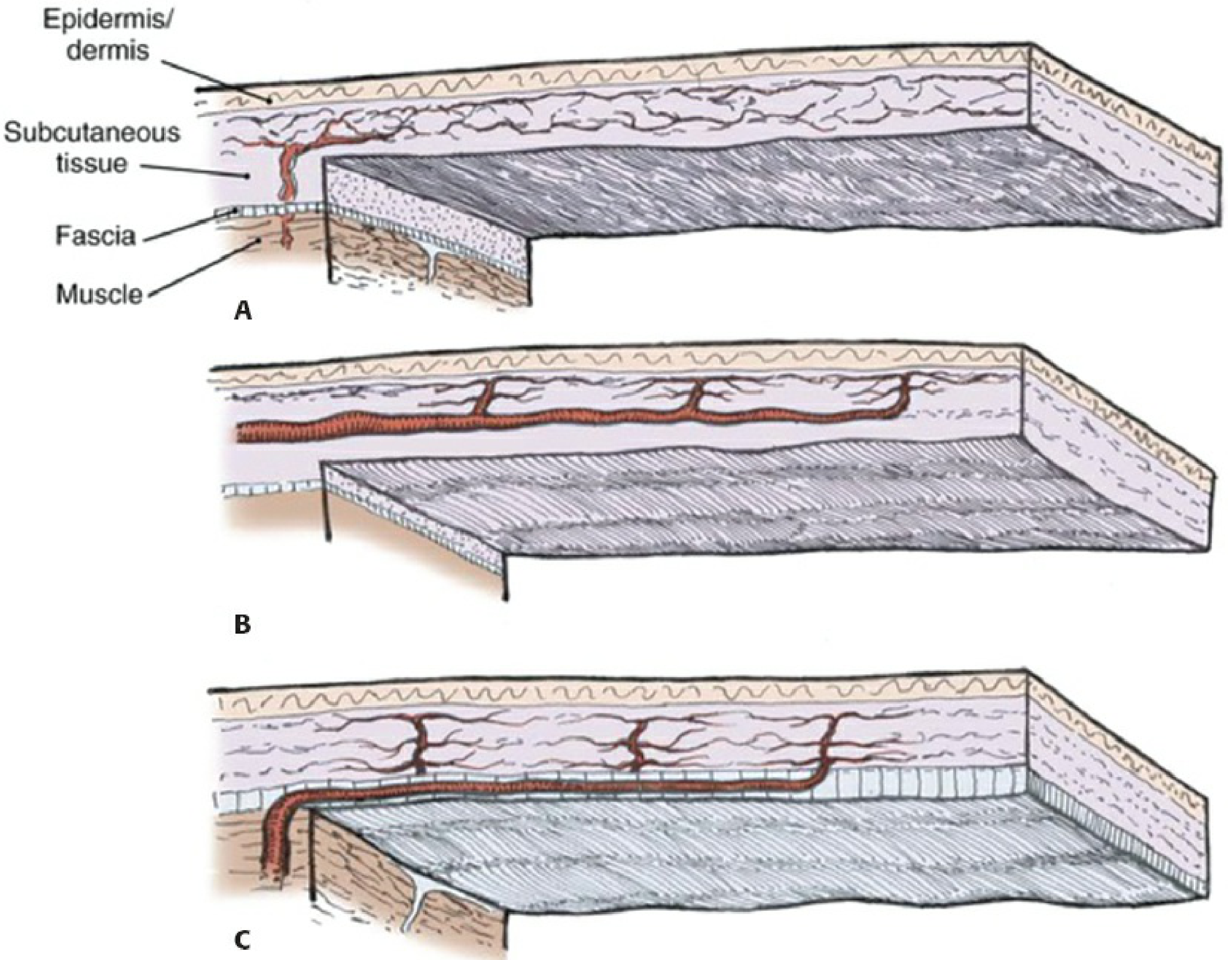
1. Random Pattern Flap
- No single named artery - relies on dermal and subdermal plexus (microcirculation)
- Blood supply unpredictable
- Length:breadth ratio must be respected:
- Upper limb/face: up to 3:1
- Lower limb: 1:1 or 2:1 (less rich blood supply)
- Examples: rotation flap, advancement flap, transposition flap
- Simple, widely available, no need to identify named vessels
2. Axial Pattern Flap
- Contains a single named arteriovenous pedicle running along its axis
- More predictable and reliable blood supply
- Greater resistance to infection
- Can have greater length:breadth ratio than random
- Can be pedicled (vessels kept intact) or used as free flap (vessels divided and reanastomosed)
- Examples: radial forearm flap (radial artery), latissimus dorsi flap (thoracodorsal artery), groin flap (superficial circumflex iliac artery)
3. Perforator Flap (Modern concept)
- Based on perforating vessels that emerge from deep vessels through muscle/fascia to skin
- Allows harvest of skin and fat alone without sacrificing the underlying muscle
- Lower donor site morbidity
- Requires Doppler marking of perforators preoperatively
- Examples: DIEAP (deep inferior epigastric artery perforator), ALT (anterolateral thigh), propeller flap
- Common perforator flaps: DIEAP, ALT, thoracodorsal artery perforator (TDAP), superior/inferior gluteal artery perforator (SGAP/IGAP)
B. By Tissue Composition (Tissue Type)
| Type | Components | Examples |
|---|---|---|
| Cutaneous | Skin + subcutaneous tissue only | Thenar flap, cross-finger flap |
| Fasciocutaneous | Fascia + overlying skin + subcutaneous | Radial forearm flap, lateral arm flap, ALT |
| Muscular | Muscle only | Gastrocnemius, soleus (covered with STSG) |
| Musculocutaneous (Myocutaneous) | Muscle + overlying skin, subcutaneous, fascia | Gracilis, TRAM flap |
| Osseous | Bone only | - |
| Osteocutaneous | Bone + overlying soft tissue | Fibular free flap, iliac crest flap |
| Fascial | Deep fascia only | Temporal fascia flap |
| Innervated (Sensate) | Nerve preserved with tissue | Sural artery flap with sural nerve |
C. By Donor Site / Location of Transfer
1. Local Flap
- Tissue immediately adjacent to defect
- Requires that adjacent tissue is healthy (outside zone of injury)
- Subtypes by movement pattern (see Section 5)
- Examples: rotation flap, advancement flap, Z-plasty
2. Regional Flap
- Tissue from same anatomical region but not immediately adjacent
- Pedicle remains in continuity
- Examples: gastrocnemius for proximal tibia, pectoralis major for chest wall
3. Distant Flap (Pedicled)
- Tissue from a different anatomical region
- Pedicle bridges across - staged procedure usually needed
- Examples: groin flap for hand (degloved hand placed in abdominal pocket), cross-leg flap
- Disadvantages: requires multiple stages, immobilization
4. Free Flap (Free Tissue Transfer)
- Tissue completely divided from donor site including its vessels
- Vessels anastomosed to recipient vessels near defect (microsurgery)
- Single-stage procedure
- Most versatile - can reach any site
- Requires microvascular expertise
D. By Method of Transfer (Movement)
1. Advancement Flap
- Tissue moved directly forward in a straight line to fill the defect
- No rotation
- Types: V-Y advancement, Moberg flap (for fingertip)
- V-Y: triangular incision → advanced → closed as Y shape
2. Rotation Flap
- Semicircular flap rotated into defect around a pivot point
- Random pattern in blood supply
- Rule: rotated flap should be no longer than width of its base
- Back-cut increases arc of rotation but reduces blood supply to base
3. Transposition Flap
- Flap transposed over an intervening bridge of tissue into adjacent defect
- Can be geometric (rhomboid/Limberg flap) or non-geometric
- May be axial or random
- Examples: Z-plasty, rhomboid flap
4. Propeller Flap (Modern)
- Islanded on a perforator pedicle, rotated up to 180° around the perforator axis
- Like a propeller spinning around its axis
- Used for pretibial/medial leg defects
- Identified with hand-held Doppler preoperatively
- Advantages: minimal donor morbidity, single stage, no need for microsurgery
5. Interpolation Flap
- Flap raised from nearby but non-adjacent area, passed over intact skin to reach defect
- Pedicle divided at second stage (3 weeks later)
- Example: cross-finger flap, forehead flap
6. Tube/Tubed Pedicle Flap
- Flap rolled into a tube to protect the pedicle
- Historical technique; rarely used today
- Example: Italian flap (Tagliacozzi)
5. Z-PLASTY - THE CLASSIC LOCAL FLAP
Definition: Two triangular transposition flaps created by a Z-shaped incision to lengthen a scar or reorient a scar in a new direction.
Principle: Exchange the two triangular flaps → scar realigns parallel to relaxed skin tension lines (RSTLs) + gain in length
Z-plasty angle vs lengthening:
| Angle of Z-plasty | % Lengthening |
|---|---|
| 30° | 25% |
| 45° | 50% |
| 60° | 75% (most commonly used) |
| 75° | 100% (rarely, poor vascularity) |
Rule: Both limbs are always equal length. Only the angle changes.
Indications:
- Release of scar contracture (web space contractures, burns, flexor surface scars)
- Reorientation of scar into RSTL
- Correction of skin web deformities
- Release of trapdoor deformities
6. MATHES AND NAHAI CLASSIFICATION OF MUSCLES (Vascular Anatomy of Muscles)
This classifies muscles by their vascular supply pattern - critical for planning muscle flaps.
| Type | Blood Supply | Examples | Clinical significance |
|---|---|---|---|
| I | One vascular pedicle | Gastrocnemius, Tensor fasciae latae (TFL) | Safe elevation on single pedicle |
| II | Dominant pedicle + minor pedicle(s) | Gracilis, Soleus, Biceps femoris | Can use either end; hemisoleus possible |
| III | Two dominant pedicles | Rectus abdominis, Gluteus maximus, Serratus anterior | Can be used from either end |
| IV | Multiple segmental pedicles | Tibialis anterior, Sartorius, External oblique | Each pedicle small; can only mobilize short distances |
| V | One dominant + secondary segmental pedicles | Latissimus dorsi, Pectoralis major | Most versatile; dominant pedicle supports whole muscle |
Key for exams:
- Type I and II = most reliable for free flap transfer
- Type IV = unreliable as free flap; too risky
- Type V (latissimus dorsi) = the workhorse free flap
7. SPECIFIC FLAPS - TIBIAL FRACTURE COVERAGE (MOST EXAM-RELEVANT)
The principle for tibial fracture coverage by level:
"Thirds Rule" for tibial coverage:
- Proximal third → Gastrocnemius flap
- Middle third → Soleus flap
- Distal third → Free flap (no reliable local muscle) OR fasciocutaneous flap OR propeller flap
A. Gastrocnemius Flap (Proximal Third)
Type: Mathes-Nahai Type I (one vascular pedicle)
Pedicle: Medial or lateral sural artery (branch of popliteal artery)
Parts: Medial head (larger, used more often) + Lateral head
Coverage: Proximal tibia + knee area (cannot reach below middle third)
Technique:
- Posterior midline incision
- Separate gastrocnemius from soleus
- Preserve sural nerve and great saphenous vein
- Pes anserinus may restrict rotation - can be released
- Muscle surface covered with split-thickness skin graft (STSG) Donor site: Primary closure of posterior calf Limitation: Cannot reach knee joint above joint line; cannot cover distal leg
B. Soleus Flap (Middle Third)
Type: Mathes-Nahai Type II (dominant + minor pedicles)
- Proximal: branches from popliteal artery
- Medial: posterior tibial artery branches
- Lateral: peroneal artery branches Unique feature: Can be used as a hemisoleus flap (split longitudinally) to cover different areas Use: Proximally based for middle third; distally based for distal third Technique: More complex dissection than gastrocnemius Limitation: Limited arc of rotation; within zone of injury for high-energy tibial fractures Donor site: Partial sacrifice acceptable (other head remains); STSG needed on muscle
C. Fasciocutaneous Flaps (All Thirds, Especially Middle and Distal)
Medial fasciocutaneous flap:
- Based on medial perforators from posterior tibial artery
- Covers medial tibial defects
- Risk: great saphenous vein and saphenous nerve must be spared
- Secondary defect requires STSG
Lateral fasciocutaneous flap:
- Based on peroneal artery perforators in lateral compartment
- Less frequently used - reduced rotational reach
- Risk to superficial peroneal nerve
Propeller flap (perforator-based):
- Island of skin rotated 180° on a single perforator (identified by Doppler)
- Used for pretibial/medial leg defects including distal third
- Modern workhorse for many leg defects
- No muscle sacrifice; single stage
Ponten's "Super Flap" (1981):
- Historical landmark: first described that including deep fascia allows longer, thinner, more reliable fasciocutaneous flaps in the lower extremity
- This observation led to the development of fasciocutaneous flap concept
8. FREE FLAPS - KEY WORKHORSE FLAPS
| Flap | Composition | Pedicle | Coverage area | Notes |
|---|---|---|---|---|
| Anterolateral Thigh (ALT) | Fasciocutaneous | Descending branch of lateral circumflex femoral | 25 × 35 cm | Workhorse; thin; synchronous harvest; low morbidity |
| Latissimus Dorsi (LD) | Musculocutaneous | Thoracodorsal artery (Type V) | 20 × 40 cm | Large coverage; fills dead space; long pedicle; seroma common |
| Gracilis | Muscular | Medial femoral circumflex (MFCA) | 6 × 20 cm | Thin; synchronous harvest; short pedicle; can be reinnervated; "Plan B" flap |
| Rectus Abdominis (TRAM/DIEAP) | Musculocutaneous/Perforator | DIEA (Type III) | Large | Breast reconstruction; can thin to perforator (DIEAP reduces donor morbidity) |
| Radial Forearm | Fasciocutaneous | Radial artery | 10-15 × 25 cm | Thin; pliable; long pedicle; sacrifice of radial artery; used for hand/head-neck |
| Fibula | Osteocutaneous | Peroneal artery | Bone + skin | Long bone defect reconstruction; mandible; up to 25 cm bone |
| Scapular | Fasciocutaneous (± bone) | Circumflex scapular | Large | Can include scapular bone; good for composite defects |
| Serratus Anterior (SA) | Muscular | Serratus branch of subscapular | Medium | Chimeric with LD; risk of scapular winging |
| Iliac Crest | Osteocutaneous | DCIA (deep circumflex iliac) | Bone + skin | Mandible, femur defects; significant donor morbidity |
ALT flap - current first choice for head-neck reconstruction and large lower limb defects
LD flap - most reliable; largest coverage; Type V muscle (best pedicle)
Gracilis - preferred for functioning muscle transfer (nerve included for reinnervation)
Fibula free flap - gold standard for long bone defects and mandible reconstruction
9. MUSCLE FLAP vs FASCIOCUTANEOUS FLAP - COMPARISON
| Feature | Muscle Flap | Fasciocutaneous Flap |
|---|---|---|
| Dead space | Fills better | Less effective |
| Infection | Better vascular defense | Slightly less |
| Bone healing | Theoretically better (evidence equivocal) | Similar in practice |
| Skin cover | Needs additional STSG | Immediate skin-to-skin closure |
| Monitoring | Difficult | Easier (skin visible) |
| Donor morbidity | Greater | Lower |
| Re-elevation for secondary surgery | Very difficult | Easier |
| Post-op shrinkage | More (atrophies with denervation) | Less |
| Scar quality | Poor (STSG over muscle) | Better |
| Cosmesis | Bulky initially, atrophies | Better final contour |
10. TIMING OF FLAP COVERAGE - CRITICAL FOR ORTHOPAEDICS
For open tibial fractures:
- Ideal: within 72 hours (before bacterial colonization becomes established)
- Definitive coverage within 7 days: significantly reduces infection rates
- Delay >7 days → higher risk of infection and flap failure
- "Fix and flap": bone fixation and soft tissue coverage at same operative setting is ideal (single-stage "orthoplastic" approach)
- Emergency fixation (external fixator) first, then formal fixation + coverage when patient stable
11. FLAP MONITORING
Post-operative monitoring is critical - especially for free flaps.
Clinical monitoring (done every 1-2 hours for 48-72h):
- Color - pink (healthy), pale (arterial problem), blue/purple (venous congestion)
- Temperature - warm = good; cool = arterial compromise
- Capillary refill - <2 seconds = healthy
- Turgor/tension - soft = good; tense/firm = venous congestion
- Pin-prick bleeding - bright red = arterial flow; dark/no bleeding = problem
- Skin paddle "monitor" - LD muscle flap can have a de-epithelialized skin island left externally for monitoring
Special monitoring:
- Implantable Doppler - monitors vessel patency (Doppler signal continuously)
- Near-infrared spectroscopy - non-invasive tissue oxygenation
- Transcutaneous oxygen tension
- Handheld Doppler - check pedicle signal through skin
Signs of flap compromise:
| Problem | Signs | Action |
|---|---|---|
| Arterial insufficiency | Pale, cool, no capillary refill, no pin-prick bleed | Urgent return to theatre |
| Venous congestion | Purple/blue, tense, brisk dark bleeding | Leech therapy (short-term) + early return to theatre |
Free flap survival rate: >95% in elective reconstructive cases (e.g., breast reconstruction)
Leech therapy:
- Used for venous congestion of flaps (arterial supply intact, venous outflow blocked)
- Hirudo medicinalis (medicinal leech) - bites and allows blood to drain; secretes hirudin (anticoagulant) and histamine
- Risk: Aeromonas hydrophila infection → must cover with ciprofloxacin/trimethoprim
12. COMPLICATIONS OF FLAPS
Early:
- Flap necrosis - partial or total
- Arterial: pale, cold flap → urgent re-exploration
- Venous: congested, purple → leeches then re-exploration
- Haematoma - compresses vessels → venous congestion → necrosis
- Infection - especially in contaminated open fractures
- Wound dehiscence
- Seroma - especially with LD flap (use drains)
Late:
- Partial necrosis requiring regrafting
- Hypertrophic/keloid scarring
- Contracture of donor site or flap
- Flap bulkiness requiring revision/thinning (liposuction or direct excision)
- Loss of sensation at donor site
- Functional deficit at donor site (e.g., shoulder function after LD harvest)
- Aesthetic issues
13. SPECIFIC REGIONAL FLAP EXAMPLES FOR ORTHOPAEDICS
For the Hand:
- Thenar flap - for fingertip defect; PIP flexion contracture risk
- Cross-finger flap - for volar fingertip; distant from amputated finger; sensory recovery
- Homodigital flag flap - based on digital artery; homodigital (same finger) or heterodigital (adjacent finger)
- Moberg advancement flap - V-Y advancement on thumb; based on bilateral digital arteries
- Radial forearm free flap - large dorsal/palmar hand defects
- Groin flap (SCIA) - degloved hand placed in groin pocket; distant pedicle flap; 3-week wait
For the Foot/Ankle:
- Sural artery flap - distally based neurocutaneous flap on sural nerve + small saphenous vein; covers heel and ankle
- Dorsalis pedis flap - covers small foot/ankle defects
- Instep flap - sensate; excellent for heel/sole coverage
- Free flap (ALT, gracilis, LD) - for large/complex defects
For Hip/Thigh/Pelvis:
- Tensor fasciae latae (TFL) - Type I muscle; covers groin/trochanteric/perineal/hip defects
- Pedicle: transverse branch of lateral femoral circumflex artery (from profunda femoris)
- 10 × 30 cm skin flap available
- Can be osteocutaneous (include iliac crest bone)
- Motor: superior gluteal nerve; sensory: T12 + lateral femoral cutaneous nerve
- Rectus femoris - covers groin/lower abdominal wall defects
- Biceps femoris - covers ischial pressure sores
- Gluteus maximus - ischiallpressure sore coverage; Type III muscle
For the Knee:
- Medial gastrocnemius - proximal tibial defects + knee defect coverage
- Patellar/knee - rotation flaps from thigh/calf
14. KEY EXAM CONCEPTS - QUICK SUMMARY
| Concept | Key Fact |
|---|---|
| Flap vs graft | Flap has its own blood supply; graft does not |
| Reconstructive ladder | Bottom to top: supportive → closure → graft → local flap → free flap |
| Random flap L:B ratio | 2:1 (upper limb/face); 1:1 to 2:1 (lower limb) |
| Axial flap advantages | Greater L:B ratio, more predictable, infection resistant |
| Most important local flap operation | Z-plasty |
| Z-plasty 60° angle gives | 75% lengthening (most commonly used) |
| Proximal 1/3 tibia coverage | Gastrocnemius flap (medial head) |
| Middle 1/3 tibia coverage | Soleus flap |
| Distal 1/3 tibia coverage | Free flap OR propeller flap |
| Mathes-Nahai Type I | One pedicle (gastrocnemius, TFL) |
| Mathes-Nahai Type V | Dominant + segmental (LD, pectoralis major) |
| Workhorse free flap for limb | ALT (anterolateral thigh) |
| Best flap for dead space filling | Muscle flap (gastrocnemius, gracilis, LD) |
| Venous congestion treatment | Leeches (Hirudo medicinalis) + prophylactic ciprofloxacin (Aeromonas) |
| Timing of coverage for open fracture | Ideally within 72 hours, maximum 7 days |
| Ponten's super flap (1981) | First described fasciocutaneous concept - fascia inclusion allows longer flaps in leg |
| Perforator flap concept | Skin + fat based on perforators; preserves underlying muscle |
| Free flap success rate | >95% in elective cases |
| LD flap pedicle | Thoracodorsal artery (Type V) |
| Gracilis flap pedicle | Medial femoral circumflex artery; short (<7 cm) |
| TFL flap pedicle | Transverse branch of LFCA from profunda femoris |
| Sural artery flap use | Heel/ankle coverage; based on sural nerve territory |
| "Orthoplastic" approach | Combined orthopaedic + plastic surgery; single-stage fix and flap |
| DIEAP vs TRAM | Both use rectus/DIEA; DIEAP spares muscle → lower donor morbidity |
Sources:
- Rockwood & Green's Fractures in Adults 10th Edition (2025) - Reconstructive ladder, tibial coverage, free flaps table
- Miller's Review of Orthopaedics 9th Edition - Classification, Z-plasty, upper extremity
- Campbell's Operative Orthopaedics 15th Edition 2026 - TFL flap, perforator flaps
- Mulholland & Greenfield's Surgery 7th Ed - Mathes-Nahai classification, workhorse flaps
This is a shared conversation. Sign in to Orris to start your own chat.