
Pigmented villonodular synovities in details according to cambell
I'll look this up in Campbell's orthopedic textbook right away.
Searching sources...
Reading File
Finding Sources
Finding Sources
Found the Campbell's section. Now let me read it in full.
Reading File
Now let me get the figure 30.14 and any surrounding content (the imaging section with the radiograph/MRI images mentioned earlier):
Reading File
Finding Sources
Now let me also read the synovial lesions table and the section just before the PVNS heading for complete context:
Reading File
Loading Image
Loading Image
I now have all the information from Campbell's. Let me compile the complete detailed answer.
Pigmented Villonodular Synovitis (PVNS)
Campbell's Operative Orthopaedics, 15th Ed. (2026) - Chapter 30: Synovial Lesions
Overview / Classification
PVNS is a benign but locally aggressive synovial proliferative disorder that occurs in two distinct forms:
| Form | Also Known As | Key Feature |
|---|---|---|
| Localized | Focal nodular synovitis | Solitary pedunculated lesion |
| Diffuse | Classic PVNS | Involves entire synovium of the affected joint |
It is closely related to Giant Cell Tumor of Tendon Sheath (GCTTS) - the localized form is histologically identical to GCTTS; the diffuse form is also histologically identical but involves the entire joint synovium. Both are now increasingly classified under the umbrella term Tenosynovial Giant Cell Tumor (TSGCT).
Epidemiology & Clinical Presentation
- Typically presents with monoarticular pain and swelling
- Most common joint: Knee (by far the most frequent)
- Other joints affected: Hip, ankle, shoulder, wrist, and others
- Intraarticular lesions may cause mechanical symptoms (locking, clicking)
- A palpable mass may be present
- Symptoms are often nonspecific, leading to delayed diagnosis
- Joint aspiration characteristically reveals serosanguineous or blood-tinged fluid - a hallmark finding
Imaging
Plain Radiographs:
- Often normal in early or soft-tissue disease
- May show bony erosions, especially when the hip is involved
- No soft-tissue abnormality may be visible on plain films (see Figure 30.14 A & B below)
MRI - The Key Diagnostic Tool:
- Shows intraarticular masses that are dark on BOTH T1-weighted and T2-weighted sequences - this is the classic and virtually diagnostic MRI finding
- The hemosiderin deposition (from repeated hemorrhage into the joint) causes low signal on both T1 and T2 due to its paramagnetic properties
- MRI also delineates the full extent of the disease process
Figure 30.14 from Campbell's - Plain radiographs (A, B), T1-weighted sagittal MRI (C), T2-weighted axial MRI (D), and intraoperative photograph (E) of PVNS of the knee:
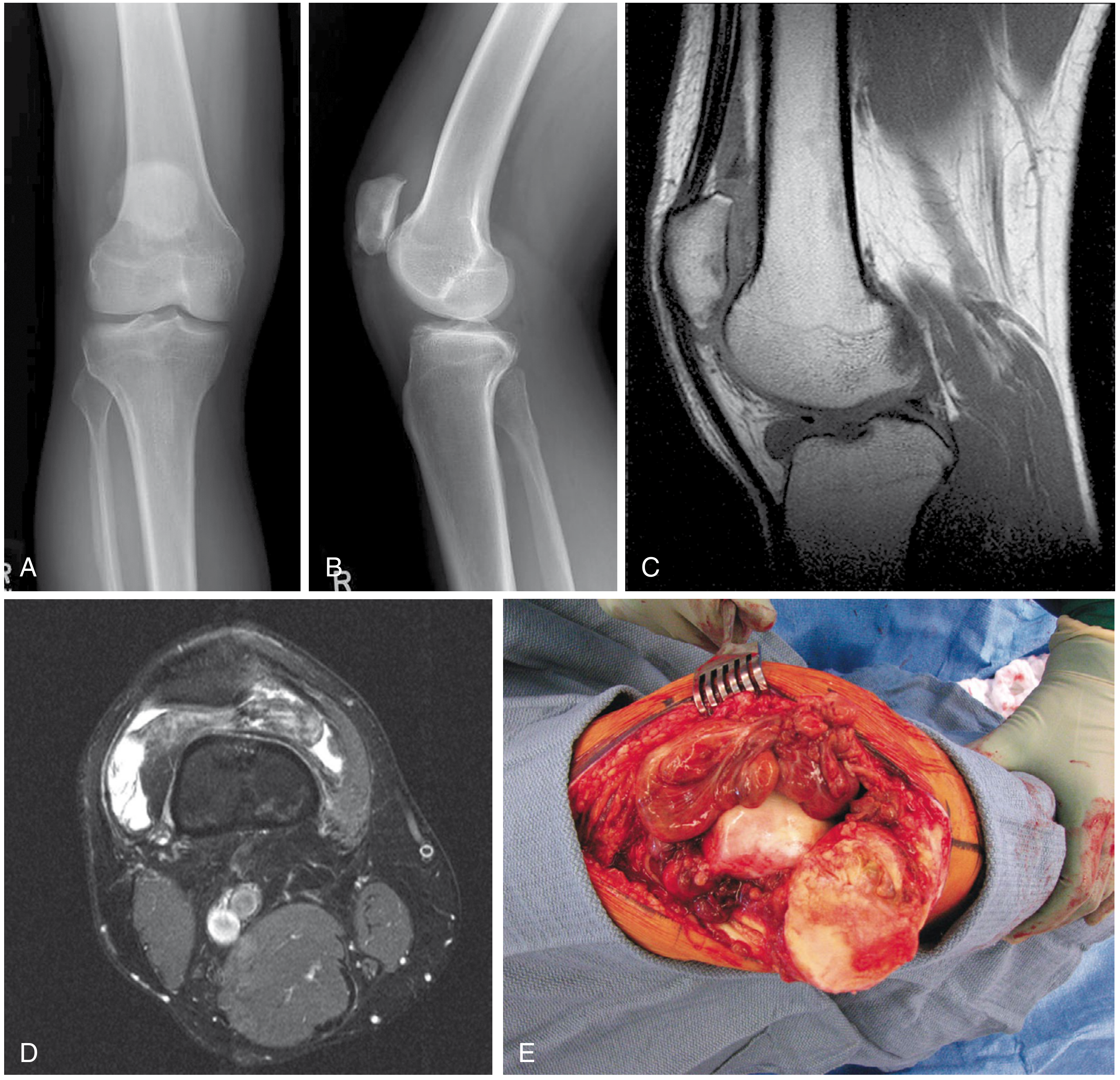
A & B: Plain radiographs show no soft-tissue abnormality. C: T1-weighted (sagittal) MRI - diffuse dark intraarticular process. D: T2-weighted (axial) MRI - also dark (hemosiderin). E: Intraoperative view of diffuse PVNS of the knee.
Histology
Localized form (Focal Nodular Synovitis):
- Solitary pedunculated lesion
- Histologically identical to Giant Cell Tumor of Tendon Sheath
Diffuse form:
- Also histologically identical to the localized form
- The distinction is that it involves the entire synovium of the affected joint
Microscopic features (Figure 30.13):
- Bland polyhedral (histiocyte-like) cells surrounded by collagen
- Giant cells (multinucleated)
- Hemosiderin deposits (the "pigmented" component, responsible for brown-yellow appearance)
- Foam cells (lipid-laden histiocytes)
- Low mitotic activity
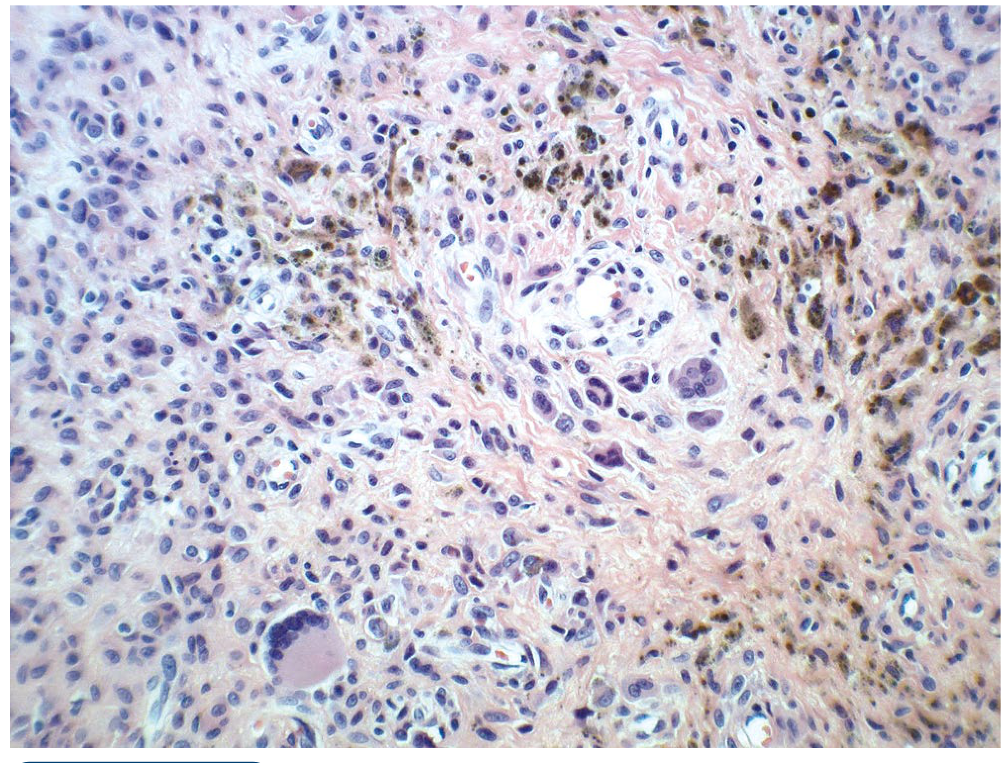
H&E ×20: Bland polyhedral cells with collagen stroma, multinucleated giant cells, and scattered hemosiderin deposits (brown granules).
Treatment
The three goals of treatment are:
- Alleviation of symptoms
- Minimizing recurrence
- Preservation of the joint
Localized Form
- Marginal excision is the recommended treatment
- Can frequently be treated by arthroscopic excision
- Reported recurrence rate with arthroscopic excision: 0% to 8%
Diffuse Form
- Total synovectomy is the recommended treatment (open or arthroscopic)
- Higher recurrence rates than the localized form
- Radiotherapy may be justified if surgery fails to control the disease process
Advanced / Refractory Disease
- If significant secondary degenerative changes of the joint surfaces have occurred, arthroplasty should be strongly considered
- Clinical trials are currently underway evaluating a potential role for CSF1R inhibitors (Colony Stimulating Factor 1 Receptor - targets the CSF1/CSF1R signaling pathway which drives synovial proliferation; pexidartinib is the approved CSF1R inhibitor)
Relationship to Adjacent Lesions (Campbell's Context)
Campbell's places PVNS within the Synovial Lesions chapter alongside:
| Lesion | Key Distinguishing Feature |
|---|---|
| Synovial chondromatosis | Cartilaginous metaplasia; multiple loose bodies; radiographically visible calcified loose bodies |
| Giant cell tumor of tendon sheath | Fingers/tendon sheaths; localized; histologically same as PVNS |
| PVNS | Joint synovium; hemosiderin; dark on T1 and T2 MRI |
Summary Table
| Feature | Detail |
|---|---|
| Nature | Benign synovial proliferative lesion |
| Forms | Localized (focal) vs. Diffuse |
| Most common joint | Knee; also hip, ankle, shoulder, wrist |
| Symptom | Monoarticular pain, swelling, mechanical symptoms |
| Joint fluid | Serosanguineous / blood-tinged |
| X-ray | Often normal; bony erosion (especially hip) |
| MRI hallmark | Dark on BOTH T1 and T2 (hemosiderin) |
| Histology | Bland polyhedral cells, giant cells, hemosiderin, foam cells |
| Localized Rx | Marginal/arthroscopic excision (recurrence 0-8%) |
| Diffuse Rx | Total synovectomy ± radiotherapy if refractory |
| End-stage joint | Arthroplasty |
| Emerging therapy | CSF1R inhibitors (e.g., pexidartinib) |
Source: Campbell's Operative Orthopaedics, 15th Edition (2026), Chapter 30 - Soft Tissue Tumors, Synovial Lesions section, pp. 1264-1268.
This is a shared conversation. Sign in to Orris to start your own chat.