
Sinus
Asking for Preferences
What aspect of 'sinus' are you asking about?
Tell me the basic about sinus anatomy and infection and how many are in total
There are 4 pairs of paranasal sinuses (8 total): maxillary, frontal, ethmoid, and sphenoid. They are air-filled cavities in the skull that drain into the nasal cavity. Sinusitis (sinus infection) occurs when these cavities become inflamed, usually after a cold or allergic reaction, causing congestion, facial pain, and thick discharge.
Searching sources...
Reading File
Finding Sources
Finding Sources
Reading File
Reading File
Reading File
Reading File
Reading File
Reading File
Finding Sources
Reading File
Loading Image
Here is a thorough overview of sinus anatomy and infection, drawn from Gray's Anatomy for Students, Robbins Pathologic Basis of Disease, and the Textbook of Family Medicine.
The Paranasal Sinuses - Anatomy
There are 4 pairs of paranasal sinuses (8 individual sinuses total), each named after the skull bone it sits in. They are air-filled cavities that develop as outgrowths from the nasal cavity, and all share three key features:
- Lined by ciliated, mucus-secreting respiratory mucosa
- Open (drain) into the nasal cavity
- Innervated by branches of the trigeminal nerve [V]
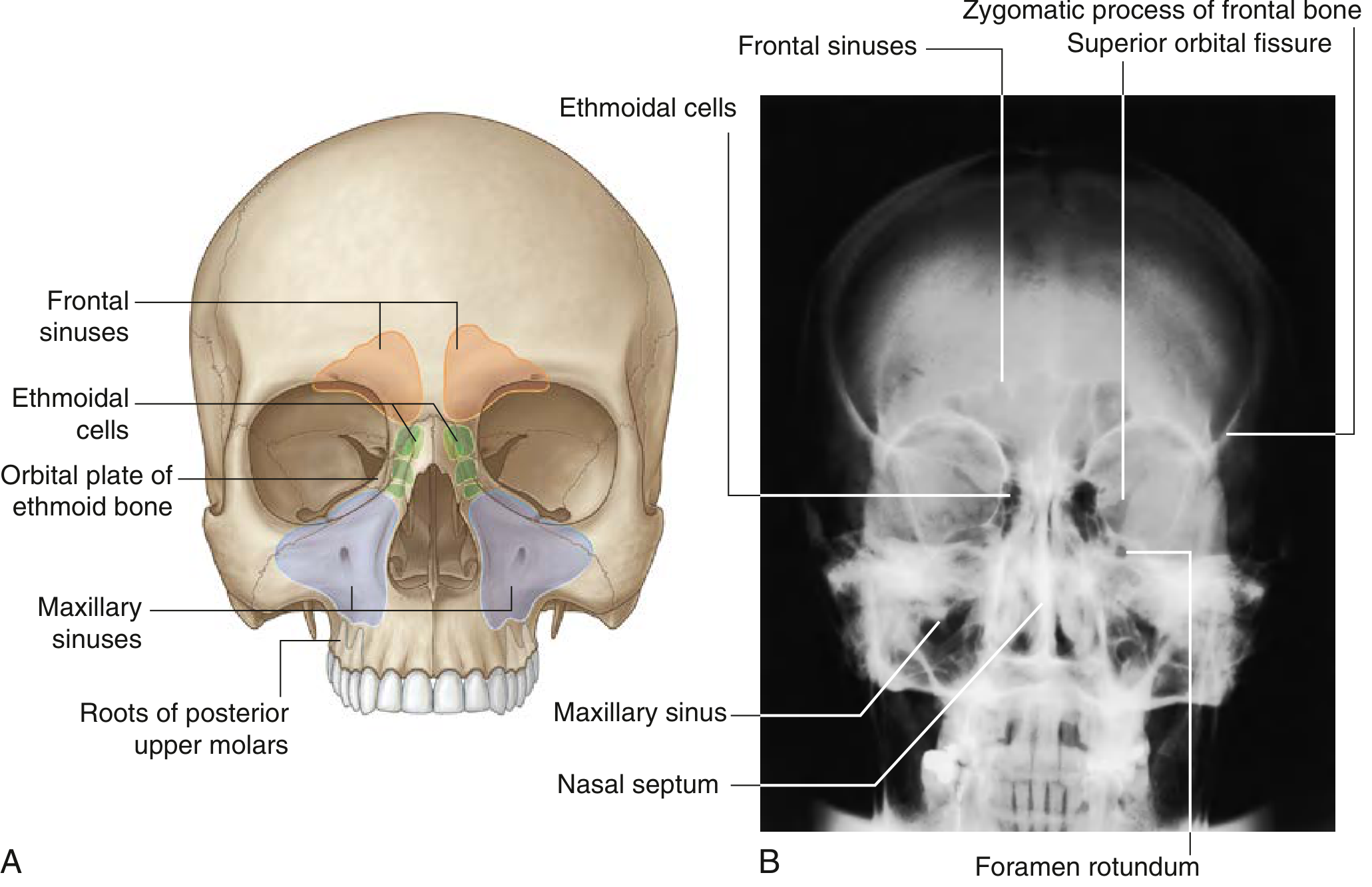
1. Frontal Sinuses (paired)
- Located in the frontal bone, just above and between the eyebrows
- Triangular in shape; variable in size between individuals
- Drain into the middle meatus via the frontonasal duct
- Innervated by the supra-orbital nerve (branch of ophthalmic nerve V1)
2. Ethmoidal Cells (paired clusters)
- Fill the ethmoidal labyrinth between the orbit and nasal cavity
- Made up of multiple small air chambers - divided into anterior, middle, and posterior groups
- Separated from the orbit by only a thin bone plate (orbital plate)
- The most complex sinus group; they can erode into neighboring bones
3. Maxillary Sinuses (paired)
- The largest of all paranasal sinuses
- Pyramid-shaped, filling the bodies of the maxillae (cheekbones)
- Their opening is positioned near the top of the medial wall - this anatomically poor drainage position makes them the most commonly infected sinus
- Closely related to the roots of the upper molar and premolar teeth (dental infections can spread here)
- Innervated by infra-orbital and alveolar branches of the maxillary nerve [V2]
4. Sphenoidal Sinuses (paired)
- Located in the body of the sphenoid bone, deep in the skull
- Drain into the roof of the nasal cavity (spheno-ethmoidal recess)
- Surrounded by critical structures: the pituitary gland above, optic chiasm above, and cavernous sinuses laterally
- Infections here can have serious intracranial consequences
Drainage - The Key Concept
Most sinuses (frontal, maxillary, anterior ethmoid) drain through small openings that converge into a channel called the ostiomeatal unit, emptying into the middle meatus under the middle turbinate. Blockage here is the central event in most sinusitis.
Sinusitis (Rhinosinusitis)
What causes it?
Sinusitis almost always starts with mucociliary dysfunction and sinus obstruction - most commonly after a viral upper respiratory infection (cold) or allergic rhinitis. Mucosal swelling clogs the narrow drainage openings, mucus stagnates, and bacteria can then take hold.
Risk factors include:
- Viral URI or allergic rhinitis (most common trigger)
- Nasal polyps, deviated septum, adenoid hypertrophy
- Smoking, cocaine use (impairs cilia)
- Immunodeficiency, cystic fibrosis, Kartagener syndrome (primary ciliary dyskinesia)
- Dental infections (maxillary sinusitis specifically)
Classification by duration
| Type | Duration |
|---|---|
| Acute | Up to 3 weeks |
| Subacute | 3 to 6 weeks |
| Chronic | > 6 weeks |
| Recurrent acute | Repeated episodes that fully clear between attacks |
Causative organisms
- Acute (viral onset): If it persists > 7-10 days, bacteria take over - mainly Streptococcus pneumoniae, Haemophilus influenzae, and Moraxella catarrhalis
- Chronic: Same bacteria plus anaerobes, Pseudomonas, and Staphylococci (including MRSA)
- Fungal: Aspergillus or Mucor - seen in diabetics and immunocompromised patients; can be life-threatening
Symptoms
Major symptoms (diagnostic weight):
- Facial pain/pressure
- Nasal congestion and obstruction
- Nasal discharge (or discolored posterior nasal drip)
- Loss of smell (anosmia/hyposmia)
- Fever (in acute cases)
- Purulent material seen on nasal exam
Minor symptoms:
- Headache, ear pressure, halitosis, dental pain, cough, fatigue
Diagnosis is probable with 2 or more major factors, or 1 major + 2 or more minor factors.
Complications (if untreated)
- Mucocele - mucus accumulation from blocked drainage
- Orbital spread - periorbital/orbital cellulitis (especially from ethmoid sinusitis, since the orbital plate is very thin)
- Osteomyelitis of surrounding bone
- Intracranial spread - septic thrombophlebitis of a dural venous sinus, meningitis, brain abscess
Quick Summary
| Sinus | Location | Size | Most common in |
|---|---|---|---|
| Maxillary | Cheekbones | Largest | Most frequently infected |
| Frontal | Forehead | Medium | Common in adults |
| Ethmoid | Between eyes/nose | Multiple cells | Kids and adults |
| Sphenoid | Deep skull | Variable | Least common, most dangerous if infected |
Total: 4 pairs = 8 sinuses
Sources: - Gray's Anatomy for Students, pp. 1214-1215 | - Robbins Pathologic Basis of Disease | - Textbook of Family Medicine, 9e
This is a shared conversation. Sign in to Orris to start your own chat.