
Prevention of radiation hazards
I'll start with a quick answer, then search the medical library for detailed content.
Radiation hazard prevention relies on three core principles: time (minimize exposure duration), distance (maximize distance from source - dose falls with the inverse square of distance), and shielding (use appropriate barriers like lead, concrete, or water). These apply to both ionizing and non-ionizing radiation in clinical, occupational, and public settings.
Searching sources...
Reading File
Now let me search the library and web in parallel for comprehensive content on radiation hazard prevention.
Finding Sources
Finding Sources
Searching the Web
prevention of radiation hazards principles ALARA shielding time distance
Good results found. Now let me read the most relevant textbook sections in parallel.
Reading File
Reading File
Reading File
Excellent - rich textbook content retrieved. Let me also get more from the Barash anesthesia text and search for images.
Reading File
Finding Sources
Searching Images
radiation protection shielding lead apron dosimeter
I now have comprehensive textbook content. Let me also fetch the electromagnetic spectrum diagram that was found in Rosen's EM.
Loading Image
Finding Sources
I now have all the content needed for a comprehensive answer. Here is the full synthesis:
Prevention of Radiation Hazards
The Electromagnetic Spectrum and Ionizing Radiation
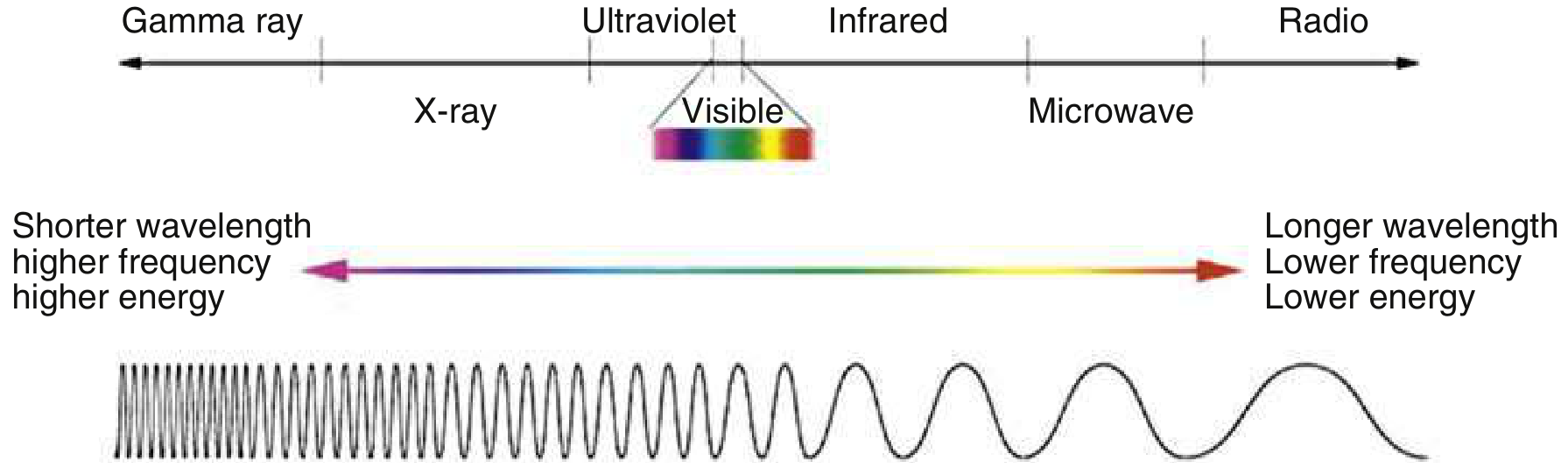
Fig. Electromagnetic radiation: higher frequency/shorter wavelength = higher energy. Ionizing radiation (gamma, X-ray) carries enough energy to displace electrons and damage biological tissue. - Rosen's Emergency Medicine
Ionizing radiation includes X-rays, gamma rays, alpha particles, beta particles, and neutrons. Radiation exposure can be external (e.g., X-rays, gamma rays from an external source) or internal (from inhalation, ingestion, or injection of radioisotopes). - Rosen's Emergency Medicine
Types of Radiation Effects
| Effect | Description |
|---|---|
| Deterministic | Dose-related; causes cell death and tissue damage (e.g., radiation burns, bone marrow suppression). Threshold dose exists |
| Stochastic | Probabilistic; cancer from DNA ionization or hydroxyl radical generation. No safe threshold - risk increases with dose |
- Barash Clinical Anesthesia, 9e
The ALARA Principle
The overriding framework for radiation protection is ALARA - "As Low As Reasonably Achievable." It is guided by three foundational principles from the International Commission on Radiological Protection (ICRP):
- Justification - Any exposure must do more good than harm
- Optimization - Doses must be kept as low as reasonably possible (ALARA)
- Dose Limitation - Total dose for any individual must not exceed established limits
The Four Core Protective Principles
1. TIME - Minimize Exposure Duration
- Reducing exposure time directly reduces absorbed dose - the relationship is linear
- Use short bursts of fluoroscopy rather than continuous exposure
- Use the "last image hold" feature on fluoroscopy units to avoid repeating acquisitions
- Leave the area during imaging when patient care allows
- Staff should work efficiently and avoid lingering near active sources
"Radiation dose during fluoroscopy is directly proportional to the time of exposure and to the number of exposures." - Campbell-Walsh-Wein Urology
2. DISTANCE - Maximize Distance from Source
This is governed by the Inverse Square Law:
Dose ∝ 1/Distance²
Doubling the distance reduces the radiation dose to one quarter. Tripling it reduces dose to one ninth.
- Staff not directly involved in a procedure should step back as far as possible
- The x-ray tube should be positioned below the table (not above) to reduce scatter reaching personnel
- The image intensifier should be positioned as close to the patient as feasible - this substantially reduces scatter radiation
"Maintaining the maximum practical distance from an active radiation source significantly decreases exposure to medical personnel." - Campbell-Walsh-Wein Urology
3. SHIELDING - Use Appropriate Barriers
Shielding effectiveness depends on the type of radiation:
| Radiation Type | Shielding Required |
|---|---|
| Alpha particles | Paper, outer layer of skin |
| Beta particles | Plastic, aluminium, thin metal |
| X-rays / Gamma rays | Lead, concrete, dense materials |
| Neutrons | Water, polyethylene, boron-containing materials |
Personal Protective Equipment (PPE) in clinical settings:
- Lead aprons (0.25-0.5 mm lead equivalent) - protect the trunk and gonads
- Thyroid collars - protect the thyroid gland (highly radiosensitive)
- Leaded eyeglasses - protect the lens (risk of radiation cataracts)
- Leaded gloves - when hands are near the primary beam
- Fixed and movable lead-lined glass shields - allow anesthesiologists and staff to maintain access to patients while being protected
"Shielding is the most reliable form of radiation protection; typical personal shielding is in the form of leaded aprons, thyroid collars, and glasses." - Morgan and Mikhail's Clinical Anesthesiology, 7e
4. QUANTITY - Limit Source Activity
- Limit the amount of radioactive material in the work area
- Use collimation - restrict the X-ray beam to the minimum required field. This reduces exposure to both patient and operator, and lowers the risk of radiation-induced malignancy
- Use pulse fluoroscopy mode rather than continuous fluoroscopy
Dose Limits (Occupational)
Established by the ICRP (International Commission on Radiological Protection) and adopted in most countries. In the United States, the NCRP (National Council on Radiation Protection and Measurements) recommends:
| Category | Limit |
|---|---|
| Occupational (single year) | 50 mSv |
| Occupational (lifetime) | 10 mSv × age in years |
| Radiation-equivalent whole body (effective dose) | Monitored via dosimeter badge |
- Barash Clinical Anesthesia, 9e
For reference, the average annual dose per person in the United States is 6.2 mSv - about 50% from background radiation and 48% from medical procedures. - Rosen's Emergency Medicine
Monitoring and Surveillance
- All radiation workers should be issued personal dosimeter badges (thermoluminescent dosimeters or film badges)
- Cumulative exposure data should be regularly reviewed by the institution
- Thyroid dose badges may be worn at collar level for separate thyroid monitoring
- Ring badges measure hand doses for those handling radioactive materials
- Dosimetry records must be maintained and provided to workers on request
Special Populations: Pregnancy
- The fetus is especially radiosensitive; teratogenic risk is greater with doses >150 mGy intrauterine
- Pregnant staff should notify their employer and be reassigned away from high-exposure areas where possible
- If imaging is necessary in a pregnant patient, radiation should be carefully justified, minimum dose used, and the uterus shielded where possible
Sources of Radiation Exposure in Healthcare
| Source | Approximate Dose |
|---|---|
| Chest X-ray | 0.02 mSv |
| CT scan (chest/abdomen) | 8-20 mSv |
| Pulmonary angiography | 20-40 mSv |
| Fluoroscopy | 100-1,000× greater than plain X-ray |
| Nuclear medicine | ~3-20 mSv depending on study |
- Rosen's Emergency Medicine, Barash Clinical Anesthesia
Radon deserves special mention: it is a naturally occurring radioactive gas (from uranium decay) that can accumulate indoors and is the second leading cause of lung cancer in the United States after tobacco. Mitigation includes measuring indoor radon levels and improving ventilation.
Summary: Hierarchy of Radiation Hazard Prevention
- Eliminate unnecessary exposures - justify every study (LNT model: no dose is entirely "safe")
- Reduce time of exposure
- Increase distance from source (inverse square law)
- Shield with appropriate materials and personal protective equipment
- Collimate the beam to the minimum required field
- Monitor with dosimeter badges and review cumulative data
- Limit source quantity in the work environment
- Protect special groups - pregnant staff, trainees, high-exposure workers
"Radiation protection for medical personnel includes: (1) limiting time of exposure, (2) maximizing distance from radiation source, and (3) shielding." - Campbell-Walsh-Wein Urology
This is a shared conversation. Sign in to Orris to start your own chat.