
Phacomatosis As a theory question for 10 marks from reference from kanski dukes and ak khurana
phacomatosis neurofibromatosis tuberous sclerosis Sturge Weber Von Hippel Lindau ophthalmology
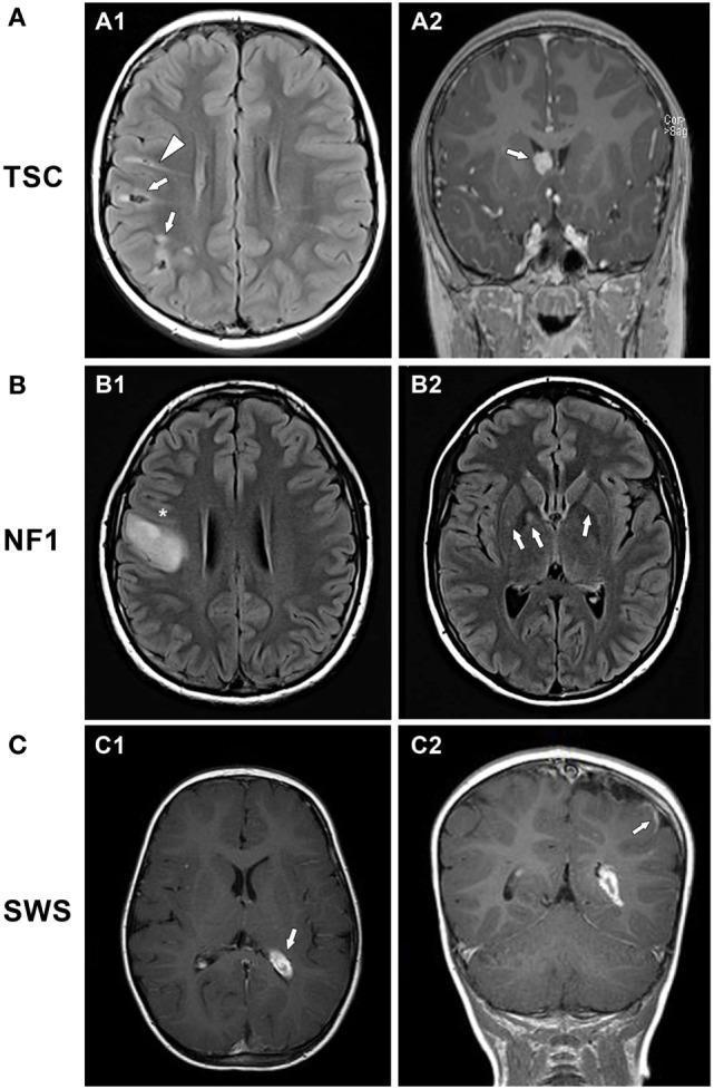
This composite of diagnostic images presents magnetic resonance imaging (MRI) findings across three major neurocutaneous syndromes: Tuberous Sclerosis Complex (TSC), Neurofibromatosis Type 1 (NF1), and Sturge–Weber Syndrome (SWS). Section A (TSC) displays axial FLAIR and coronal contrast-enhanced T1 images showing multiple cortical and subcortical tubers in the right frontoparietal lobes, characterized by mixed signal intensity and linear FLAIR-hyperintense migration lines. A round, enhancing subependymal giant cell astrocytoma (SEGA) is visible at the right foramen of Monro. Section B (NF1) includes axial FLAIR and T1 images demonstrating a large, hyperintense, expansile low-grade glioma in the right precentral gyrus, along with focal T2/FLAIR hyperintensities (unidentified bright objects or UBOs) in the basal ganglia, specifically the globus pallidus and putamen. Section C (SWS) utilizes post-contrast T1-weighted axial and coronal views to illustrate left hemispheric atrophy, prominent enhancement of the choroid plexus glomus, and classic leptomeningeal angiomatosis enhancement in the parieto-occipital region. The collection serves as an educational comparison of hallmark intracranial manifestations and pathognomonic lesions associated with phakomatoses.
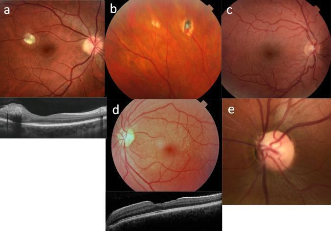
A composite medical visual containing five fundus photographs (a-e) and two Optical Coherence Tomography (OCT) scans, illustrating ocular manifestations of systemic diseases. (a) Fundus image of tuberous sclerosis featuring a semi-translucent, elevated white-yellow retinal astrocytoma near the optic disc, with a corresponding OCT showing an intraretinal mass. (b) Peripheral fundus view in von Hippel-Lindau syndrome showing atrophic, treated retinal hemangioblastomas. (c) Fundus photograph illustrating severe congenital vascular tortuosity of the retinal arteries and veins, typical of HANAC syndrome. (d) X-linked Alport syndrome presentation showing diffuse yellowish-white fleck retinopathy (fleck dystrophy) sparing the macula; the accompanying OCT demonstrates characteristic temporal retinal thinning and an altered foveal profile. (e) Close-up of the optic nerve head in renal coloboma syndrome, showing an excavated optic disc with retinal vessels emerging eccentrically from the disc periphery rather than the center. These images serve as educational resources for identifying ophthalmic markers in pediatric and adult renal ciliopathies.
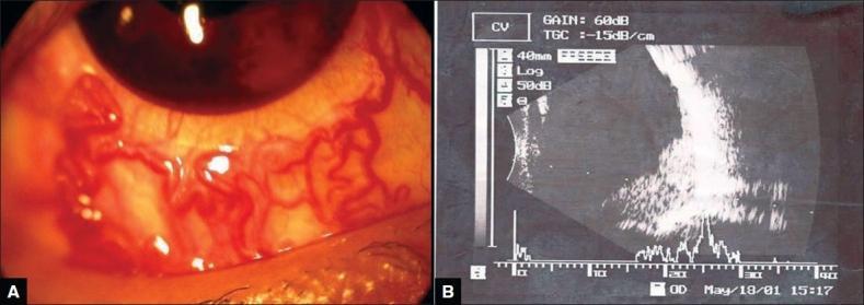
Educational medical visual presenting ocular manifestations of Von Hippel-Lindau (VHL) disease. Panel A is a clinical photograph of the right eye demonstrating significant racemose dilatation and tortuosity of the bulbar conjunctival blood vessels. A cosmetic contact lens is visible over the cornea. Panel B is a B-scan ocular ultrasonography (USG) displaying a large, hyperechoic mass lesion originating from the posterior pole. The mass is localized primarily in the inferior and infero-temporal quadrants, obscuring the optic disc and macula. The imaging illustrates complications associated with bilateral angiomatous retinal lesions, including potential secondary closed-angle glaucoma and posterior segment tumor extension. This material is suitable for ophthalmology students and residents focusing on retinal hemangioblastomas and their secondary clinical effects on ocular surface and internal structures.
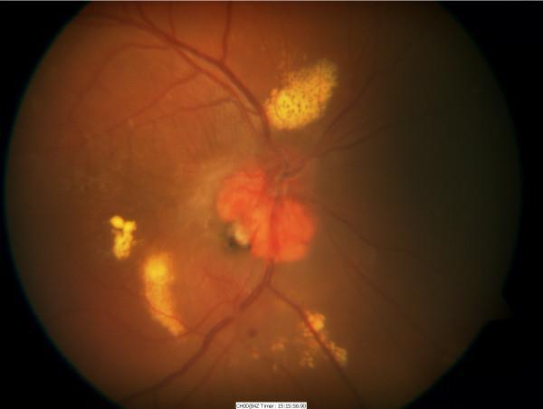
This clinical photograph displays a color fundus image of a right eye showing advanced manifestations of Von Hippel-Lindau (VHL) disease. The primary finding is a large, elevated, reddish-orange exophytic haemangioblastoma situated at the optic nerve head (peripapillary region). The lesion is associated with significant vascular alterations, including dilated and tortuous feeding vessels. Extensive, dense yellow hard exudates are visible in a circinate-like pattern and scattered clusters, particularly superior and temporal to the optic disc, indicating chronic lipid leakage. There is evidence of secondary serous retinal detachment evidenced by the hazy, elevated appearance of the peripapillary and macular retina. The image illustrates the progressive nature of retinal capillary hemangioblastomas and their potential to cause severe exudative maculopathy and visual impairment in patients with VHL syndrome. The photograph is an essential educational tool for ophthalmology trainees to identify peripapillary tumors and the associated secondary retinal changes.
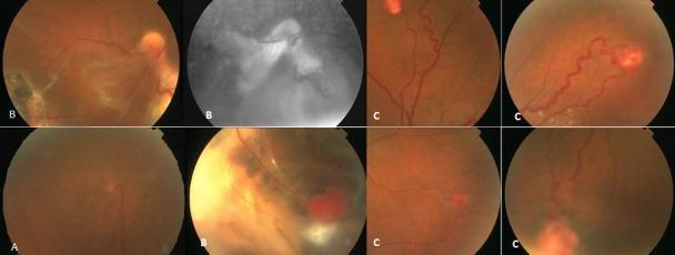
This set of fundus photographs illustrates various clinical presentations of retinal capillary hemangiomas, commonly associated with Von Hippel-Lindau disease. Panel A displays a small, isolated superior retinal capillary hemangioma in the right eye, characterized by a circumscribed reddish lesion. Panels labeled B demonstrate a large, complex hemangioma in the right eye, complicated by an extensive serous retinal detachment and a visible gray-white cryotherapy scar. These images include a red-free or grayscale view highlighting the vascular alterations and exudative changes. Panels labeled C show multiple, multifocal retinal capillary hemangiomas in the left eye, featuring characteristic dilated, tortuous feeder and drainer vessels leading to the orange-red tumors. The educational focus is on the spectrum of disease severity, from small asymptomatic lesions to large tumors with vision-threatening complications like exudative detachment, and the typical vascular morphology used for diagnostic identification in ophthalmology.
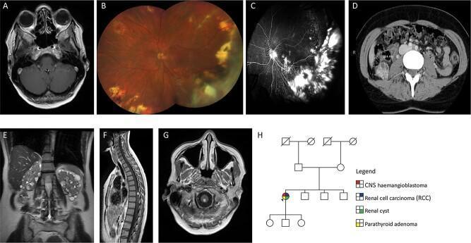
This composite figure illustrates the multi-system manifestations of Von Hippel-Lindau (VHL) disease through various imaging modalities. (A) Axial T1-weighted post-contrast MRI of the orbits demonstrates an intensely enhancing lesion in the left globe, consistent with a retinal hemangioblastoma. (B) Color fundus photograph of the left eye reveals peripheral chorioretinal scars from prior laser/cryotherapy, dragging of the optic nerve vessels, and active hemangiomas in the macula and nasal quadrants. (C) Fluorescein angiogram shows hyperfluorescence in areas of exudation and tractional retinal detachment. (D, E) Abdominal imaging via axial CT and coronal T2-weighted MRI displays a 29 mm right renal cell carcinoma (RCC) and bilateral numerous renal cysts. (F, G) Sagittal and axial post-contrast MRI of the spine and cervicomedullary junction highlight solid enhancing hemangioblastomas with associated feeding vessels on the dorsal spinal cord. (H) A family pedigree indicates the clinical history of a sporadic VHL case, including central nervous system hemangioblastomas, RCC, renal cysts, and parathyroid adenoma. This collection highlights the diagnostic features of VHL disease across ophthalmology, radiology, and genetics.
PHACOMATOSIS (Phakomatosis)
Definition
Classification
| # | Condition | Also Known As | Inheritance |
|---|---|---|---|
| 1 | Neurofibromatosis Type 1 | Von Recklinghausen disease | AD |
| 2 | Neurofibromatosis Type 2 | Bilateral acoustic neuroma syndrome | AD |
| 3 | Tuberous Sclerosis | Bourneville disease | AD |
| 4 | Sturge-Weber Syndrome | Encephalotrigeminal angiomatosis | Sporadic |
| 5 | Von Hippel-Lindau Disease | Retinocerebellar angiomatosis | AD |
| 6 | Ataxia Telangiectasia | Louis-Bar syndrome | AR |
| 7 | Wyburn-Mason Syndrome | Racemose angiomatosis | Sporadic |
1. NEUROFIBROMATOSIS TYPE 1 (NF1 / Von Recklinghausen Disease)
- Six or more café-au-lait macules (>5 mm prepubertal; >15 mm postpubertal)
- Two or more neurofibromas, or one plexiform neurofibroma
- Axillary or inguinal freckling (Crowe's sign - pathognomonic)
- Two or more Lisch nodules
- Optic nerve glioma
- Distinctive osseous lesion (sphenoid wing dysplasia)
- First-degree relative with NF1
Systemic Features:
- Skin: Café-au-lait macules (appear in 1st year of life), axillary/inguinal freckles, cutaneous neurofibromas, plexiform neurofibromas (diffuse, "bag of worms" texture)
- Skeletal: Short stature, facial hemiatrophy, scoliosis, sphenoid wing dysplasia
- Neurological: Intracranial tumours (meningiomas, gliomas), learning difficulties, seizures
- Associations: Malignant peripheral nerve sheath tumours, gastrointestinal stromal tumours, hypertension, autism spectrum disorder (nearly 50%)
Ophthalmic Features:
- Eyelid: Plexiform neurofibroma → characteristic S-shaped deformity of upper lid
- Iris: Bilateral Lisch nodules (iris hamartomas, ≥95% of patients by 3rd decade) - tiny pigmented nodular lesions protruding above iris surface; pathognomonic for NF1
- Cornea: Prominent corneal nerves
- Orbit: Optic nerve glioma (pilocytic astrocytoma, 15-40%) - fusiform nerve enlargement, presents with painless proptosis, visual impairment, optic atrophy. Spheno-orbital encephalocoele causes pulsating proptosis
- Fundus: Choroidal hyper-reflective nodules (>80%, highly specific for NF1 on OCT), retinal "corkscrew" vessels (~one-third, do not leak), choroidal naevi (increased melanoma risk)
- Glaucoma: Rare; usually unilateral, congenital; associated with ipsilateral plexiform neurofibroma of upper lid; mechanism - congenital angle anomaly ± ectropion uveae
- Congenital ectropion uveae
2. NEUROFIBROMATOSIS TYPE 2 (NF2)
Ophthalmic Features (often the FIRST manifestation):
- Cataract: ~Two-thirds of patients; posterior subcapsular, capsular, cortical, or mixed; onset before 30 years - characteristic finding
- Fundus: Epiretinal membrane (frequent); combined hamartoma of retina and RPE (relatively common)
- Ocular motor defects: ~10%
- Less common: Optic nerve sheath meningioma, optic nerve glioma, unilateral Lisch nodules, abnormal ERG
3. TUBEROUS SCLEROSIS (Bourneville Disease)
Cutaneous Features:
- Adenoma sebaceum (angiofibromas): Fibroangiomatous red papules with butterfly distribution around nose and cheeks - universal finding
- Ash leaf spots: Hypopigmented macules on trunk, limbs, scalp - best seen under Wood's lamp (UV) in fair-skinned infants
- Shagreen patches: Diffuse thickening over lumbar region (collagenoma)
- Subungual/periungual hamartomas (fibromata)
- Skin tags (molluscum fibrosum pendulum)
- Café-au-lait spots
Neurological Features:
- Subependymal nodules ("candle guttering" on CT)
- Paraventricular giant cell astrocytomas
- Learning difficulties
- Seizures (infantile spasms → West syndrome)
Visceral Features:
- Renal angiomyolipomas and cysts
- Cardiac rhabdomyomas
- Pulmonary lymphangiomatosis
Ophthalmic Features:
- Retinal astrocytoma (phakoma): The hallmark. Three types:
- Type 1 (most common): Small, flat, semitransparent grey-white lesion near disc
- Type 2: Calcified, mulberry-like (nodular), yellowish-white, located near disc ("mulberry lesion" / Fig. 20.45A in Kanski)
- Type 3: Mixed type - transitional
- Patchy iris hypopigmentation
- Atypical iris colobomas
4. STURGE-WEBER SYNDROME (Encephalotrigeminal Angiomatosis)
Classic Features:
- Port-wine stain (Naevus flammeus): Unilateral facial capillary haemangioma extending over the distribution of one or more branches of the trigeminal nerve (typically V1 ± V2)
- Leptomeningeal haemangioma: Involving ipsilateral parietal/occipital region → contralateral focal or generalized seizures, hemiparesis, hemianopia; "tram-track" calcification on skull X-ray (gyriform calcification on CT)
Ophthalmic Features:
- Glaucoma: Ipsilateral to facial haemangioma in ~30%; in 60% of these, IOP elevation occurs before age 2 years → buphthalmos. Mechanism: trabeculodysgenesis in infants; raised episcleral venous pressure (arteriovenous communication in episcleral haemangioma) in older patients
- Episcleral haemangioma
- Iris heterochromia
- Diffuse choroidal haemangioma: "Tomato ketchup fundus" - causes secondary exudative retinal detachment
- Angioma of conjunctiva and iris
Treatment of Glaucoma in SWS:
- Medical treatment may be adequate
- Goniotomy for angle anomalies (early onset)
- Combined trabeculotomy-trabeculectomy for early cases (risk: choroidal effusion, suprachoroidal haemorrhage)
5. VON HIPPEL-LINDAU DISEASE (Retinocerebellar Angiomatosis)
Systemic Features:
- CNS haemangioblastoma: Cerebellum (most common, ~25% of patients with retinal tumours), spinal cord, medulla, pons
- Phaeochromocytoma
- Renal cell carcinoma (clear cell type)
- Pancreatic islet cell carcinoma
- Cysts: Kidneys, testes, ovaries, lungs, liver, pancreas
- Polycythaemia (erythropoietin from tumour)
- Endolymphatic sac tumours (~10%) → hearing/balance difficulties
Ocular Features - Retinal Capillary Haemangioblastoma:
- Prevalence ~60% in VHL patients (often the first manifestation, age ~25 years)
- Signs:
- Early: Small red oval/round lesion between arteriole and venule
- Established: Round orange-red mass (typically superotemporal periphery) with dilated, tortuous feeder artery and draining vein extending from disc - pathognomonic appearance
- Advanced: Exudates, retinal detachment, vitreous haemorrhage
Treatment of Retinal Haemangioblastoma:
- Laser photocoagulation (small, peripheral tumours)
- Cryotherapy (peripheral lesions)
- Brachytherapy (large lesions)
- PDT and anti-VEGF (juxtapapillary tumours - avoid radiation damage)
- Vitreoretinal surgery (for vitreous haemorrhage, ERM, tractional RD)
Screening Protocol (VHL patients and at-risk relatives):
- Annual retinal examination from age 2-5 years (6-monthly from ages 10-30)
- Renal ultrasonography from age 15
- 24-hour urine catecholamines/VMA from age 2-5 (for phaeochromocytoma)
- 2-yearly abdominal and brain MRI from age 15
- Audiometry if hearing/balance issues
- Genetic testing in all suspected cases and first/second-degree relatives (mutation detection approaches 100% with modern techniques)
- Screening may be discontinued ~age 60 if no abnormality found
6. ATAXIA-TELANGIECTASIA (Louis-Bar Syndrome)
Features:
- Ocular: Conjunctival and eyelid telangiectasias (appears age 3-6 years); oculomotor apraxia; nystagmus
- Neurological: Progressive cerebellar ataxia
- Skin: Telangiectasias on ears, face, neck, antecubital/popliteal fossae
- Systemic: Immunodeficiency (IgA deficiency), recurrent sinopulmonary infections, high risk of lymphoreticular malignancies (leukemia, lymphoma), radiosensitivity, premature aging
7. WYBURN-MASON SYNDROME (Racemose Angiomatosis)
Features:
- Ocular: Racemose (grape-like, tortuous) AVM of retina and optic nerve - grossly dilated, tortuous arteries and veins with direct arteriovenous communications (no capillary interposition)
- CNS: Ipsilateral AVM of midbrain, optic tract, optic chiasm
- Skin: Facial naevus (in some)
- Usually asymptomatic unless complications (haemorrhage, thrombosis) occur
Summary Comparison Table
| Feature | NF1 | NF2 | TSC | SWS | VHL |
|---|---|---|---|---|---|
| Inheritance | AD | AD | AD | Sporadic | AD |
| Gene | Chr 17 (Neurofibromin) | Chr 22 (Merlin) | Chr 9/16 (Hamartin/Tuberin) | GNAQ | Chr 3 (VHL) |
| Skin | Café-au-lait, neurofibromas | - | Adenoma sebaceum, ash leaf spots | Port-wine stain | - |
| Iris | Lisch nodules | Rare Lisch | Hypopigmentation | Heterochromia | - |
| Retina | Corkscrew vessels | Combined hamartoma | Astrocytoma (phakoma) | Choroidal haemangioma | Haemangioblastoma |
| Glaucoma | Rare | No | No | Yes (~30%) | No |
| Optic nerve | Glioma (15-40%) | Sheath meningioma | - | - | Juxtapapillary haemangioma |
| CNS | Gliomas, meningiomas | Bilateral acoustic schwannoma | Subependymal nodules, SEGA | Leptomeningeal angioma | Cerebellar haemangioblastoma |
Key Points for Examination
- Term coined by Van der Hoeve (1932); phacomatoses = hamartomatoses
- NF1 is the most common phacomatosis (1:4,000); NF2 is less common
- Lisch nodules are pathognomonic for NF1 (present in >95%); NOT seen in NF2 significantly
- Bilateral acoustic schwannoma is pathognomonic for NF2
- SWS is the only sporadic phacomatosis in the main group (no genetic inheritance)
- VHL gene on chromosome 3 - the "3p25 mnemonic": 3 letters in VHL, chromosome 3
- Choroidal haemangioma in SWS produces "tomato ketchup fundus"
- Retinal astrocytomas in TSC = "mulberry lesion" (calcified type) or grey-white translucent disc lesions
- Dilated tortuous feeder vessel (artery + vein) to peripheral retinal tumour = VHL pathognomonic finding on fundoscopy
- Ataxia telangiectasia - only AR phacomatosis; ATM gene defect with radiosensitivity