
anterior and posterior ankle scopy portals with diagram
Reading File
Ankle arthroscopy uses standard anterior portals (anteromedial, anterolateral, anterocentral) and posterior portals (posteromedial, posterolateral). The anterolateral and anteromedial portals are most commonly used. Posterior portals are used for posterior impingement, loose bodies, and FHL pathology.
Searching sources...
Finding Sources
Searching the Web
anterior posterior ankle arthroscopy portals anatomy diagram
Finding Sources
Loading Image
Loading Image
Reading File
Loading Image
Loading Image
Reading File
Reading File
Loading Image
Reading File
Now let me get the posterior arthroscopy portal details and any textbook images for the posterior view:
Reading File
Reading Web Page
https://pmc.ncbi.nlm.nih.gov/articles/PMC12125997
Now let me get the posterior ankle portal section from Campbell's:
Finding Sources
Finding Sources
Reading File
Reading File
Loading Image
Loading Image
I now have comprehensive diagrams and content from authoritative textbooks. Here is the complete answer:
Ankle Arthroscopy Portals - Anterior and Posterior
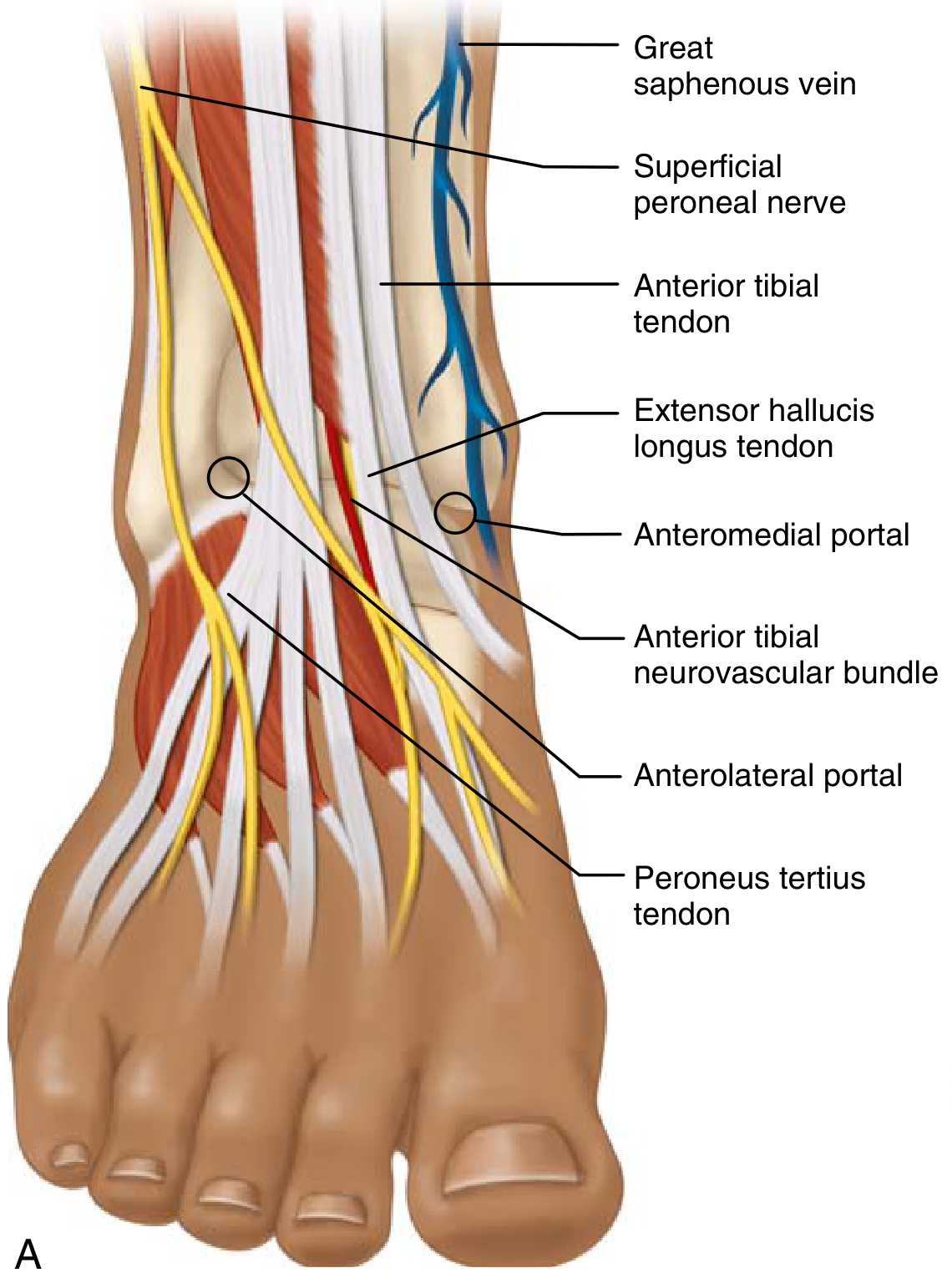
ANTERIOR PORTALS
The anterior ankle is approached with the patient supine, hip and knee flexed in a leg holder, foot hanging free (gravity-assisted distraction).
1. Anteromedial Portal (established first)
- Location: Just lateral to the medial malleolus, immediately medial to the tibialis anterior tendon, at the joint line
- Technique: Dorsiflex the ankle to open the joint space; insufflate with saline via 18G spinal needle first; use "nick-and-spread" blunt dissection
- Structures at risk: Saphenous nerve and great saphenous vein
- Established first because fewer structures are at risk than the anterolateral
2. Anterolateral Portal
- Location: Anterior to the fibula, lateral to the extensor digitorum longus tendon, at the joint line
- Technique: Mark the superficial peroneal nerve before making this portal - visualize it by plantarflexing and inverting the foot; it moves laterally when foot goes to neutral/dorsiflexion
- Structures at risk: Intermediate dorsal cutaneous branch of the superficial peroneal nerve (most commonly injured nerve in ankle arthroscopy; 68% of cadavers had a branch near this portal); peroneus tertius tendon
- The nerve crosses near this portal in 83% of specimens
3. Anterocentral Portal (accessory/seldom used)
- Location: Between the two main anterior portals, along the extensor hallucis longus tendon
- Structures at risk: Anterior tibial neurovascular bundle (deep peroneal nerve + dorsalis pedis artery) - generally avoided for this reason
Anterior portal diagram (Campbell's Operative Orthopaedics 15th Ed 2026):
POSTERIOR PORTALS
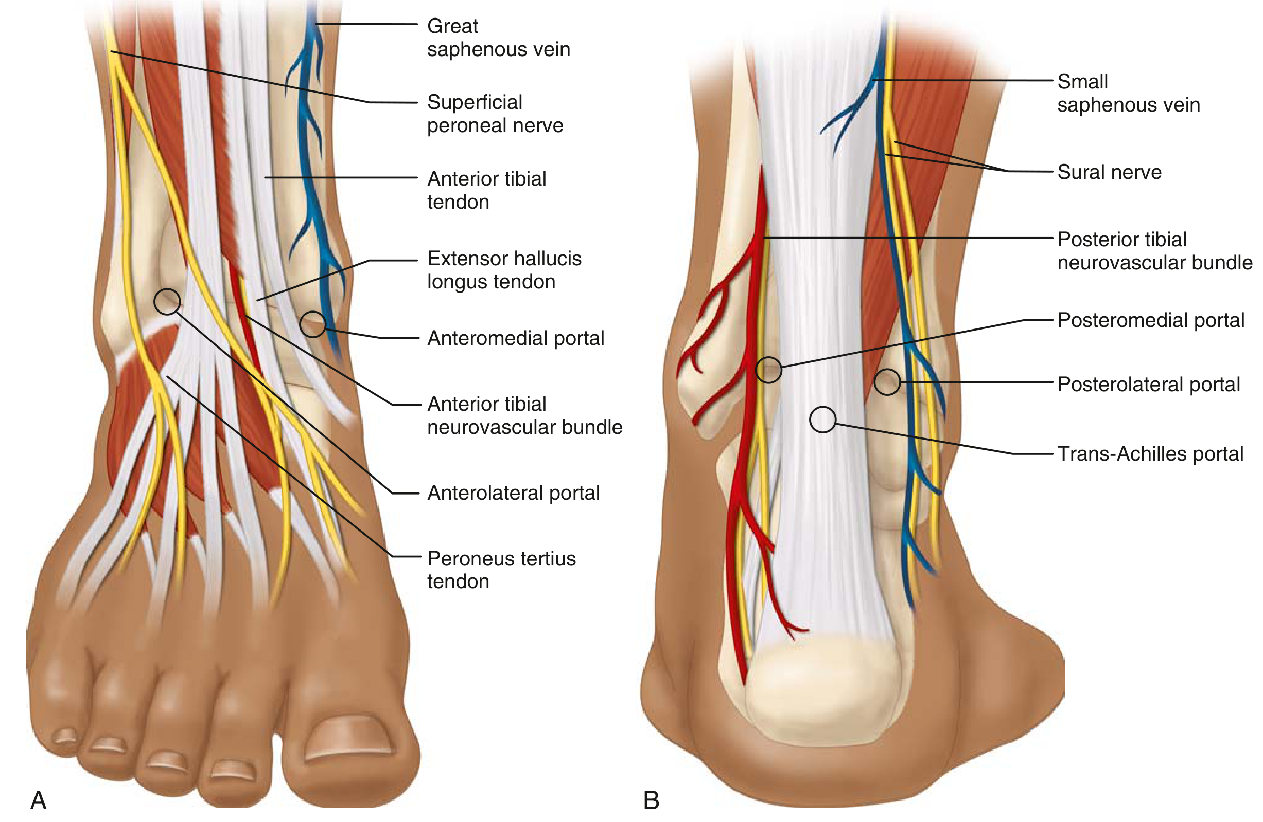
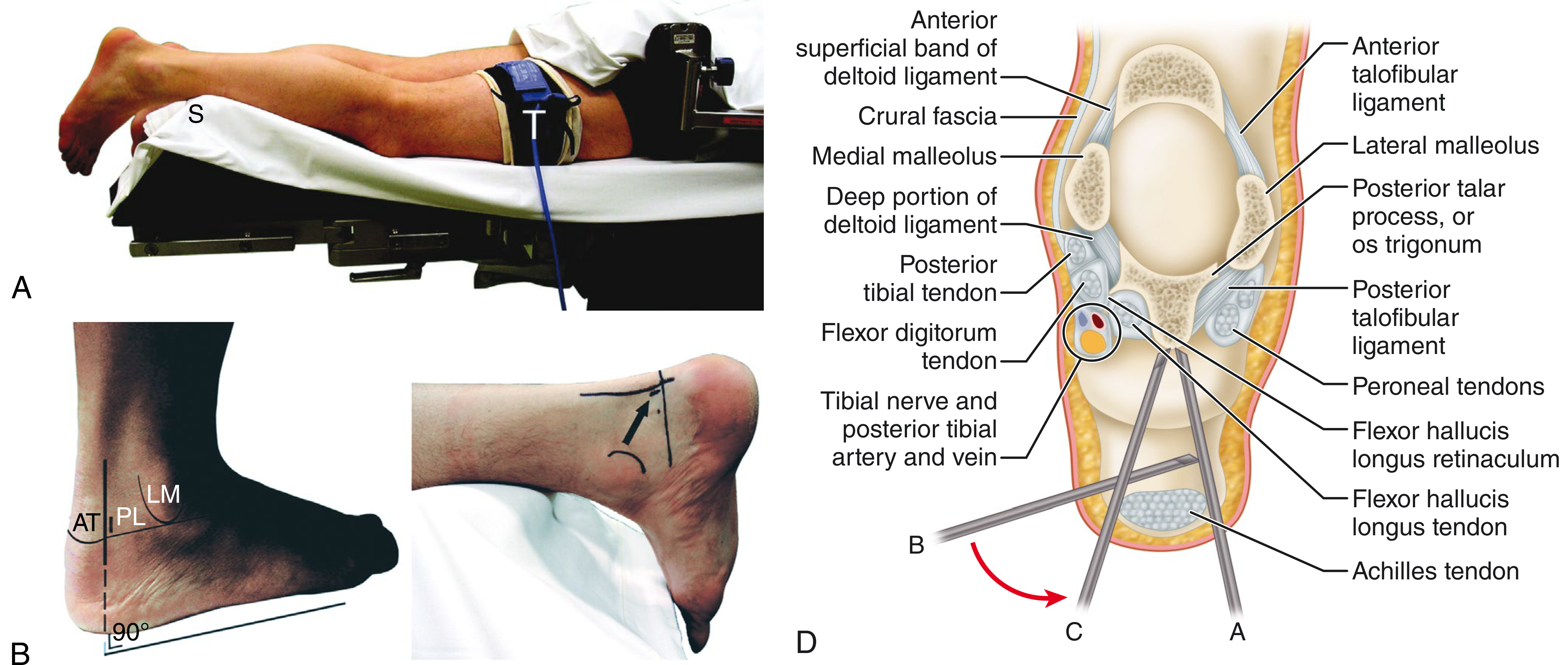
The posterior ankle is approached with the patient prone. Key surface landmarks are marked: medial/lateral malleoli, Achilles tendon borders, and intermalleolar line.
1. Posterolateral Portal (established first / arthroscope portal)
- Location: 1 mm anterior to the lateral border of the Achilles tendon, at the intermalleolar line (level of the ankle joint)
- Technique: A blunt trocar is inserted directed toward the lateral border of the 3rd metatarsal (aim toward 1st web space) to avoid the neurovascular bundle; stay lateral to the FHL tendon; use "nick-and-spread" for skin
- Structures at risk: Branches of the sural nerve and small saphenous vein - placing the portal too laterally risks the sural nerve
- The arthroscope is inserted through this portal
2. Posteromedial Portal (working/instrument portal)
- Location: 1 mm anterior to the medial border of the Achilles tendon, at the same level as the posterolateral portal
- Technique: Establish after the arthroscope is in the posterolateral portal; instruments (shaver, probes) inserted here
- Structures at risk: Posterior tibial neurovascular bundle (tibial nerve + posterior tibial artery and vein); these must be avoided by staying medial to the FHL tendon
- Key rule: keep all instruments lateral to the FHL tendon - FHL is the "lighthouse" protecting the neurovascular bundle
3. Trans-Achilles Portal (accessory, rarely used)
- Location: Directly through the Achilles tendon
- Occasionally used for specific posterior procedures
Posterior portal diagram - both views (Miller's Review of Orthopaedics 9th Ed + Campbell's 15th Ed):
Cross-sectional posterior anatomy showing instrument trajectory:
Summary Table
| Portal | Position | Structures at Risk | Notes |
|---|---|---|---|
| Anteromedial | Medial to tibialis anterior, lateral to medial malleolus | Saphenous nerve, great saphenous vein | Established first |
| Anterolateral | Anterior to fibula, lateral to EDL | Superficial peroneal nerve (most common injury) | Mark SPN before portal |
| Anterocentral | Between EHL & EDL | Anterior tibial NV bundle (deep peroneal n. + dorsalis pedis a.) | Generally avoided |
| Posterolateral | 1 mm anterior to lateral Achilles border | Sural nerve, small saphenous vein | Arthroscope portal; aim toward 3rd metatarsal |
| Posteromedial | 1 mm anterior to medial Achilles border | Posterior tibial NV bundle (tibial n., post. tibial a.) | Instrument portal; stay lateral to FHL |
| Trans-Achilles | Through Achilles tendon | Achilles tendon itself | Rarely used |
Key Operative Pearls
- SPN identification: Plantarflex + invert the 4th toe to visualize the superficial peroneal nerve before making the anterolateral portal; 49% of ankle arthroscopy complications involve nerve injury
- Nick-and-spread technique is essential at all portals to avoid nerve laceration
- FHL as a landmark: In posterior arthroscopy, the flexor hallucis longus (FHL) tendon is the key safe-zone boundary - instruments must stay lateral to it to protect the posterior tibial NV bundle
- Anteromedial before anterolateral (fewer at-risk structures anteriorly)
- Posterolateral before posteromedial (arthroscope in PL, instruments in PM)
- Posterior third of talar dome is inaccessible from anterior approach in ~55% of cases - use posterior portals for posterior talar pathology
Sources: Campbell's Operative Orthopaedics 15th Ed 2026, Ch. 55; Miller's Review of Orthopaedics 9th Ed; Basics of Ankle Arthroscopy Part 4 - PMC
This is a shared conversation. Sign in to Orris to start your own chat.