
Development of adrenal gland
I'll start by reading the medical library skill, then search for adrenal gland development content.
Reading File
Now I'll search the library for adrenal gland development content in parallel.
Finding Sources
Finding Sources
Good, found a relevant block. Let me read it and also search for more specific embryology content:
Reading File
Finding Sources
I can see the file has the development section. Let me read more of the relevant content and also fetch key images:
Reading File
Finding Sources
Excellent content found. Let me now read the Bailey & Love surgery embryology section and the Histology textbook section, plus fetch the key development diagram:
Reading File
Reading File
Loading Image
Now let me fetch the development diagram and also get the adrenal cortex zonation diagram:
Loading Image
Reading File
I now have comprehensive content from multiple authoritative sources. Let me compile a thorough answer.
Development of the Adrenal Gland
The adrenal gland is unique in that its two functional components - the cortex and medulla - arise from completely different embryological origins and develop on distinct timelines before fusing into a single organ.
1. Dual Embryological Origins
| Component | Embryological Origin |
|---|---|
| Adrenal cortex | Mesoderm (coelomic/mesothelial epithelium) |
| Adrenal medulla | Ectoderm (neural crest cells - chromaffin cells) |
- Bailey and Love's Short Practice of Surgery, 28th Ed.
2. Timeline of Development
Weeks 3-4: Initial Condensation
Development begins at approximately the 3rd-4th week of fetal life, when columns of coelomic epithelium start to condense. Over the next 2 weeks, these cells proliferate and migrate cranially to the upper end of the mesonephros.
Weeks 5-6: Adrenogonadal Ridge Formation
The migrating mesothelial cells form the adrenogonadal ridge - the common precursor to both the adrenals and the gonads:
- The adrenal cortex arises from the cephalic (cranial) portion of the ridge
- Gonadal precursor cells arise from the caudal portion of the ridge
This explains why ectopic adrenal tissue (adrenal rests) can be found along the path of gonadal descent.
At around week 5, the fetal cortex develops first. It is then surrounded by a second wave of mesothelial cells that will eventually form the definitive (permanent) cortex.
Weeks 8-10: Zone Differentiation
Morphologic differentiation into the definitive zone and fetal zone occurs between 8-10 weeks. Key events:
- Mesenchymal cells surrounding the fetal cortex condense to form the adrenal capsule
- Neural crest-derived chromaffin cells migrate medially into the gland to form the future adrenal medulla
- Blood vessels and nerves of the adrenal gland develop
At this stage the fetal adrenal gland lacks a definitive medulla - chromaffin cells are present but scattered among the cells of the fetal zone in small clusters.
- Histology: A Text and Atlas (Pawlina), 9th Ed.
The Fetal-Placental Unit
The fetal adrenal gland functions as part of a fetal-placental steroidogenic unit:
- The fetal adrenal lacks certain enzymes needed for complete steroid synthesis that are present in the placenta
- The placenta lacks certain enzymes present in the fetal adrenal
- Precursor molecules are shuttled back and forth between the two organs, enabling synthesis of glucocorticoids, aldosterone, androgens, and estrogens
3. The Fetal Zone vs. Definitive Zone
| Feature | Fetal Zone | Definitive (Permanent) Zone |
|---|---|---|
| Cell size | Considerably larger | Smaller |
| Mitochondria | Spherical with tubular cristae | Small with shelf-like (lamellar) cristae |
| sER | Extensive (accounts for eosinophilia) | Less prominent |
| Lipid droplets | Small | Larger stores |
| Significance | Steroid synthesis (fetal-placental unit) | Forms adult cortex zones |
4. Postnatal Changes
At Birth
- The adrenal glands are relatively large - they weigh twice the weight of the adult glands
- The fetal zone begins rapid involution - within the first postnatal month the gland shrinks to ~1/4 of its previous size
First Year of Life
- The fetal zone involutes and is replaced by the definitive zone
- The definitive zone differentiates (under ACTH stimulation) into:
- Zona glomerulosa (ZG) - outer layer
- Zona fasciculata (ZF) - middle, largest layer
- As the fetal zone regresses, the scattered chromaffin cells aggregate centrally to form the adrenal medulla
- The medulla is homologous to a sympathetic ganglion without postganglionic processes
Ages 6-8 Years: Adrenarche
- The zona reticularis (ZR) remains a relatively minor component until early childhood
- Between 6 and 8 years of life, the ZR expands to its adult volume
- This drives the rise in adrenal androgen (DHEA/DHEAS) production known as adrenarche
5. Adult Adrenal Architecture (End Result)
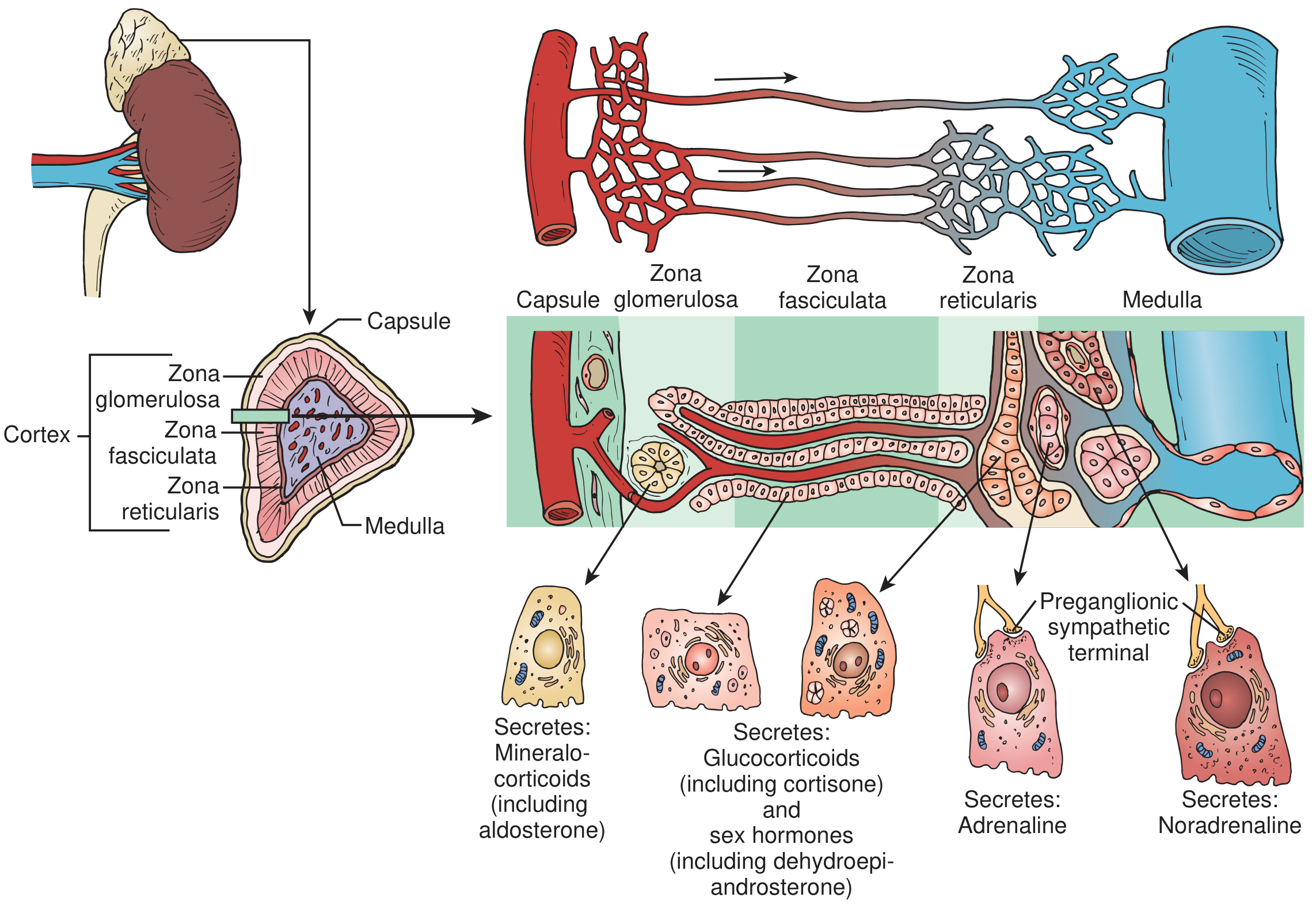
Adrenal gland structure showing the cortical zones, vascular sinusoids, and secretory products. - Campbell-Walsh-Wein Urology
The adult gland is surrounded by a fibrous capsule, with the cortex forming 90% of the gland mass:
| Zone | % of Cortex | Cell Appearance | Hormone |
|---|---|---|---|
| ZG (outer) | ~15% | Small polyhedral cells in ovoid clusters; vacuolated, high nuclear-to-cytoplasmic ratio | Aldosterone (mineralocorticoid) |
| ZF (middle) | ~75-80% | Large pale "clear cells" in straight radial columns; lipid-rich | Cortisol + sex hormones (glucocorticoids + androgens) |
| ZR (inner) | 5-7% | Small cells in anastomosing cords; compact and pigmented | Androgens (DHEA, androstenedione) |
| Medulla | ~10% | Large epithelioid chromaffin cells in clusters; stain yellow with chromium salts | Epinephrine and norepinephrine |
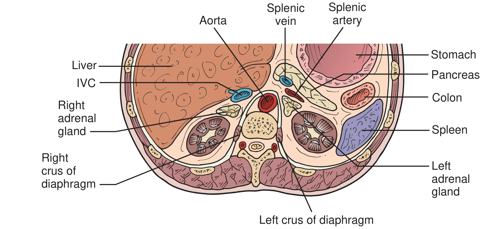
Cross-sectional anatomy of the adrenal glands and their relationships to nearby structures:
6. Developmental Anomalies
Adrenal rests (ectopic adrenal tissue)
- Found in ~1% of adults
- Typically located near the celiac axis or along the path of gonadal descent
- Found in 7.5%-15% of neonates; usually regress over time
- Clinically significant in congenital adrenal hyperplasia (CAH), where they can present as testicular masses, or during inadvertent surgical excision
Renal agenesis
- The developing kidneys ascend from the pelvis to unite with the adrenal glands at ~8 weeks
- In renal agenesis, the adrenal glands remain in their orthotopic position but may be discoid rather than their normal triangular/crescent shape
Congenital Adrenal Hyperplasia (CAH)
- Results from failure of proper adrenal gland development
- An autosomal recessive group of disorders characterized by enzyme deficiency in the steroid synthesis pathway
- Histology: A Text and Atlas (Pawlina), 9th Ed.
Summary Timeline
Weeks 3-4  → Coelomic epithelial condensation begins
Weeks 5-6  → Adrenogonadal ridge forms; fetal cortex appears
             (mesoderm origin, cephalic part = adrenal; caudal = gonads)
Weeks 8-10 → Definitive zone + fetal zone differentiation
             Neural crest cells migrate in → future medulla
             Adrenal capsule forms from mesenchyme
At birth   → Gland is twice adult size; fetal zone involution begins
Year 1     → Fetal zone regresses; ZG + ZF form; chromaffin cells aggregate → medulla
Ages 6-8   → Zona reticularis expands → adrenarche
Sources: Campbell-Walsh-Wein Urology (11th Ed.); Bailey and Love's Short Practice of Surgery (28th Ed.); Histology: A Text and Atlas with Correlated Cell and Molecular Biology (Pawlina)
This is a shared conversation. Sign in to Orris to start your own chat.