
Tapping site
Which 'tapping site' are you asking about?
paracentesis tapping site ascites abdomen
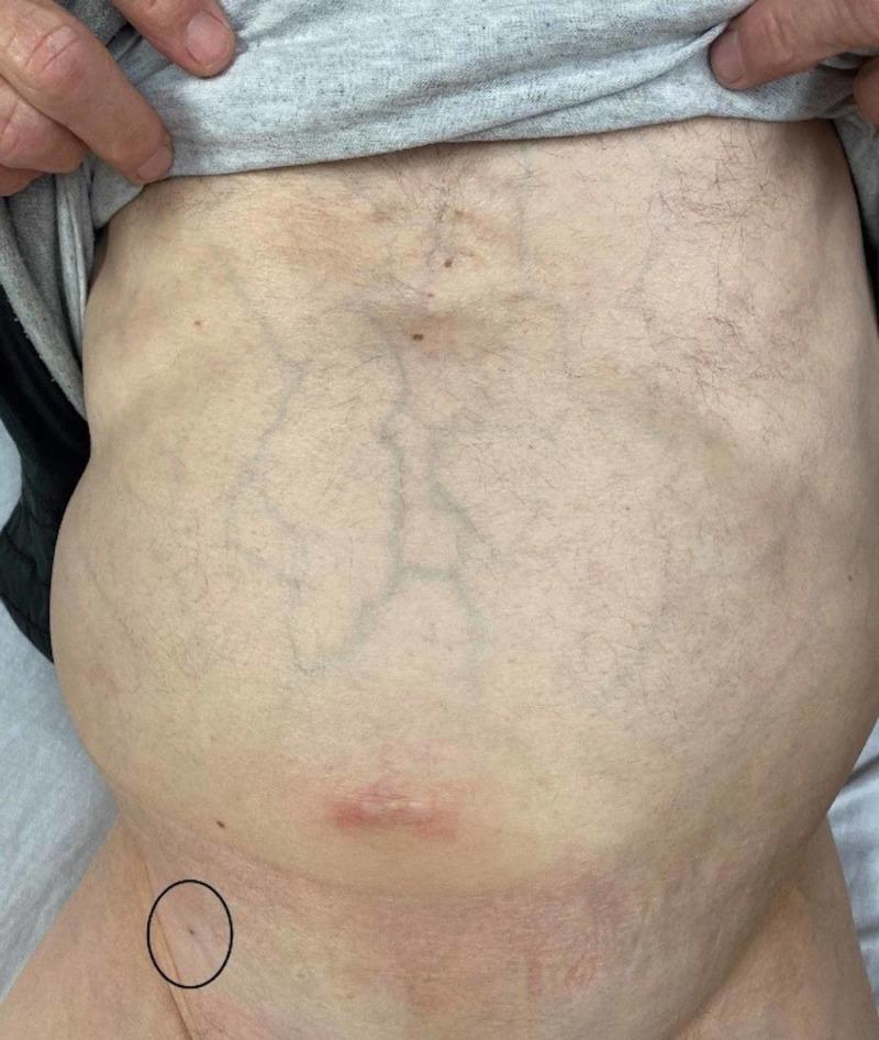
This clinical photograph shows a distended adult abdomen with several notable physical examination findings. Centrally, the abdomen displays prominent, dilated superficial veins, characteristic of caput medusae or collateral venous circulation often associated with portal hypertension. A red, linear scar or healing wound is visible in the lower midline (periumbilical region). A black circle in the patient's right lower quadrant highlights a small puncture site on the skin. This marked area represents a paracentesis site from three days prior, located medial to the right anterior inferior iliac spine. The skin within the circle shows a clean, dry puncture wound without evidence of active infection, such as surrounding erythema, swelling, or discharge. The overall clinical presentation illustrates a patient with likely ascites (suggested by the distention and venous patterns) and the follow-up appearance of a diagnostic or therapeutic paracentesis procedure. This image is relevant for gastroenterology and internal medicine education regarding the physical signs of chronic liver disease and post-procedural site assessment.
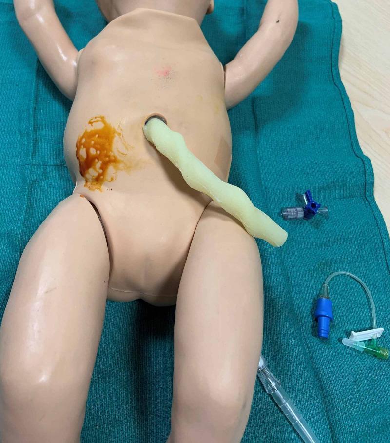
This clinical photograph displays a neonatal medical manikin positioned for a simulation of abdominal paracentesis. The manikin is placed in a supine position on a green sterile drape. The right lower quadrant (RLQ) of the abdomen is visibly prepped with a brown-orange povidone-iodine solution, indicating the intended puncture site for ascitic fluid drainage. A pale, tube-like anatomical simulator protrudes from the umbilical region. Adjacent to the manikin on the right side, procedural equipment is staged, including a blue three-way stopcock, intravenous extension tubing with a blue Luer lock adapter, and a clear plastic syringe or catheter component. The image serves as an educational tool for demonstrating neonatal procedural skills, specifically the sterile preparation and positioning required for paracentesis in the management of neonatal ascites.
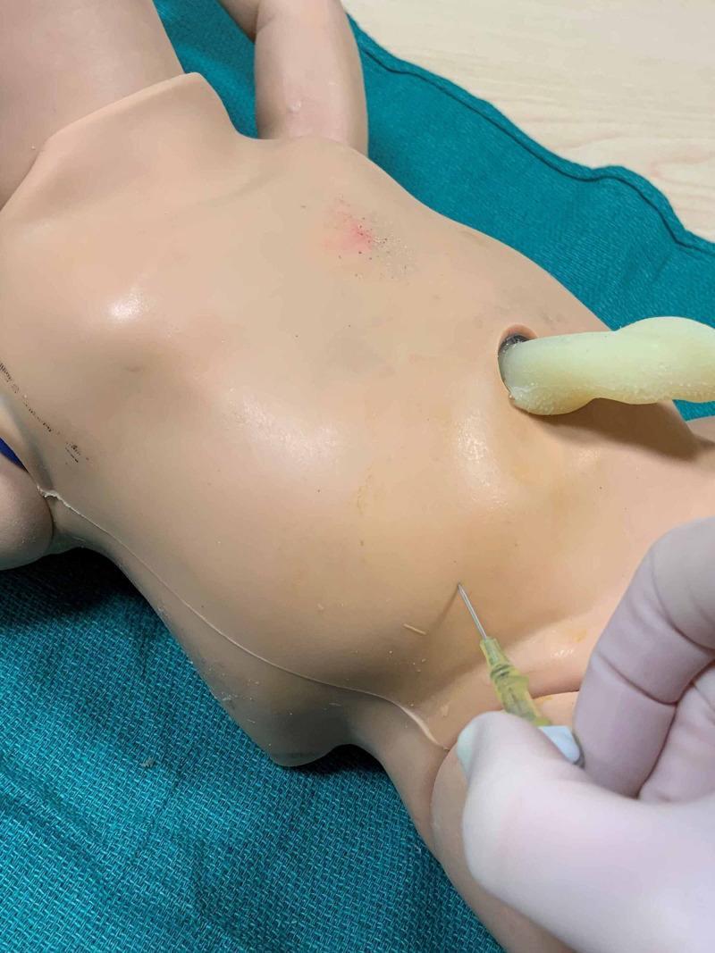
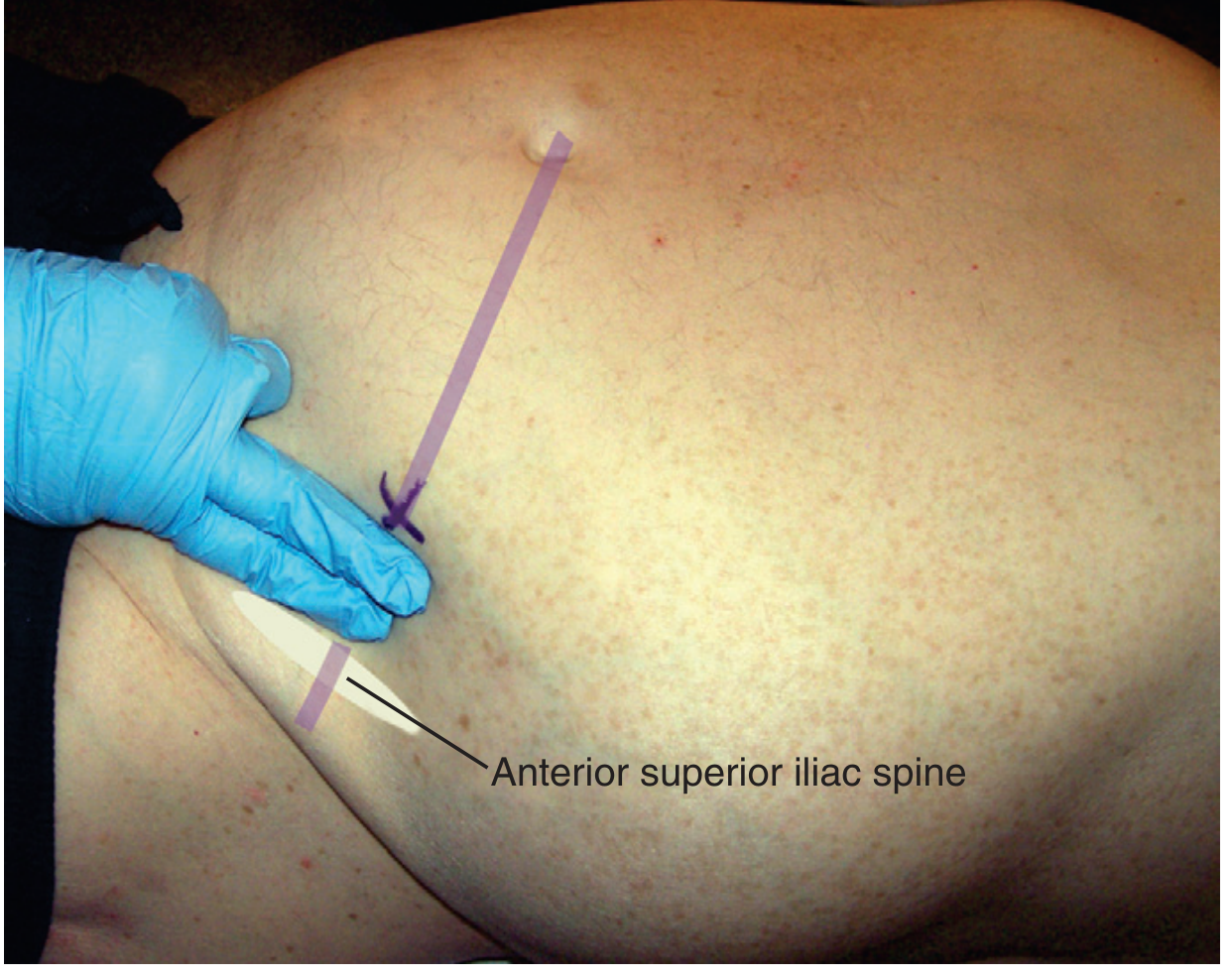
A close-up clinical photograph showing a procedural demonstration of a neonatal abdominal paracentesis on a medical training manikin. The manikin is placed in a supine position on a green surgical drape. A gloved hand is visible, holding a 20- or 22-gauge angiocatheter inserted at a 45-degree angle into the lower quadrant of the manikin's abdomen. The site of insertion is approximately two-thirds the distance along a line drawn from the umbilicus to the anterior superior iliac spine, which is a key clinical landmark for safe paracentesis to avoid internal structures. Near the center of the abdomen, an open hole represents the umbilicus, with a yellowish tube-like structure protruding. The task trainer is used to teach neonatology procedures, specifically the drainage of abdominal ascites. The syringe connected to the catheter contains a yellowish fluid, simulating the appearance of serous ascitic fluid or povidone-iodine solution used in simulation training.
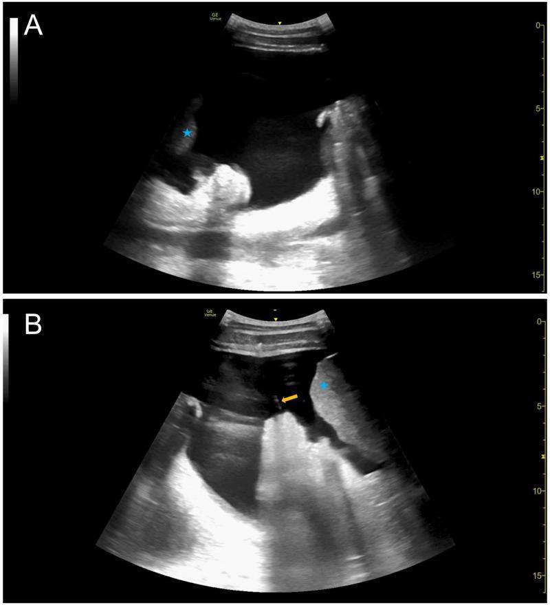
This diagnostic imaging composite displays two ultrasound frames (A and B) showing Point-of-Care Ultrasound (POCUS) of the abdomen in a patient with malignant ascites. Image A shows a large, anechoic (black) fluid pocket representing significant ascites in the right lower quadrant. A blue asterisk identifies the liver parenchyma, which appears echogenic relative to the surrounding fluid. Image B captures an intraprocedural view where a paracentesis catheter is visualized as a linear, hyperechoic (bright) structure (yellow arrow). The catheter is positioned within the ascitic fluid, in close proximity to the liver (blue asterisk) and bowel/omentum. These images illustrate the utility of ultrasound for both pre-procedural identification of fluid pockets and real-time guidance during therapeutic paracentesis to avoid injury to adjacent abdominal viscera.
Ascitic Tap (Paracentesis) - Tapping Site
Two Accepted Sites
1. Midline (Traditional) Approach
- Site: 2 cm below the umbilicus, through the linea alba
- Patient position: Semiupright (45°)
- The linea alba is avascular, reducing bleeding risk
- Bowel floats away from the midline when supine/semiupright
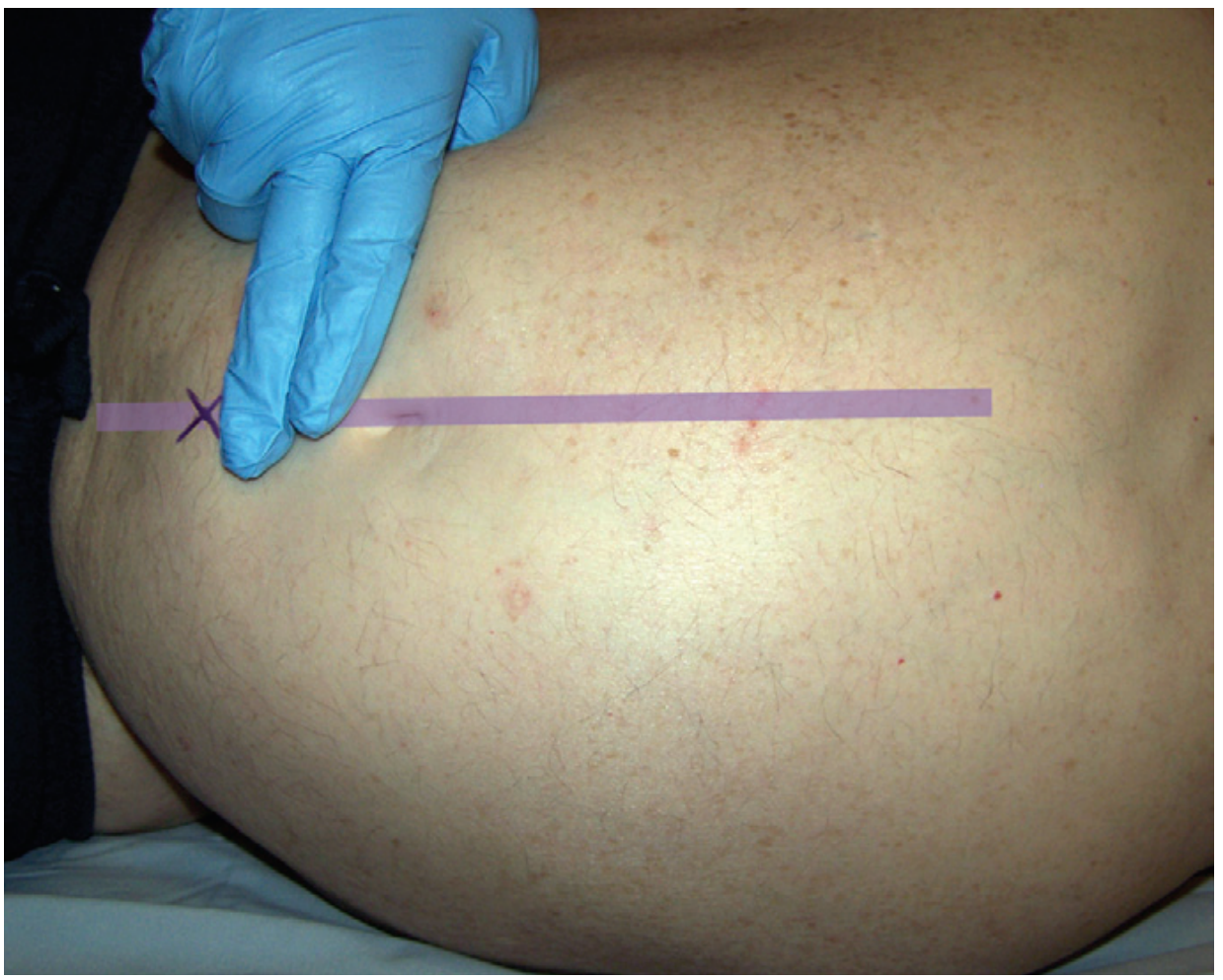
2. Lateral (Preferred / Increasingly Advocated) Approach
- Site: 3 to 5 cm medial and cranial to the anterior superior iliac spine (ASIS) - in the left or right lower quadrant
- Left lower quadrant is preferred over right, because:
- The cecum (right side) is relatively fixed and less mobile than the sigmoid colon
- Bowel perforation risk is higher on the right
- Must remain lateral to the rectus sheath to avoid the inferior epigastric artery
- Patient position: Supine (can be tilted slightly toward the side of collection)
Key Anatomical Points to Remember
| Feature | Detail |
|---|---|
| Preferred side | Left lower quadrant (sigmoid is mobile; cecum on right is fixed) |
| Midline entry | 2 cm below umbilicus through linea alba |
| Lateral entry | 3-5 cm medial and cranial to ASIS |
| Structure to avoid | Inferior epigastric artery (stay lateral to rectus sheath) |
| Other structures to avoid | Distended bladder (decompress with Foley if needed), gravid uterus |
| Ascitic fluid behavior | Air-filled bowel floats anteriorly and toward midline in supine position |
Important Precautions for Site Selection
- Avoid prior surgical scars - bowel may be adherent to the wall
- Avoid sites with cellulitis or abscess
- Use ultrasound guidance when site selection is uncertain, in obesity, post-surgery, or in pregnancy (mandatory after first trimester)
- Prior surgical scars risk adherent bowel loops - choose a site away from them or use ultrasound to find a safe fluid pocket