
Teach me everything I need to know and treat pts with hypertension get information from all the reliable sources includung harrisons and even the other differential causes and the investigations to be done Thankyou
"hypertension"[MeSH Terms] AND "guideline"[pt]
hypertension pathophysiology mechanism diagram blood pressure regulation
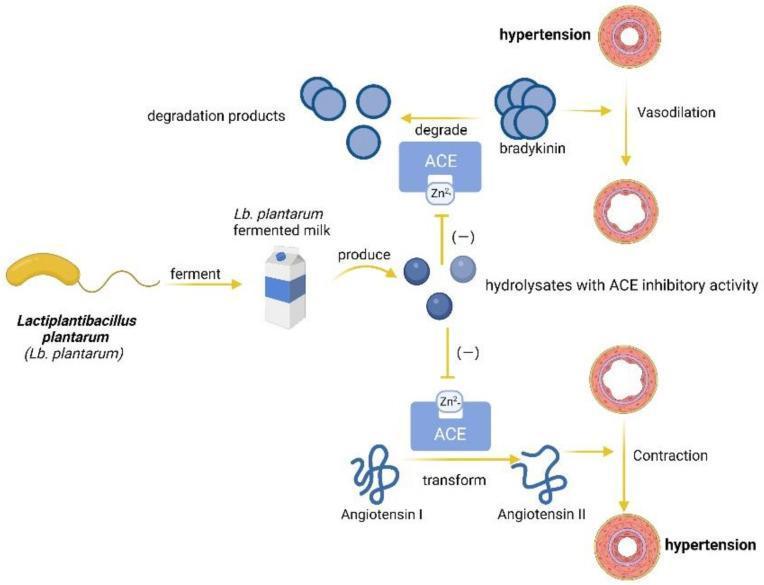
This pathophysiology diagram illustrates the anti-hypertensive mechanism of Lactiplantibacillus plantarum (Lb. plantarum) in functional foods. The process begins with Lb. plantarum fermenting milk, which produces bioactive hydrolysates. These hydrolysates function as Angiotensin-Converting Enzyme (ACE) inhibitors, targeting the zinc-binding (Zn2+) site of the ACE protein. The diagram depicts two physiological pathways influenced by this inhibition: 1) The Bradykinin Pathway: Typically, ACE degrades the vasodilator bradykinin into inactive products. By inhibiting ACE, hydrolysates prevent bradykinin degradation, promoting vasodilation and relieving hypertension. 2) The Renin-Angiotensin Pathway: Normally, ACE transforms Angiotensin I into the potent vasoconstrictor Angiotensin II, leading to vascular contraction and hypertension. The hydrolysates inhibit this transformation, preventing pathological vasoconstriction. The visual contrast emphasizes the transition from constricted, hypertensive blood vessel states to relaxed, dilated states through the modulation of enzymatic activity. This diagram serves as a model for how probiotic-derived peptides can mitigate cardiovascular risk factors by interfering with the biochemical pathways responsible for systemic blood pressure regulation.
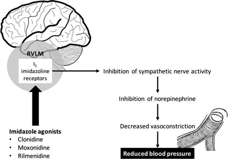
This pathophysiology diagram illustrates the mechanism of action of imidazoline receptor agonists on blood pressure regulation. The visual is divided into a sagittal brain anatomical diagram and a secondary physiological flowchart. On the left, the diagram highlights the 'I1 imidazoline receptors' located within the Rostral Ventrolateral Medulla (RVLM) of the brainstem, situated inferior to the cerebrum and anterior to the cerebellum. Below this, three pharmacological agents—Clonidine, Moxonidine, and Rilmenidine—are listed as imidazole agonists that target these receptors. To the right, a downward flowchart depicts the systemic consequences of receptor activation: stimulation of the RVLM leads to the 'Inhibition of sympathetic nerve activity,' followed by the 'Inhibition of norepinephrine' release. This progression results in 'Decreased vasoconstriction,' visually represented by an illustration of a dilated blood vessel with reduced smooth muscle tension. The final clinical outcome is identified as 'Reduced blood pressure.' The content is designed for intermediate medical education, focusing on neuropharmacology and cardiovascular physiology.
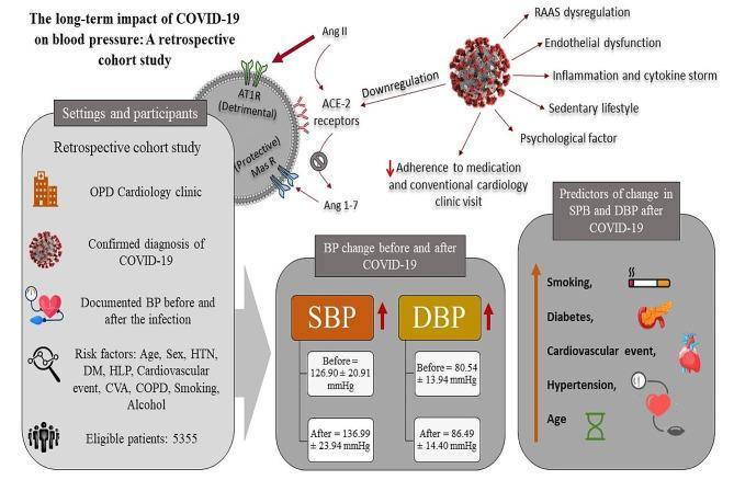
A medical infographic and pathophysiology diagram summarizing the long-term impact of COVID-19 on blood pressure based on a retrospective cohort study. The upper portion illustrates the cellular mechanism where SARS-CoV-2 leads to the downregulation of ACE-2 receptors, causing RAAS dysregulation (increased Ang II binding to AT1R and decreased Ang 1-7 binding to Mas receptors), endothelial dysfunction, and inflammation. The left panel details the 'Settings and participants' section, noting a study of 5,355 eligible patients with documented BP pre- and post-infection. The central 'BP change' panel shows quantitative increases in systolic blood pressure (SBP from 126.90 to 136.99 mmHg) and diastolic blood pressure (DBP from 80.54 to 86.49 mmHg), highlighted with red upward arrows. The right panel lists clinical predictors of BP changes, including smoking, diabetes, cardiovascular events, hypertension, and age, accompanied by illustrative icons. The graphic integrates clinical data with biological mechanisms to demonstrate the cardiovascular sequelae of COVID-19.
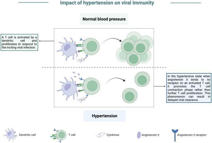
A pathophysiology diagram titled 'Impact of hypertension on viral immunity' comparing cellular immune responses under normal and hypertensive conditions. The top panel, 'Normal blood pressure,' illustrates a dendritic cell presenting antigens to a T cell via receptor-ligand interactions and cytokine release. This stimulation leads to robust T-cell activation and proliferation, resulting in a large cluster of effector T cells essential for viral clearance. The bottom panel, 'Hypertension,' depicts the same initial dendritic cell-T cell interaction but highlights the presence of elevated Angiotensin II. Angiotensin II binds to Angiotensin II receptors expressed on the activated T cell. This signaling inhibits sustained proliferation and instead promotes a T-cell contraction phase, resulting in a significantly reduced population of T cells. The diagram indicates that this hypertensive state leads to an impaired effector response and delayed viral clearance. Key components labeled include dendritic cells, T cells, cytokines, Angiotensin II, and Angiotensin II receptors.
hypertension investigations workup ECG LVH fundoscopy target organ damage
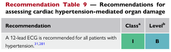
Recommendation Table 9 — Recommendations for assessing cardiac hypertension-mediated organ damage <table><thead><tr><th>Recommendation</th><th>Class<sup>a</sup></th><th>Level<sup>b</sup></th></tr></thead><tbody><tr><td>A 12-lead ECG is recommended for all patients with hypertension.<sup>31,281</sup></td><td>I</td><td>B</td></tr></tbody></table>
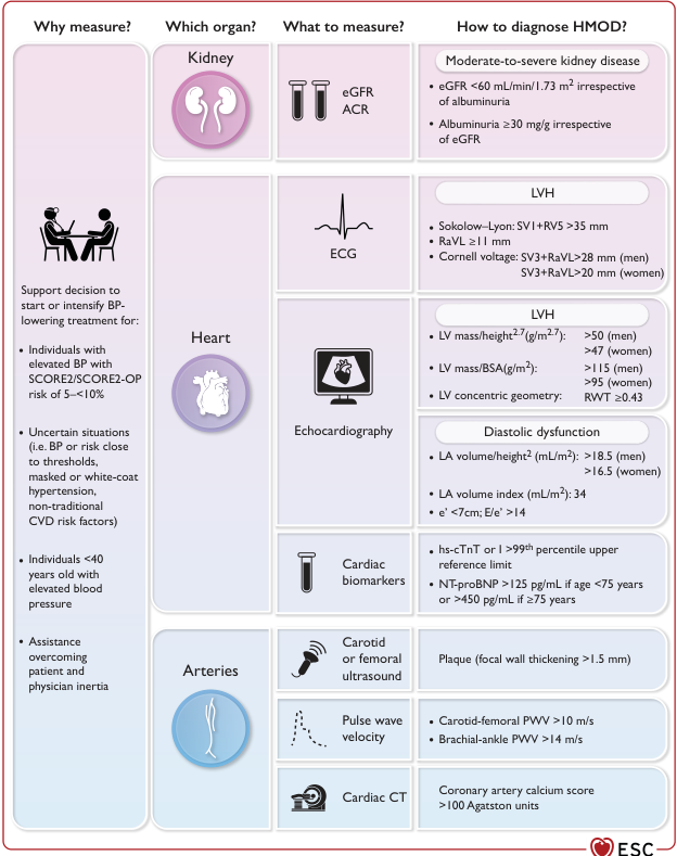
Summary : This figure provides a structured overview of how to measure and diagnose hypertension-mediated organ damage (HMOD) in the kidney, heart, and arteries, including what to measure, diagnostic criteria, and clinical indications for assessment. flowchart/table hybrid: # Why measure? : • Support decision to start or intensify blood pressure (BP)-lowering treatment for: – Individuals with elevated BP and SCORE2/SCORE2-OP risk of 5–<10% – Uncertain situations (BP/risk close to thresholds, masked/white-coat hypertension, non-traditional CVD risk factors) – Individuals <40 years old with elevated BP – Assistance overcoming patient/physician inertia # Which organ? : • Kidney • Heart • Arteries # What to measure? : ## Kidney : • eGFR (estimated glomerular filtration rate) • ACR (albumin-to-creatinine ratio) ## Heart : • ECG • Echocardiography • Cardiac biomarkers ## Arteries : • Carotid or femoral ultrasound • Pulse wave velocity • Cardiac CT # How to diagnose HMOD? : ## Kidney : • Moderate-to-severe kidney disease: – eGFR <60 mL/min/1.73 m² (irrespective of albuminuria) – Albuminuria ≥30 mg/g (irrespective of eGFR) ## Heart : • LVH (left ventricular hypertrophy) by ECG: – Sokolow–Lyon: SV1+RV5 >35 mm – RaVL ≥11 mm – Cornell voltage: SV3+RaVL >28 mm (men), >20 mm (women) • LVH by echocardiography: – LV mass/height².⁷ (g/m².⁷): >50 (men), >47 (women) – LV mass/BSA (g/m²): >115 (men), >95 (women) – LV concentric geometry: RWT ≥0.43 • Diastolic dysfunction: – LA volume/height² (mL/m²): >18.5 (men), >16.5 (women) – LA volume index (mL/m²): 34 – e’ <7cm; E/e’ >14 • Cardiac biomarkers: – hs-cTnT or >99th percentile upper reference limit – NT-proBNP >125 pg/mL (<75 years) or >450 pg/mL (≥75 years) ## Arteries : • Carotid/femoral ultrasound: Plaque (focal wall thickening >1.5 mm) • Pulse wave velocity: – Carotid-femoral PWV >10 m/s – Brachial-ankle PWV >14 m/s • Cardiac CT: Coronary artery calcium score >100 Agatston units # Design Encodings : • Organ sections are colour-coded: kidney (purple), heart (light purple), arteries (blue). • Icons represent each organ and measurement modality. • Diagnostic criteria are listed in shaded boxes for each organ. # Layout : • Four main columns: Why measure?, Which organ?, What to measure?, How to diagnose HMOD? • Rows for kidney, heart, and arteries, each with corresponding measurements and criteria. # Analysis : • The figure provides a comprehensive, organ-based approach to diagnosing HMOD, with clear measurement modalities and diagnostic thresholds for kidney, heart, and arterial damage. • It highlights the importance of tailored assessment based on clinical context and risk, and provides actionable criteria for each organ system. • The use of colour and icons aids rapid identification of organ systems and measurement types.
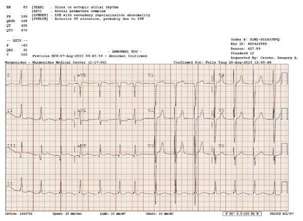
A standard 12-lead electrocardiogram (ECG) demonstrating several diagnostic abnormalities relevant to cardiovascular pathology. The tracing shows a rhythm characterized as ectopic atrial rhythm with visible atrial premature complexes (APCs). Voltage criteria for left ventricular hypertrophy (LVH) are met, notably evidenced by deep S waves in the right precordial leads (V1-V3) and tall R waves in the left precordial leads (V4-V6). Associated secondary repolarization abnormalities are present, including ST-segment elevation in the anterior leads (V1-V3) and T-wave inversions in the lateral leads, which are commonly associated with LVH-related strain patterns. The PR interval appears borderline prolonged, and the QTc is noted as 470 ms. This diagnostic image illustrates the electrical manifestations of structural heart disease, specifically left ventricular hypertrophy and atrial ectopy, in a clinical context of secondary hypertension and end-stage renal disease.
antihypertensive drug classes first line treatment algorithm ACE inhibitor ARB calcium channel blocker diuretic
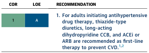
Summary : This figure presents a clinical guideline recommendation for first-line antihypertensive drug therapy in adults, including the class of recommendation (COR) and level of evidence (LOE). table: COR & LOE : • COR: 1 (Class of Recommendation 1, indicating strong recommendation). • LOE: A (Level of Evidence A, indicating high-quality evidence). Recommendation : • For adults initiating antihypertensive drug therapy, the following are recommended as first-line therapy to prevent cardiovascular disease (CVD): – Thiazide-type diuretics – Long-acting dihydropyridine calcium channel blockers (CCB) – Angiotensin-converting enzyme inhibitors (ACEi) or angiotensin receptor blockers (ARB) • Recommendation is supported by references 1 and 2. Analysis : • The figure provides a clear, evidence-based recommendation for initial antihypertensive therapy, prioritizing specific drug classes based on strong evidence for CVD prevention. • The use of COR 1 and LOE A signals that these recommendations are both strongly endorsed and supported by robust clinical data.
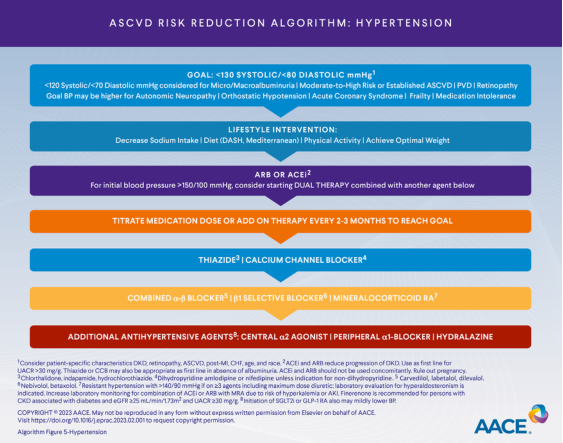
Summary : This flowchart presents the ASCVD (Atherosclerotic Cardiovascular Disease) risk reduction algorithm for hypertension management, outlining stepwise goals, interventions, and medication options. flowchart: # Nodes : • GOAL: <130 SYSTOLIC/<80 DIASTOLIC mmHg (rectangle, blue) • LIFESTYLE INTERVENTION: Decrease Sodium Intake | Diet (DASH, Mediterranean) | Physical Activity | Achieve Optimal Weight (rectangle, blue) • ARB OR ACE: For initial blood pressure >150/100 mmHg, consider starting DUAL THERAPY combined with another agent below (rectangle, purple) • TITRATE MEDICATION DOSE OR ADD ON THERAPY EVERY 2-3 MONTHS TO REACH GOAL (rectangle, orange) • THIAZIDE | CALCIUM CHANNEL BLOCKER (rectangle, blue) • COMBINED α-β BLOCKER | β1 SELECTIVE BLOCKER | MINERALOCORTICOID RA (rectangle, orange) • ADDITIONAL ANTIHYPERTENSIVE AGENTS: CENTRAL α2 AGONIST | PERIPHERAL α1-BLOCKER | HYDRALAZINE (rectangle, red) # Connectors : • Top-down linear flow from goal setting, lifestyle intervention, initial medication choice, titration, and escalation to additional agents. • ARB/ACE node includes a branch for dual therapy if initial BP is >150/100 mmHg. • Medication escalation proceeds in order: thiazide/calcium channel blocker → combined blockers/mineralocorticoid RA → additional antihypertensive agents. # Layout : • Vertically stacked rectangles, each representing a step in the algorithm. • Colour-coded sections: blue for goals/lifestyle/first-line agents, purple for ARB/ACE, orange for titration/combination, red for additional agents. • Footnotes and references are included at the bottom for clinical context and special considerations. # Analysis : • The algorithm emphasizes a stepwise approach: starting with lifestyle changes, then progressing to medication based on blood pressure severity and response. • Dual therapy is recommended for very high initial BP (>150/100 mmHg). • Medication titration and escalation are structured, with clear options for combination and additional agents. • Colour coding visually separates intervention types and escalation steps. • The flowchart provides a comprehensive, guideline-based pathway for hypertension management in ASCVD risk reduction.
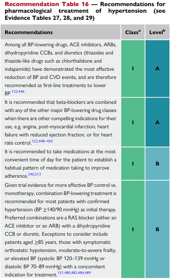
Recommendation Table 16 — Recommendations for pharmacological treatment of hypertension (see Evidence Tables 27, 28, and 29) <table><thead><tr><th>Recommendations</th><th>Class<sup>a</sup></th><th>Level<sup>b</sup></th></tr></thead><tbody><tr><td>Among all BP-lowering drugs, ACE inhibitors, ARBs, dihydropyridine CCBs, and diuretics (thiazides and thiazide-like drugs such as chlorthalidone and indapamide) have demonstrated the most effective reduction of BP and CVD events, and are therefore recommended as first-line treatments to lower BP.<sup>122,446</sup></td><td>I</td><td>A</td></tr><tr><td>It is recommended that beta-blockers are combined with any of the other major BP-lowering drug classes when there are other compelling indications for their use, e.g. angina, post-myocardial infarction, heart failure with reduced ejection fraction, or for heart rate control.<sup>122,448-450</sup></td><td>I</td><td>A</td></tr><tr><td>It is recommended to take medications at the most convenient time of day for the patient to establish a habitual pattern of medication taking to improve adherence.<sup>246,513</sup></td><td>I</td><td>B</td></tr><tr><td>Given trial evidence for more effective BP control vs. monotherapy, combination BP-lowering treatment is recommended for most patients with confirmed hypertension (BP ≥140/90 mmHg) as initial therapy. Preferred combinations are a RAS blocker (either an ACE inhibitor or an ARB) with a dihydropyridine CCB or diuretic. Exceptions to consider include patients aged ≥85 years, those with symptomatic orthostatic hypotension, moderate-to-severe frailty, or elevated BP (systolic BP 120–139 mmHg or diastolic BP 70–89 mmHg) with a concomitant indication for treatment.<sup>131,480,483,484,489</sup></td><td>I</td><td>B</td></tr></tbody></table>
Hypertension: A Complete Clinical Guide
Sources: Harrison's Principles of Internal Medicine 22E (2025), Goldman-Cecil Medicine, Fuster & Hurst's The Heart 15th Ed., Brenner & Rector's The Kidney, Comprehensive Clinical Nephrology 7th Ed., 2024 ESC Guidelines (PMID 39210715), 2025 AHA/ACC Guidelines (PMID 40815242).
1. DEFINITION AND CLASSIFICATION
| Category | Systolic (mmHg) | Diastolic (mmHg) | |
|---|---|---|---|
| Normal | < 120 | AND | < 80 |
| Elevated BP | 120-129 | AND | < 80 |
| Stage 1 Hypertension | 130-139 | OR | 80-89 |
| Stage 2 Hypertension | ≥ 140 | OR | ≥ 90 |
| Hypertensive Crisis | > 180 | AND/OR | > 120 |
When SBP and DBP fall in different categories, use the higher classification. Requires averaging ≥2 readings on ≥2 separate occasions. - Harrison's 22E, Ch. 288
- White-coat hypertension: Office BP meets criteria, but out-of-office BP is normal. Prevalence ~15-25%. CVD risk is closer to normal - manage with lifestyle + monitoring.
- Masked hypertension: Normal office BP but elevated out-of-office readings. CVD risk resembles sustained hypertension - consider antihypertensive treatment.
- Urgency: SBP > 180 or DBP > 120 without acute target-organ damage
- Emergency (Crisis): Same BP levels WITH evidence of acute end-organ damage (encephalopathy, AKI, NSTEMI, aortic dissection, pulmonary edema, eclampsia)
2. EPIDEMIOLOGY
- ~1.3 billion adults worldwide have hypertension; it is the leading modifiable risk factor for cardiovascular disease and stroke.
- Approximately 50% of U.S. adults have hypertension by the ACC/AHA definition.
- Prevalence increases sharply with age: >70% of adults over 65 are hypertensive.
- Non-Hispanic Black adults have among the highest prevalence and severity globally.
- BP tends to rise with age; isolated systolic hypertension predominates in the elderly due to reduced arterial compliance.
3. PATHOPHYSIOLOGY
Primary (Essential) Hypertension (~90-95% of cases)
- Polygenic inheritance; rare monogenic forms (e.g., Liddle syndrome, Gordon syndrome, apparent mineralocorticoid excess) exist but are uncommon.
- Diet quality - Low potassium, high sodium, unhealthy fats
- Body weight - Overweight/obesity activates RAAS, sympathetic nervous system
- Excessive dietary sodium - Nearly all adults worldwide exceed WHO limits
- Insufficient dietary potassium - Potassium lowers BP and reduces stroke risk
- Physical inactivity - Reduces vascular compliance and cardiac efficiency
- Excess alcohol consumption - Raises BP in a dose-dependent manner
- RAAS activation: Renin → Angiotensin I → ACE → Angiotensin II → vasoconstriction + aldosterone release → sodium and water retention → increased BP.
- Sympathetic nervous system overactivity: Increases heart rate, cardiac output, and peripheral resistance.
- Endothelial dysfunction: Reduced nitric oxide (vasodilator) production; increased endothelin (vasoconstrictor).
- Vascular remodeling: Smooth muscle hypertrophy, increased stiffness - raises SBP particularly in the elderly.
- Renal sodium handling: Impaired pressure-natriuresis relationship - kidneys retain sodium at higher BP levels.
4. SECONDARY HYPERTENSION AND DIFFERENTIAL CAUSES
- Treatment-resistant hypertension (uncontrolled on ≥3 drugs including a diuretic)
- Abrupt worsening of previously stable hypertension
- Onset before age 30 in a non-obese patient
- Disproportionate target organ damage for the level of BP
- Unprovoked hypokalemia, proteinuria, or LVH
- Hypertension before puberty
Complete Table of Secondary Causes
| Category | Specific Cause | Key Clinical Clues | Diagnostic Tests |
|---|---|---|---|
| Renal Parenchymal | CKD, glomerulonephritis, pyelonephritis, polycystic kidney disease, diabetic nephropathy | eGFR < 60, proteinuria, hematuria, family history of PKD | Serum creatinine, urine ACR, renal ultrasound, kidney biopsy |
| Renovascular | Renal artery stenosis (atherosclerotic or fibromuscular dysplasia) | Flank/abdominal bruit, flash pulmonary edema, creatinine rise with ACE/ARB | Renal duplex Doppler, CT/MR angiography |
| Primary Aldosteronism | Adrenal adenoma (Conn's) or bilateral adrenal hyperplasia | Resistant HTN, hypokalemia, metabolic alkalosis, adrenal adenoma on imaging | Plasma aldosterone:renin ratio (ARR), 24-hr urinary aldosterone, adrenal vein sampling |
| Pheochromocytoma | Adrenal medullary tumor (90%), extra-adrenal paraganglioma (10%) | Paroxysmal HTN, headache + palpitations + sweating triad, pallor, orthostatic hypotension | Serum/plasma metanephrines, 24-hr fractionated urinary metanephrines, CT/MRI abdomen |
| Cushing's Syndrome | Excess cortisol - pituitary (ACTH), adrenal adenoma, ectopic ACTH | Central obesity, purple striae, easy bruising, moon face, buffalo hump, proximal muscle weakness | 24-hr urine cortisol, overnight 1 mg dexamethasone suppression test, adrenal CT, ACTH level |
| Coarctation of Aorta | Congenital narrowing of aorta | Higher BP in arms than legs, weak femoral pulses, chest bruits, rib notching on CXR | MR/CT angiography, echocardiography, invasive angiography |
| Hypothyroidism | Low thyroid hormone → increased PVR | Weight gain, cold intolerance, constipation, raised DBP predominantly | TSH, free T4 |
| Hyperthyroidism | Elevated T3/T4 → increased cardiac output | Tachycardia, tremor, weight loss, raised SBP predominantly | TSH (suppressed), free T4, free T3 |
| Obstructive Sleep Apnea | Hypoxemia triggers sympathetic surges | Obesity, snoring, daytime somnolence, resistant HTN | Polysomnography (sleep study) |
| Acromegaly | Excess growth hormone | Enlarged hands/feet/jaw, soft tissue swelling | IGF-1, GH suppression test |
| Hyperparathyroidism | Elevated PTH → hypercalcemia | Kidney stones, bone pain, fatigue, "groans and moans" | Serum calcium, PTH level |
| Drug-induced | NSAIDs, OCPs, glucocorticoids, cyclosporine, tacrolimus, sympathomimetics (cocaine, amphetamines), erythropoietin, licorice, MAOIs + tyramine | Medication review | Withdrawal trial |
| Pregnancy-related | Preeclampsia/eclampsia, gestational HTN | HTN after 20 weeks, proteinuria, edema, headache | Urine protein, LFTs, CBC, uric acid |
| Neurogenic | Raised ICP (brain tumors, encephalitis), Guillain-Barré | Neurological signs, Cushing's triad (bradycardia + HTN + irregular breathing) | CT/MRI brain |
| Renoprival | Loss of kidney tissue (bilateral nephrectomy, ESRD) | Dialysis history | Clinical history |
5. CLINICAL EVALUATION AND HISTORY
History
- Duration and previous BP readings
- Symptoms of end-organ damage: headache, visual changes, chest pain, dyspnea, nocturia, palpitations
- Symptoms suggesting secondary cause: sweating attacks, flank pain, weight changes, snoring
- Medications: NSAIDs, OCPs, decongestants, supplements, herbal remedies
- Family history: HTN, stroke, CKD, pheochromocytoma, PKD
- Lifestyle: salt intake, alcohol, tobacco, physical activity, diet
- Psychosocial: stress, socioeconomic status
Physical Examination
- Accurate BP measurement: Patient seated, feet flat, arm at heart level, after 5 min rest, 2-3 readings, average
- Measure both arms (>15 mmHg difference suggests vascular disease)
- Ankle-brachial index if peripheral vascular disease suspected
- BMI and waist circumference
- Fundoscopy: Keith-Wagener-Barker (KWB) classification:
- Grade I: Arteriolar narrowing, silver wiring
- Grade II: AV nicking (venous compression at arteriovenous crossings)
- Grade III: Flame hemorrhages, cotton-wool spots, hard exudates
- Grade IV: Papilledema (hypertensive emergency)
- Neck: Thyroid enlargement, carotid bruits, JVP
- Heart: S3/S4 (LV dysfunction), displaced apex (LVH)
- Abdomen: Renal artery bruits (systolic-diastolic = renovascular), masses, palpable kidneys (PKD)
- Pulses: Radio-femoral delay (coarctation), absent peripheral pulses
- Skin: Striae, ecchymoses, acne (Cushing's), neurofibromas (NF1 + pheo), café-au-lait spots
6. INVESTIGATIONS
Routine (All Hypertensive Patients)
| Investigation | What it detects | Key findings |
|---|---|---|
| Fasting blood glucose / HbA1c | Diabetes (major CV risk factor, target organ) | Diabetes or pre-diabetes |
| Fasting lipid profile | Dyslipidemia (CV risk stratification) | Calculate ASCVD 10-year risk |
| Serum electrolytes (Na, K) | Hypokalemia (primary aldosteronism, diuretic effect), hyperkalemia (CKD, ACE/ARB use) | K < 3.5 mEq/L → screen for aldosteronism |
| Serum creatinine + eGFR | CKD (cause AND consequence of HTN) | eGFR < 60 suggests CKD |
| Serum uric acid | Associated with HTN, gout risk if using diuretics | Elevated in metabolic syndrome |
| Urine dipstick / urinalysis | Proteinuria, hematuria (renal parenchymal disease) | Proteinuria = renal damage |
| Urine albumin:creatinine ratio (ACR) | Microalbuminuria = early renal damage | ACR ≥ 30 mg/g is significant |
| 12-lead ECG | LVH, arrhythmia, ischemia | Sokolow-Lyon: SV1+RV5 > 35mm; Cornell voltage criteria |
| Thyroid function (TSH) | Hypo/hyperthyroidism as secondary cause | Abnormal TSH |
| CBC | Anemia (CKD), polycythemia (sleep apnea) |
Cardiac Assessment for Hypertension-Mediated Organ Damage (HMOD)
- Echocardiogram: LVH, LV mass index, diastolic dysfunction, EF - criteria: LV mass/height^2.7 > 50 g/m^2.7 (men), > 47 g/m^2.7 (women)
- ECG findings in LVH: ECG showing LVH with strain pattern:
- Cardiac biomarkers: NT-proBNP > 125 pg/mL (<75 yr) or > 450 pg/mL (≥75 yr); hs-cTnT elevated
- Carotid-femoral pulse wave velocity (PWV): > 10 m/s = arterial stiffness
- Carotid ultrasound: Plaque or focal wall thickening > 1.5 mm
- Coronary artery calcium (CAC) score: > 100 Agatston units = significant subclinical atherosclerosis
Investigations for Suspected Secondary HTN (Targeted)
| Suspected Cause | Investigations |
|---|---|
| Primary aldosteronism | Plasma aldosterone:renin ratio (ARR); confirmatory test (saline infusion or fludrocortisone); adrenal CT; adrenal vein sampling |
| Pheochromocytoma | Serum/plasma free metanephrines (sensitivity ~99%); 24-hr fractionated urinary catecholamines + metanephrines; CT/MRI abdomen; MIBG scan |
| Renovascular HTN | Renal duplex Doppler ultrasound; CT angiography (gold standard); MR angiography; captopril renography |
| Cushing's syndrome | Midnight salivary cortisol; overnight 1 mg DST; 24-hr urinary free cortisol; ACTH level; adrenal CT; pituitary MRI |
| CKD | Creatinine, eGFR, urinalysis, urine ACR, renal ultrasound |
| Coarctation | Echocardiography; MR/CT angiography |
| Sleep apnea | Epworth Sleepiness Scale; polysomnography |
| Thyroid disease | TSH, free T4, free T3 |
| Hyperparathyroidism | Serum calcium, PTH |
Ambulatory Blood Pressure Monitoring (ABPM) / Home BP Monitoring
- ABPM is gold standard for diagnosing white-coat and masked hypertension.
- Daytime average ≥ 135/85 mmHg or 24-hr average ≥ 130/80 mmHg = hypertension on ABPM.
- Nocturnal dipping: BP should fall 10-20% during sleep. Non-dippers and reverse-dippers have worse CV outcomes.
7. TARGET ORGAN DAMAGE (TOD) Assessment
| Organ | Manifestation |
|---|---|
| Heart | LVH, coronary artery disease, heart failure, AF |
| Brain | Lacunar infarcts, white matter lesions, stroke, TIA, vascular dementia |
| Kidney | CKD, microalbuminuria/proteinuria |
| Eye | Hypertensive retinopathy (Grade I-IV), retinal artery occlusion |
| Arteries | Peripheral artery disease, aortic aneurysm, aortic dissection |
8. TREATMENT
Management Framework
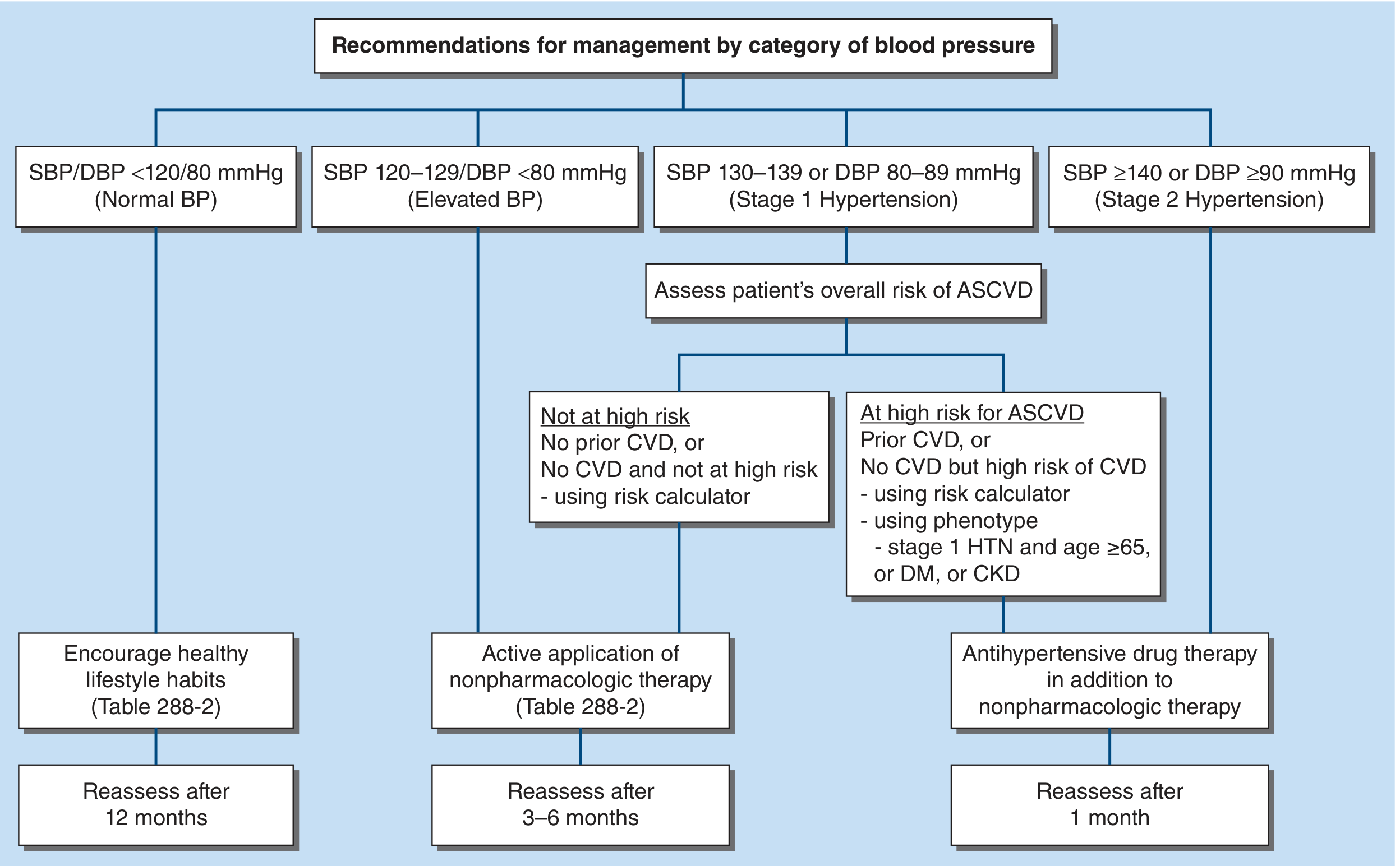
| BP Category | Action |
|---|---|
| Normal (<120/80) | Encourage healthy lifestyle. Reassess in 12 months. |
| Elevated (120-129 / <80) | Active nonpharmacologic therapy. Reassess in 3-6 months. |
| Stage 1 (130-139 / 80-89) + NOT high ASCVD risk | Nonpharmacologic therapy. Add drug if target not met in 6 months. |
| Stage 1 + HIGH ASCVD risk (CVD, DM, CKD, age ≥65) | Nonpharmacologic + antihypertensive drug therapy. |
| Stage 2 (≥140/90) | Nonpharmacologic + antihypertensive drug therapy (usually combination). Reassess in 1 month. |
A. Non-Pharmacological (Lifestyle) Treatment
| Intervention | Expected SBP Reduction | Key Recommendations |
|---|---|---|
| DASH diet (Dietary Approaches to Stop Hypertension) | 8-14 mmHg | Rich in fruits, vegetables, low-fat dairy; limits saturated fat |
| Sodium restriction | 4-9 mmHg | < 2.3 g/day (AHA recommendation); WHO: < 2 g/day |
| Potassium supplementation / dietary increase | 4-5 mmHg | Fruits, vegetables, legumes; supplements if unable to meet dietary goals |
| Aerobic exercise | 4-9 mmHg | ≥ 150 min/week moderate intensity (brisk walking, cycling, swimming) |
| Weight loss | ~1 mmHg per kg | Target BMI < 25 kg/m² |
| Alcohol restriction | 2-4 mmHg | ≤ 1 drink/day for women, ≤ 2 drinks/day for men |
| Smoking cessation | Indirect | Lowers overall CV risk significantly |
| Stress reduction | Variable | Mindfulness, CBT, sleep hygiene |
B. Pharmacological Treatment
- Thiazide/thiazide-like diuretics
- Long-acting dihydropyridine calcium channel blockers (CCBs)
- ACE inhibitors (ACEi)
- Angiotensin receptor blockers (ARBs)
Drug Class Summary Table (from Harrison's 22E, Ch. 288)
| Class | Examples | Dose (mg/day) | Mechanism | SBP Reduction vs Placebo | Key Side Effects | Contraindications/Notes |
|---|---|---|---|---|---|---|
| Thiazide-like diuretics | Chlorthalidone 12.5-25; Indapamide 1.5; HCTZ 12.5-25 | Once daily | Block Na reabsorption in distal convoluted tubule | ~12 mmHg | Hypokalemia, hyperuricemia, hyperglycemia, dyslipidemia | Chlorthalidone preferred (longer half-life, better outcome data) |
| ACE Inhibitors | Lisinopril 10-40; Ramipril 2.5-20; Enalapril 5-40; Benazepril 10-40 | Once/twice daily | Inhibit ACE, block Ang II production | ~12 mmHg | Dry cough (10-15%), angioedema (rare), hyperkalemia | Contraindicated in pregnancy, bilateral RAS; do NOT combine with ARB |
| ARBs | Losartan 50-100; Valsartan 80-320; Olmesartan 20-40; Candesartan 8-32; Azilsartan | Once/twice daily | Block Ang II (AT1) receptors | ~12 mmHg | Rare (no cough); angioedema (very rare); hyperkalemia | Contraindicated in pregnancy; do NOT combine with ACEi |
| CCB (Dihydropyridine) | Amlodipine 2.5-10; Nifedipine LA 30-90; Felodipine 2.5-10 | Once daily | Block Ca2+ entry → inhibit vasoconstriction | ~10 mmHg | Peripheral edema (dose-dependent), flushing, headache, gingival hyperplasia | Preferred in Black patients; safe in pregnancy |
| CCB (Non-dihydropyridine) | Diltiazem ER 120-360; Verapamil ER 100-300 | Once daily | Block Ca2+ entry, slow heart rate | ~9 mmHg | Bradycardia, constipation (verapamil), nausea | Avoid in HFrEF; use in HTN + rate control for AF |
| Beta-blockers | Metoprolol succinate 25-200; Bisoprolol 5-20; Carvedilol 12.5-50 (non-selective) | Once/twice daily | Block β1-adrenergic receptors → reduce HR + CO | ~9 mmHg | Fatigue, bradycardia, bronchospasm, sexual dysfunction, masking hypoglycemia | Preferred in angina, HFrEF, post-MI, AF. Avoid in asthma, severe COPD |
| Aldosterone antagonists | Spironolactone 25-100; Eplerenone 50-100 | Once daily | Block mineralocorticoid receptors | 8-12 mmHg | Hyperkalemia, gynecomastia (spironolactone) | Excellent for resistant HTN, heart failure, primary aldosteronism |
| Alpha-blockers | Doxazosin 1-16; Prazosin 2-20 | Once/twice daily | Block α1-adrenergic receptors → vasodilation | ~8 mmHg | First-dose hypotension, postural hypotension | Useful in BPH + HTN |
| Central alpha-2 agonists | Clonidine 0.1-0.8; Methyldopa 500-2000 | Twice daily | Reduce sympathetic outflow | ~8 mmHg | Dry mouth, sedation, rebound HTN (clonidine) | Methyldopa: drug of choice in pregnancy |
| Direct vasodilators | Hydralazine 25-100; Minoxidil 5-40 | Twice/thrice daily | Direct arterial vasodilation | ~10 mmHg | Reflex tachycardia, fluid retention, lupus-like syndrome (hydralazine) | Minoxidil: severe resistant HTN |
C. Treatment Algorithm (Stepwise Approach)
D. Compelling Indications: Drug Choice by Comorbidity
| Comorbidity | Preferred Agent(s) | Avoid |
|---|---|---|
| Heart failure with reduced EF (HFrEF) | ACEi/ARB + beta-blocker + spironolactone (the "triple threat") + thiazide/loop diuretic | Non-DHP CCBs (verapamil, diltiazem) |
| Post-MI / Coronary artery disease | Beta-blocker + ACEi/ARB | |
| Diabetes mellitus | ACEi or ARB (nephroprotective), GLP-1 agonists if ASCVD risk; avoid HCTZ (worsen glucose) | |
| CKD with proteinuria | ACEi or ARB (reduce proteinuria, slow CKD progression) | K-sparing agents if hyperkalemia risk |
| Pregnancy | Methyldopa (1st line), Nifedipine, Labetalol | ACEi, ARB, direct renin inhibitors (teratogenic) |
| Black patients | Thiazide-like diuretics or DHP CCBs (more effective); CCB + thiazide combination particularly effective | ACEi/ARB monotherapy less effective in this population |
| Atrial fibrillation (rate control) | Non-DHP CCB (diltiazem, verapamil) or beta-blocker | |
| Angina pectoris | Beta-blocker, DHP CCB, long-acting nitrates | |
| BPH (prostate enlargement) | Alpha-blocker (doxazosin) + antihypertensive | |
| Osteoporosis | Thiazide diuretics (reduce urine calcium loss) | |
| Gout | Losartan (uricosuric effect) | Thiazides (raise uric acid) |
| Hyperthyroidism + HTN | Beta-blocker (also controls tremor and palpitations) |
E. Hypertensive Emergency - Management
| Drug | Use Case | Mechanism | Notes |
|---|---|---|---|
| Labetalol IV | Most hypertensive emergencies | α + β blockade | Avoid in acute decompensated HF, asthma |
| Nicardipine IV | Stroke, perioperative HTN | DHP CCB | Safe in pregnancy, no reflex tachycardia |
| Clevidipine IV | Perioperative, stroke | Ultra-short-acting DHP CCB | Titrate easily |
| Sodium nitroprusside | Hypertensive encephalopathy, HF | Direct vasodilator (NO donor) | Risk of cyanide toxicity with prolonged use |
| Nitroglycerin IV | ACS + HTN, pulmonary edema | Venodilator + coronary vasodilator | |
| Esmolol IV | Aortic dissection, perioperative | Ultra-short beta-blocker | Use with vasodilator in dissection |
| Hydralazine IV/IM | Eclampsia/preeclampsia | Direct vasodilator | Drug of choice in pregnancy emergencies |
| Fenoldopam | Renal protection in HTN emergency | Dopamine-1 agonist | Increases renal perfusion |
| Phentolamine | Pheochromocytoma crisis | Alpha-adrenergic blocker | Block alpha BEFORE beta in pheo crisis |
⚠️ In pheochromocytoma crisis: Always give an alpha-blocker (phentolamine or phenoxybenzamine) FIRST before beta-blockers. Giving beta-blockers first causes unopposed alpha-adrenergic stimulation → paradoxical severe hypertension.
F. Special Populations
- BP target: < 130/80 mmHg if tolerated; use clinical judgment in frail patients.
- Beware orthostatic hypotension - assess standing BP.
- Start low, go slow with drug doses.
- Avoid alpha-blockers as first-line (postural hypotension risk, ALLHAT trial).
- Gestational hypertension: HTN after 20 weeks without proteinuria.
- Preeclampsia: HTN after 20 weeks + proteinuria ≥ 300 mg/24 hr or other end-organ damage.
- Preeclampsia with severe features: SBP ≥ 160 or DBP ≥ 110, or end-organ damage.
- Eclampsia: Preeclampsia + seizures.
- Treatment: Methyldopa, Nifedipine (preferred), Labetalol. Magnesium sulfate for seizure prevention.
- ACEi and ARBs are strictly contraindicated in pregnancy.
- ACEi or ARB first-line (reduce proteinuria and slow GFR decline).
- Target BP < 130/80 mmHg.
- Avoid NSAIDs, be cautious with potassium-sparing agents.
- Defined as uncontrolled BP on ≥3 full-dose antihypertensive drugs (including a diuretic).
- Check: non-adherence, white-coat effect, secondary causes.
- Add spironolactone (most evidence-based add-on based on PATHWAY-2 trial).
- Other options: doxazosin, bisoprolol, renal denervation (selected patients).
9. FOLLOW-UP AND MONITORING
- Recheck BP 1 month after starting/changing treatment.
- Once stable, monitor every 3-6 months.
- Repeat BMP (creatinine, potassium) 2-4 weeks after starting ACEi/ARB or diuretic.
- Annual: urinalysis, urine ACR, fasting glucose, lipids, ECG.
- Periodically reassess for new secondary causes, medication side effects, and CV risk.
- Educate patient on home BP monitoring (target < 130/80 mmHg at home).
10. KEY TAKEAWAYS SUMMARY
| Topic | Key Point |
|---|---|
| Definition | ACC/AHA: ≥ 130/80 mmHg = hypertension |
| Most common type | Primary/essential (~90-95%) - polygenic + environmental |
| Screen secondary causes when | Resistant HTN, onset < 30 yrs, hypokalemia, abrupt worsening |
| Most common secondary cause | Primary aldosteronism (commoner than once thought) |
| First-line drugs | Thiazide, DHP-CCB, ACEi or ARB |
| Beta-blockers | NOT first-line unless compelling indication |
| Pregnancy safe drugs | Methyldopa, Nifedipine, Labetalol |
| Pheo crisis rule | Alpha-blocker BEFORE beta-blocker |
| Hypertensive emergency target | Reduce MAP by ≤ 25% in first hour |
| Resistant HTN add-on | Spironolactone |
- The 2024 ESC Guidelines for Hypertension (McEvoy et al., Eur Heart J 2024) recommend combination therapy as initial treatment for most patients with confirmed hypertension, using RAS blocker + DHP-CCB or diuretic.
- The 2025 AHA/ACC Hypertension Guideline (Jones et al., J Am Coll Cardiol 2025) maintains BP target < 130/80 mmHg for most adults and reinforces thiazides, DHP-CCBs, ACEi/ARBs as first-line.
- The 2025 Endocrine Society Primary Aldosteronism Guideline updates screening recommendations - ARR should be performed in all patients with resistant hypertension, hypokalemia, or adrenal incidentaloma.