
Pelvis short notes
Reading File
Now let me search the medical library for pelvis content across anatomy textbooks.
Finding Sources
Finding Sources
Gray's Anatomy for Students is the best source. Let me read the key pelvis sections in parallel.
Reading File
Reading File
Finding Sources
Finding Sources
Good. Now let me read more pelvis content - the bones section, pelvic floor, and joints/ligaments from Gray's Anatomy for Students.
Reading File
Reading File
Now let me read the pelvis joints, diameters, and vessels sections.
Reading File
Reading File
Now let me read the sections on pelvic vessels, pelvic inlet/outlet, and obstetric aspects.
Reading File
Finding Sources
Now I have enough comprehensive content from Gray's Anatomy for Students. Let me compose the complete short notes on the pelvis.
Loading Image
Loading Image
Loading Image
Excellent - I now have rich textbook content and diagrams. Let me compile comprehensive short notes on the pelvis.
Pelvis - Short Notes
Source: Gray's Anatomy for Students
1. Overview
The pelvis is the region of the body between the abdomen and the lower limbs. It is a bony ring formed by the two pelvic (hip) bones, the sacrum, and the coccyx.
- The pelvic inlet (brim) opens directly into the abdominal cavity - no clear boundary separates them. Infections, fluids, and organs (e.g., gravid uterus, full bladder) move freely between the two cavities.
- The pelvis is divided into the false (greater) pelvis (above the pelvic brim - part of the abdomen) and the true (lesser) pelvis (below the pelvic brim - contains the pelvic cavity).
2. Bones of the Pelvis
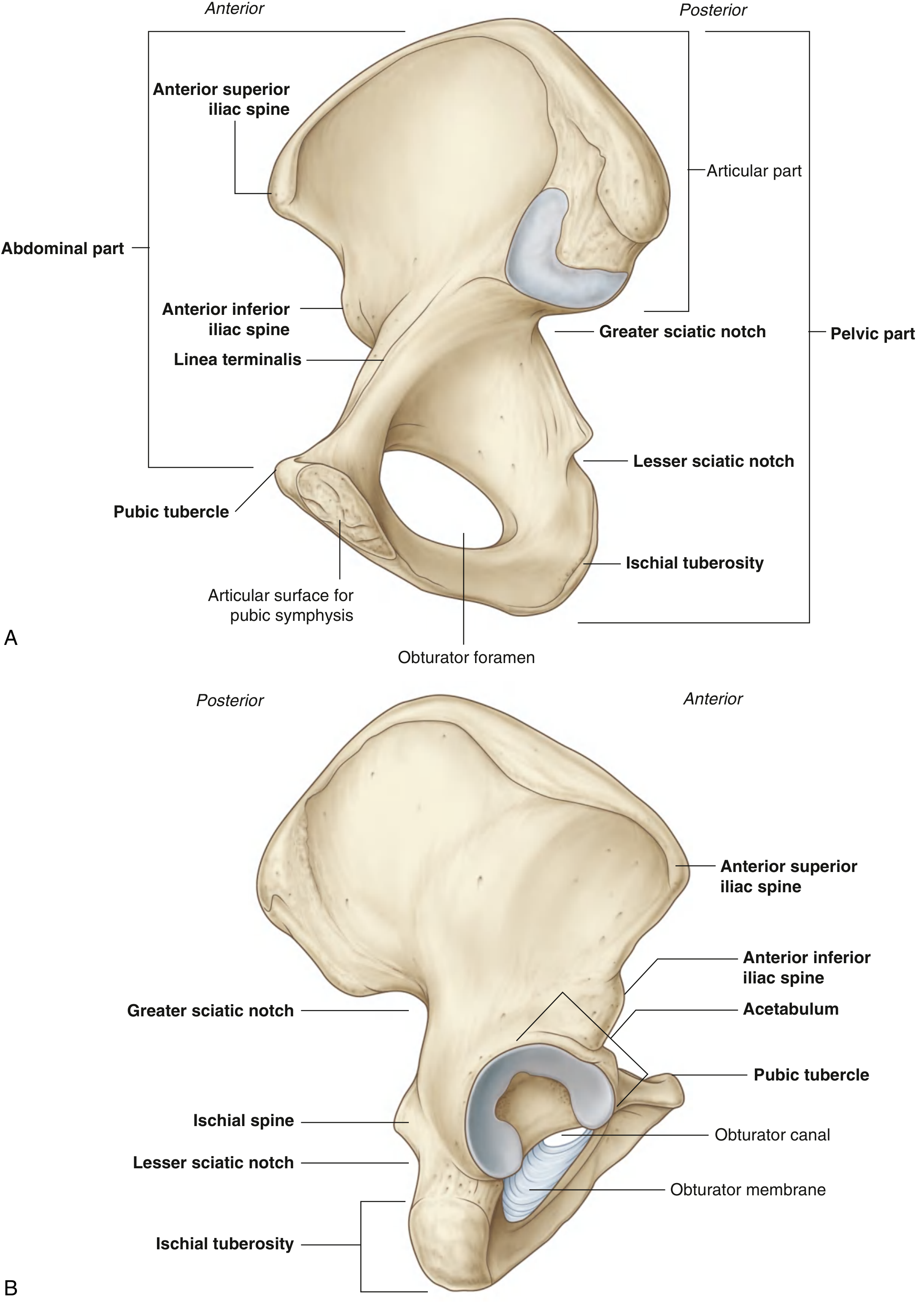
Each pelvic bone is formed by fusion of three elements - the ilium, pubis, and ischium - which fuse at the acetabulum between 16-18 years of age.
Ilium
- Most superior component; upper wing = flat fan-shaped structure supporting the false pelvis
- Key landmarks: iliac crest (ASIS, PSIS, tuberculum), iliac fossa (anteromedial), arcuate line (part of linea terminalis), greater/lesser sciatic notches, AIIS (attachment for rectus femoris and iliofemoral ligament)
- Arcuate line separates abdominal (iliac fossa) part from pelvic part
Pubis
- Anterior and inferior part; has a body + superior and inferior pubic rami
- Pubic crest and pubic tubercle on superior surface; body articulates at the pubic symphysis
Ischium
- Posteroinferior part; made of body + ramus
- Ischial tuberosity (weight-bearing when seated) and ischial spine (important landmark separating greater and lesser sciatic notches)
Sacrum & Coccyx
- Sacrum articulates with L5 at the lumbosacral joint and with pelvic bones at sacroiliac joints
- Promontory = anterior projection of S1, the most anterior point of sacrum - key obstetric landmark
3. Pelvic Inlet (Brim) and Outlet
| Feature | Boundaries |
|---|---|
| Pelvic Inlet | Sacral promontory (posteriorly), arcuate line of ilium, pecten pubis, pubic crest, upper edge of pubic symphysis |
| Pelvic Outlet | Coccyx (posteriorly), sacrotuberous ligaments, ischial tuberosities, inferior pubic rami, pubic arch |
Linea terminalis = arcuate line of ilium + pecten pubis + pubic crest - forms the boundary of the pelvic inlet.
Obstetric Diameters (True Pelvis)
| Diameter | Measurement |
|---|---|
| Anteroposterior (conjugate vera / true conjugate) | ~11 cm |
| Transverse diameter of inlet | ~13 cm |
| Oblique diameter | ~12 cm |
| Interspinous diameter (outlet) | ~10-11 cm |
4. Pelvic Wall
The walls of the pelvic cavity consist of: sacrum, coccyx, pelvic bones below linea terminalis, two ligaments, and two muscles.
Ligaments of the Pelvic Wall
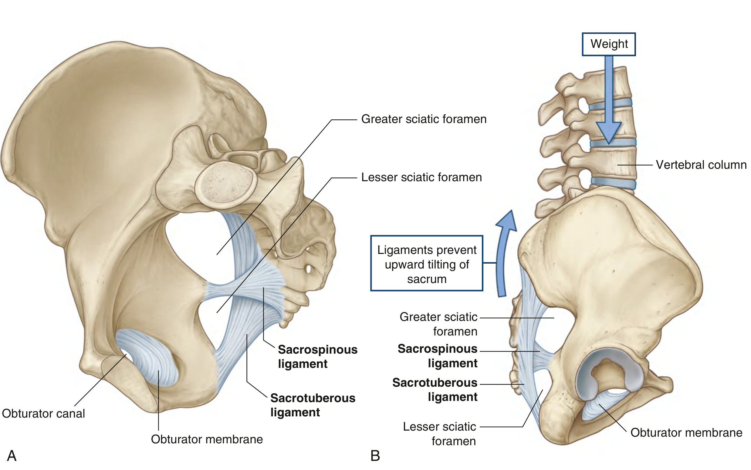
| Ligament | Attachment | Function |
|---|---|---|
| Sacrospinous | Ischial spine → sacrum/coccyx | Triangular; converts greater sciatic notch into foramen |
| Sacrotuberous | PSIS + dorsal sacrum/coccyx → ischial tuberosity | Superficial to sacrospinous; converts lesser sciatic notch into foramen |
Both ligaments resist upward tilting of the inferior sacrum (nutation-counternutation control). Together they form:
- Greater sciatic foramen = superior to sacrospinous ligament and ischial spine
- Lesser sciatic foramen = between sacrospinous and sacrotuberous ligaments
Muscles of the Pelvic Wall
| Muscle | Origin | Insertion | Nerve | Action |
|---|---|---|---|---|
| Obturator internus | Deep surface of obturator membrane + surrounding pelvic bone | Greater trochanter (medial surface) via lesser sciatic foramen | Nerve to obturator internus (L5, S1) | Lateral rotation of thigh |
| Piriformis | Anterior surface of sacrum (S2-S4) | Greater trochanter | Branches from S1, S2 | Lateral rotation + abduction of thigh |
5. Pelvic Floor (Pelvic Diaphragm)
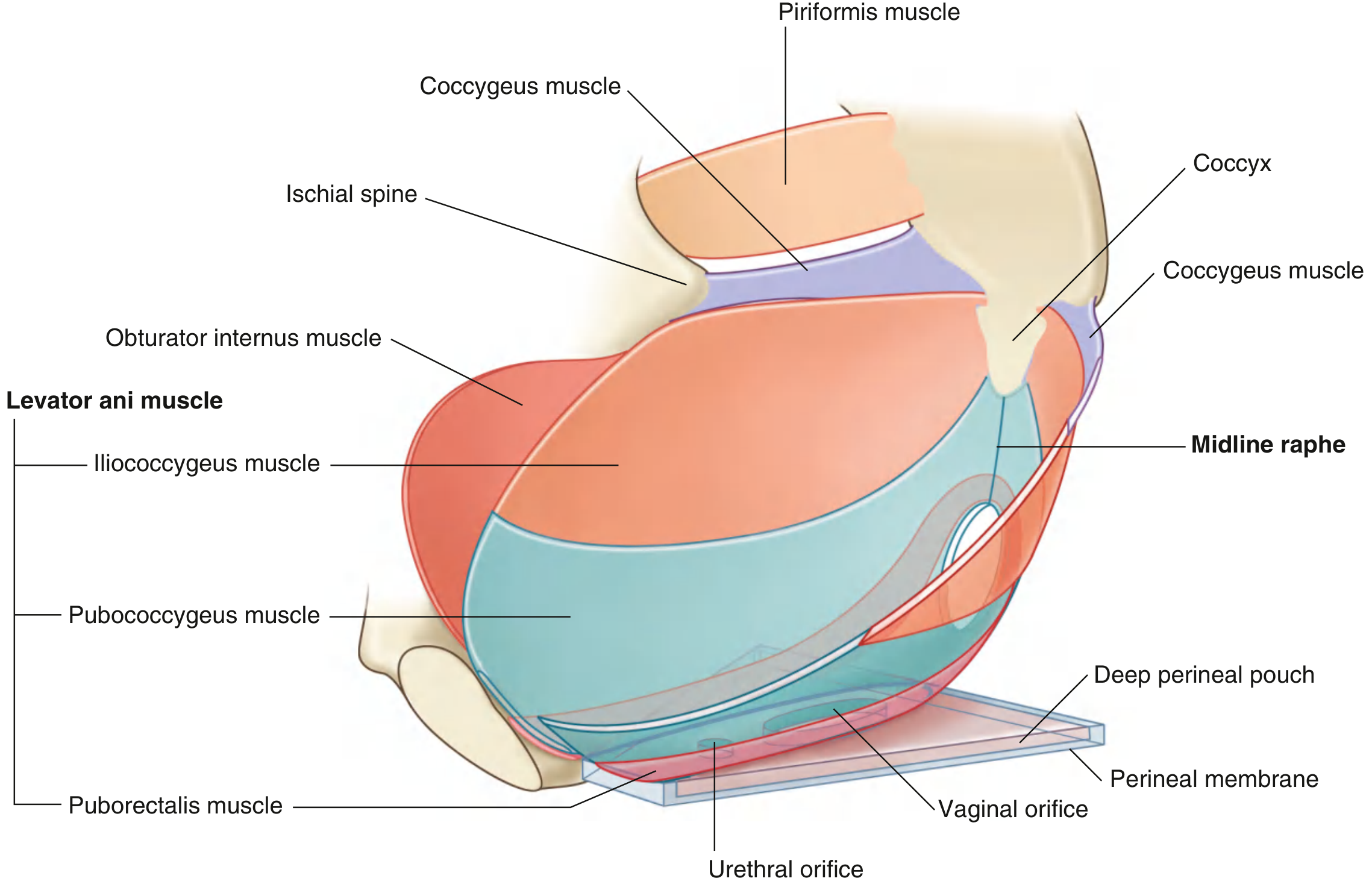
The pelvic floor separates the pelvic cavity from the perineum. It is a bowl/funnel-shaped structure formed by:
Levator Ani (main component)
Three parts:
| Part | Origin | Function |
|---|---|---|
| Pubococcygeus | Body of pubis → coccyx | Subdivided into puboprostaticus (levator prostatae), pubovaginalis, puboanalis |
| Puborectalis | Pubis → sling around anorectal junction | Forms perineal flexure (~90°); acts as pinch valve; angle increases to 130-140° during defecation |
| Iliococcygeus | Tendinous arch of obturator internus fascia → coccyx | Forms anococcygeal raphe with contralateral muscle |
- Innervation: branches of S4 anterior ramus + pudendal nerve (S2-S4)
- Contains a U-shaped urogenital hiatus anteriorly - urethra (both sexes) and vagina (females) pass through it
Coccygeus (ischiococcygeus)
- Triangular; overlies sacrospinous ligament
- Ischial spine → lateral sacrum and coccyx
- Innervation: S3, S4
- Completes the posterior part of the pelvic diaphragm
Anterior Support
- Perineal membrane: thick triangular fascial sheet between arms of pubic arch
- Deep perineal pouch: narrow space superior to perineal membrane
6. Pelvic Cavity Contents
The pelvic cavity is lined by peritoneum continuous with abdominal peritoneum (peritoneum does not reach the pelvic floor in most areas).
Contents - Midline Viscera
| Structure | Position |
|---|---|
| Bladder | Most anterior |
| Uterus (in females) | Between bladder and rectum |
| Rectum | Most posterior |
Peritoneal folds between viscera create pouches:
- Rectouterine pouch (Pouch of Douglas) - deepest in females; site of fluid accumulation
- Vesicouterine pouch - between bladder and uterus
- Rectovesical pouch - in males, between bladder and rectum
7. Communications of the Pelvic Cavity
| Aperture | Structures Passing Through |
|---|---|
| Pelvic inlet | Continuous with abdominal cavity |
| Greater sciatic foramen | Piriformis, sciatic nerve, superior/inferior gluteal nerves and vessels, pudendal nerve, internal pudendal artery |
| Lesser sciatic foramen | Obturator internus tendon, pudendal nerve + internal pudendal artery (re-entering perineum) |
| Obturator canal | Obturator nerve + vessels (to thigh) |
| Urogenital hiatus | Urethra, vagina (females) |
- The pudendal nerve exits via the greater sciatic foramen, wraps around the ischial spine/sacrospinous ligament, and re-enters through the lesser sciatic foramen into the perineum
8. Neurovascular Supply
Arteries
- Internal iliac artery (from bifurcation of common iliac at L5/S1 level) - principal supply to pelvic viscera
- Anterior division: obturator, inferior vesical, uterine, vaginal, middle rectal, internal pudendal, inferior gluteal
- Posterior division: iliolumbar, lateral sacral, superior gluteal
- Superior rectal (from inferior mesenteric artery) - supplies upper rectum
Veins
- Internal iliac vein → common iliac vein → inferior vena cava
- Pelvic venous plexuses are extensive and valveless - allow spread of infection/malignancy
Nerve Supply
- Sacral plexus (L4-S4): formed on anterior surface of piriformis; gives sciatic nerve (largest peripheral nerve in body), superior/inferior gluteal nerves, pudendal nerve, nerve to obturator internus, nerve to quadratus femoris
- Pelvic splanchnic nerves (nervi erigentes) - S2, S3, S4: parasympathetic to pelvic viscera; also carry pain from pelvic organs
- Sympathetic supply: hypogastric plexuses from lumbar sympathetics
9. Male vs. Female Pelvis
| Feature | Male | Female |
|---|---|---|
| Shape | Heart-shaped inlet | Oval/round inlet |
| Pubic arch angle | <70° (narrow, subpubic angle) | >80° (wide, subpubic arch) |
| Ischial spines | Prominent, project medially | Less prominent |
| Greater sciatic notch | Narrow | Wide |
| Pelvic cavity | Deep, narrow, funnel-shaped | Shallow, wide, cylindrical |
| Sacrum | Long, narrow, more curved | Short, wide, less curved |
| Ischial tuberosity | More vertical/medial | More everted |
Female pelvis is adapted for childbirth - wider inlet, larger cavity, wider outlet.
10. Clinical Points
- Bone marrow biopsy: iliac crest is the preferred site - superficial, easily palpated
- Obturator hernia: can occur through the obturator canal - more common in elderly, thin females; may compress obturator nerve
- Ischial tuberosity fracture in adolescents: avulsion at hamstring origin
- Pudendal nerve block: infiltrated at ischial spine (accessible transvaginally or transperineally)
- Pouch of Douglas: first site of fluid/pus accumulation in the female peritoneal cavity; drained via posterior fornix
- Piriformis syndrome: compression of sciatic nerve by piriformis muscle - mimics sciatica
- Levator ani tears: occur during difficult vaginal delivery - predispose to pelvic organ prolapse and incontinence
- Defecation: puborectalis relaxation increases anorectal angle from ~90° to ~130-140°, combined with increased intraabdominal pressure
Gray's Anatomy for Students (9780323934237), Chapter 5: Pelvis and Perineum
3rd cranial and 6th cranial nerves
Finding Sources
Finding Sources
Reading File
Reading File
Reading File
Reading File
Reading File
Reading File
Finding Sources
Finding Sources
Reading File
Finding Sources
Loading Image
I now have comprehensive content from Gray's Anatomy for Students, Harrison's, and Adams & Victor's. Let me compile detailed short notes.
CN III - Oculomotor Nerve & CN VI - Abducens Nerve
Sources: Gray's Anatomy for Students, Harrison's Principles of Internal Medicine 22E, Adams & Victor's Principles of Neurology 12E
CRANIAL NERVE III - OCULOMOTOR NERVE
Fiber Types
| Fiber | Type | Target |
|---|---|---|
| General Somatic Efferent (GSE) | Motor | Extraocular muscles |
| General Visceral Efferent (GVE) | Parasympathetic (preganglionic) | Sphincter pupillae + ciliary muscle |
Nucleus
- Located in the rostral midbrain (at the level of the superior colliculus), in the periaqueductal grey
- Composed of multiple subnuclei, one for each muscle it supplies
- Edinger-Westphal nucleus = parasympathetic nucleus; provides preganglionic fibers for pupil constriction and accommodation
- Key nuclear feature: levator palpebrae superioris is controlled by a single midline subnucleus - so a nuclear lesion causes bilateral ptosis
- Superior rectus subnucleus is crossed - it supplies the contralateral superior rectus
Course
1. Intramedullary (fascicular): Nerve fibers pass anteriorly through the red nucleus and cerebral peduncle → exits the anterior surface of the midbrain in the interpeduncular fossa (between the two cerebral peduncles)
2. Subarachnoid space: Runs between the posterior cerebral artery (above) and the superior cerebellar artery (below) - vulnerable to posterior communicating artery (PCoA) aneurysm here; travels forward toward the cavernous sinus
3. Cavernous sinus: Runs in the lateral wall of the cavernous sinus (superior position, above CN IV)
4. Superior orbital fissure: Passes through the superior orbital fissure within the common tendinous ring → enters the orbit and divides into superior and inferior branches
Branches in the Orbit
| Branch | Muscles Supplied |
|---|---|
| Superior branch | Levator palpebrae superioris, Superior rectus |
| Inferior branch | Medial rectus, Inferior rectus, Inferior oblique |
| Parasympathetic root to ciliary ganglion | From branch to inferior oblique → synapses in ciliary ganglion → short ciliary nerves → sphincter pupillae + ciliary muscles |
Muscles supplied (mnemonic: LR₆SO₄, rest CN III):
- Levator palpebrae superioris
- Superior rectus
- Medial rectus
- Inferior rectus
- Inferior oblique
Parasympathetic Pathway
Preganglionic: Edinger-Westphal nucleus → CN III → ciliary ganglion (in orbit)
Postganglionic: Short ciliary nerves → sphincter pupillae (miosis) + ciliary muscle (accommodation)
Complete CN III Palsy - Clinical Features
| Feature | Explanation |
|---|---|
| Ptosis | Levator palpebrae superioris paralysis |
| "Down and out" eye position | Unopposed lateral rectus (CN VI) and superior oblique (CN IV) |
| Diplopia | False image projected up and medially from affected eye |
| Dilated, fixed pupil (mydriasis) | Loss of parasympathetic constriction; iridoplegia |
| Loss of accommodation (cycloplegia) | Ciliary muscle paralysis |
Partial vs. Complete Palsy - Pupil Rule (High-Yield)
| Lesion Type | Pupil | Reason |
|---|---|---|
| Compressive (aneurysm, tumor, herniation) | Pupil INVOLVED (dilated) - early | Parasympathetic fibers run on the outer surface of the nerve - compressed first |
| Ischemic/microvascular (diabetes, HTN) | Pupil SPARED | Ischemia affects the inner core first; superficial parasympathetics relatively spared |
A painful CN III palsy with pupil involvement = compressive cause until proven otherwise - urgent neuroimaging + CT/MR angiogram
Midbrain Syndromes (Fascicular CN III Palsies)
| Syndrome | Lesion | CN III Palsy + |
|---|---|---|
| Weber's syndrome | Cerebral peduncle | Contralateral hemiparesis |
| Benedikt's syndrome | Red nucleus | Contralateral tremor, chorea, athetosis |
| Nothnagel's syndrome | Superior cerebellar peduncle | Contralateral cerebellar ataxia |
| Claude's syndrome | Red nucleus + SCP | Features of both Benedikt + Nothnagel |
All produce ipsilateral CN III palsy + contralateral long tract/cerebellar signs (crossed deficits).
Causes of CN III Palsy by Location
| Location | Causes |
|---|---|
| Nuclear/fascicular | Brainstem infarct, hemorrhage, tumor, demyelination |
| Subarachnoid space | PCoA aneurysm, meningitis, uncal herniation, trauma |
| Cavernous sinus | Carotid aneurysm, carotid-cavernous fistula, pituitary adenoma, thrombosis, Tolosa-Hunt syndrome, herpes zoster |
| Orbit/superior orbital fissure | Tumor, trauma |
| Microvascular (anywhere along course) | Diabetes mellitus, hypertension (pupil-sparing; spontaneous recovery in weeks-months) |
Aberrant Regeneration (Post-CN III Palsy)
After compression or trauma (NOT microvascular), aberrant re-wiring can occur:
- Eyelid elevates on downgaze or adduction (lid-gaze dyskinesis)
- Pupil constricts on attempted adduction, elevation, or depression
- Suggests previous CN III compression - primary aberrant regeneration without prior palsy suggests cavernous sinus meningioma
CRANIAL NERVE VI - ABDUCENS NERVE
Fiber Type
- General Somatic Efferent (GSE) only - purely motor
Nucleus
- Located in the caudal pons, beneath the floor of the fourth ventricle (forms the facial colliculus with the looping fibers of CN VII)
- Contains two types of neurons:
- Motor neurons → lateral rectus muscle (ipsilateral)
- Interneurons → cross to the contralateral medial longitudinal fasciculus (MLF) → reach the contralateral CN III nucleus (medial rectus) - this mediates conjugate horizontal gaze
A lesion of the abducens nucleus causes an ipsilateral conjugate gaze palsy (not just lateral rectus palsy), because both the motor neurons and the interneurons are destroyed.
Course
1. Intramedullary (fascicular): Fibers pass ventrally through the pons → exits the brainstem at the pontomedullary junction (between the pons and medulla) - the longest intracranial course of any cranial nerve
2. Subarachnoid space: Passes over the petrous apex of the temporal bone - where it takes a sharp bend over the dorsum sellae (Gruber's/petrosphenoidal ligament holds it here - Dorello's canal)
3. Cavernous sinus: Runs within the cavernous sinus (not in the lateral wall like CN III, IV) - travels lateral to the internal carotid artery - the only CN to pass through the body of the sinus
4. Superior orbital fissure: Enters through the superior orbital fissure within the common tendinous ring → passes laterally to supply the lateral rectus muscle
Action
Lateral rectus = abducts the eye (turns it outward/laterally)
CN VI Palsy - Clinical Features
| Feature | Detail |
|---|---|
| Eye deviation | Affected eye deviated medially (esotropia) - unopposed medial rectus |
| Diplopia | Horizontal, uncrossed (images side by side) - maximal on ipsilateral gaze |
| Limitation | Cannot abduct (laterally deviate) the affected eye |
| Compensation | Patient turns head toward the side of palsy to reduce diplopia |
Diplopia Fields
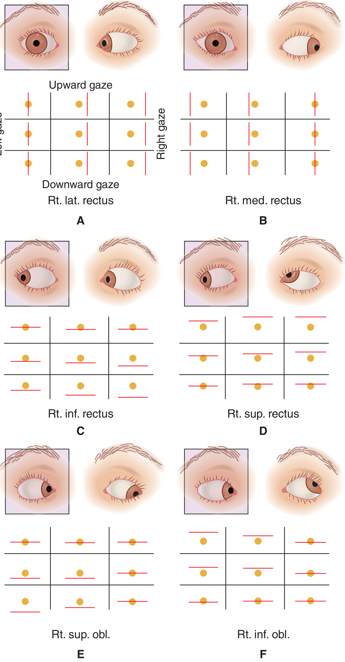
A: Right lateral rectus palsy - images maximally separated on right gaze
Causes of CN VI Palsy
| Level | Causes |
|---|---|
| Nuclear/fascicular (pons) | Pontine infarct, Möbius syndrome, Wernicke's encephalopathy, tumor, demyelination |
| Subarachnoid/radicular | Trauma, meningitis, aneurysm, raised ICP (false localizing sign) |
| Petrous apex | Gradenigo's syndrome (mastoiditis/petrous osteitis, CN VI palsy + deep facial pain - CN V + ipsilateral deafness) |
| Cavernous sinus | Carotid aneurysm, cavernous sinus thrombosis, pituitary tumor, Tolosa-Hunt syndrome, diabetes, herpes zoster |
| Orbit | Tumor, granuloma |
| Uncertain | Migraine, viral, raised ICP (bilateral CN VI palsy = false localizing sign in raised ICP) |
CN VI as a "False Localizing Sign"
CN VI has the longest intracranial course and passes over the sharp petrous apex - it is vulnerable to downward displacement from raised intracranial pressure. Bilateral CN VI palsies with papilloedema suggest raised ICP rather than a pontine lesion.
Internuclear Ophthalmoplegia (INO) - CN VI Nucleus Connection
The MLF connects the CN VI nucleus (in pons) to the contralateral CN III nucleus (in midbrain). Damage to this pathway causes INO:
- Impaired adduction of the ipsilateral eye on lateral gaze (medial rectus does not fire properly)
- Nystagmus of the contralateral (abducting) eye
- Most common cause: MS (bilateral INO); also brainstem infarct
- One-and-a-half syndrome: ipsilateral gaze palsy + ipsilateral INO = lesion in PPRF + MLF on same side
Summary Comparison Table
| Feature | CN III (Oculomotor) | CN VI (Abducens) |
|---|---|---|
| Nucleus | Rostral midbrain (periaqueductal grey) | Caudal pons (floor of 4th ventricle) |
| Exits brainstem | Anterior midbrain (interpeduncular fossa) | Pontomedullary junction |
| Fiber type | GSE + GVE (parasympathetic) | GSE only |
| Muscles supplied | Levator palpebrae, SR, MR, IR, IO | Lateral rectus only |
| Cavernous sinus | Lateral wall (superior) | Inside the sinus (lateral to ICA) |
| SOF entry | Within common tendinous ring | Within common tendinous ring |
| Palsy position | "Down and out" + ptosis + mydriasis | Medial deviation (esotropia) |
| Diplopia | Vertical + horizontal | Horizontal (uncrossed) |
| Key causes | PCoA aneurysm, DM (pupil-sparing), herniation | Raised ICP (false localizing), Gradenigo's, DM |
| Midbrain syndromes | Weber's, Benedikt's, Nothnagel's, Claude's | Foville's syndrome (pons) |
| Gaze palsy | No (nuclear lesion: rare bilateral ptosis) | Yes (nuclear lesion = ipsilateral gaze palsy) |
Gray's Anatomy for Students (9780323934237); Harrison's Principles of Internal Medicine 22E (9781265977061); Adams & Victor's Principles of Neurology 12E (9781264264520)
Temporomendibular joint short notes
Finding Sources
Reading File
Reading File
Finding Sources
Reading File
Finding Sources
Reading File
Reading File
Reading File
Loading Image
This is a shared conversation. Sign in to Orris to start your own chat.