Hey i am a first year student in kusms in nepal and i have a pbl session tomorrrow. I have some learnining objective and my pbl tutuor me to learn about the topics from a medical book i will ask you qustion and the answer you give everything must be from a medical book. also give me the source . tell me tell me about why zinc is given to baby in diarrhoea? also tell me about layer of abdomen and blood cs, its important and about blood culture sensitivity with a diagram remeber every answer must be with respect to a book
PBL Study Notes - KUSMS First Year
TOPIC 1: Why Is Zinc Given to Babies in Diarrhoea?
Source: Park's Textbook of Preventive and Social Medicine (p. 721 & Diarrhoeal Disease Control chapter)
"Zinc deficiency is common in children from developing countries due to lack of intake of animal food, high dietary phytate content, inadequate food intake and increased faecal losses during diarrhoea."
- Park's Textbook of Preventive and Social Medicine, p. 721
"Zinc supplementation in combination with oral rehydration therapy has been shown to significantly reduce the duration and severity of acute and persistent diarrhoea and to increase survival in a number of randomized control trials."
- Park's Textbook of Preventive and Social Medicine, p. 721
"Adequate zinc intake is essential for maintaining the integrity of the immune system."
- Park's Textbook of Preventive and Social Medicine, p. 721
"When a zinc supplement is given during an episode of acute diarrhoea, it reduces the episode's duration and severity. In addition, zinc supplements given for 10 to 14 days lower the incidence of diarrhoea in the following 2 to 3 months."
- Park's Textbook of Preventive and Social Medicine, Diarrhoeal Disease Control chapter
| Age | Dose | Duration |
|---|---|---|
| Infants under 6 months | 10 mg/day | 10-14 days |
| Children older than 6 months | 20 mg/day | 10-14 days |
"WHO and UNICEF therefore recommend daily 10 mg of zinc for infants under 6 months of age, and 20 mg for children older than 6 months for 10-14 days."
- Park's Textbook of Preventive and Social Medicine, p. 885
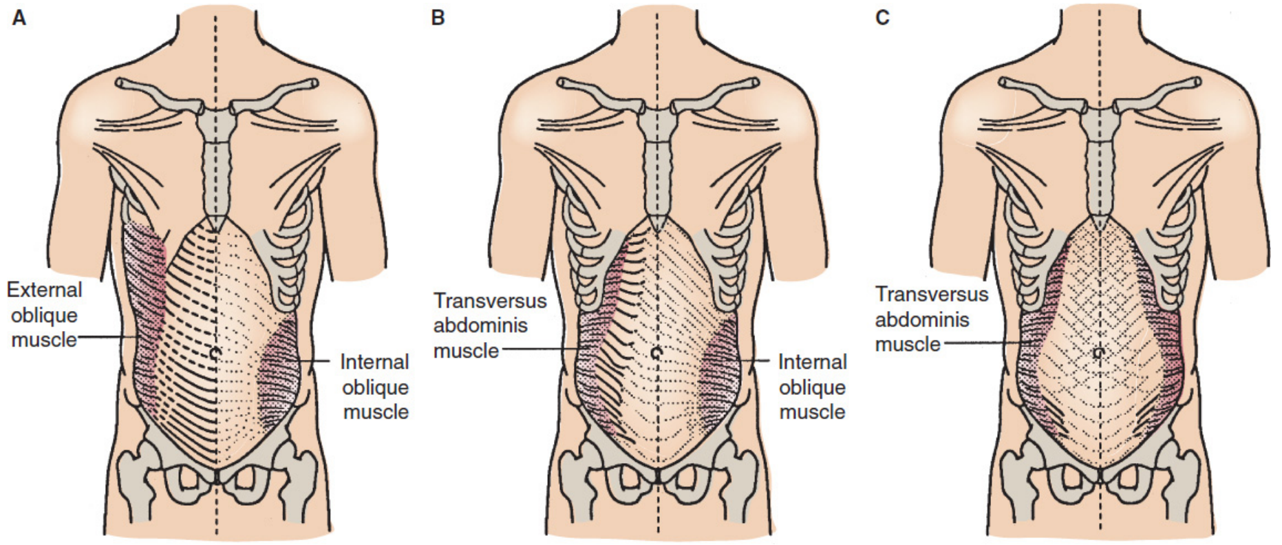
TOPIC 2: Layers of the Abdomen (Anterior Abdominal Wall)
Source: Mulholland and Greenfield's Surgery - Scientific Principles and Practice, 7e (p. 3632-3639) & THIEME Atlas of General Anatomy
Layers (Superficial to Deep):
| # | Layer | Details |
|---|---|---|
| 1 | Skin | Segmentally innervated (dermatome pattern, 7th-12th intercostal nerves) |
| 2 | Superficial fascia (Camper's fascia) | Contains fat (panniculus adiposus); continuous with perineal fascia; contains superficial epigastric and superficial circumflex iliac vessels |
| 3 | Deep fascia (Scarpa's fascia) | Compressed fibrous layer; forms fundiform ligament of penis |
| 4 | External abdominal oblique muscle & aponeurosis | Most superficial of three flat muscles; fibres run obliquely downward and medially; aponeurosis forms anterior rectus sheath and inguinal ligament |
| 5 | Internal abdominal oblique muscle & aponeurosis | Fibres run perpendicular to external oblique; aponeurosis splits to enclose rectus abdominis |
| 6 | Transversus abdominis muscle & aponeurosis | Deepest flat muscle; fibres run transversely; forms arcuate line (line of Douglas) |
| 7 | Transversalis fascia | Thin fibrous layer lining the inner surface of transversus abdominis |
| 8 | Extraperitoneal (preperitoneal) fat | Loose fatty connective tissue between transversalis fascia and peritoneum |
| 9 | Peritoneum (parietal) | Innermost layer |
"The three muscles of the lateral aspect of the anterior abdominal wall are composed of a variable amount of muscle with a large aponeurosis. The aponeurosis is the tendon of insertion for the lateral muscles, and it also forms the sheath of the rectus abdominis. The midline decussation of the three aponeuroses forms the linea alba."
- Mulholland and Greenfield's Surgery, p. 3634
Central Muscle - Rectus Abdominis:
"The rectus abdominis forms the central and anchoring muscle mass of the anterior abdomen. The rectus muscle arises from the fifth to the seventh costal cartilages and inserts on the pubic symphysis and pubic crest. Each rectus muscle is segmented by tendinous intersections at the levels of the xiphoid process and the umbilicus and at a point midway between these two. The principal blood supply reaches the muscle from the superior and inferior epigastric arteries."
- Mulholland and Greenfield's Surgery, p. 3639
Diagram of Abdominal Wall Layers:
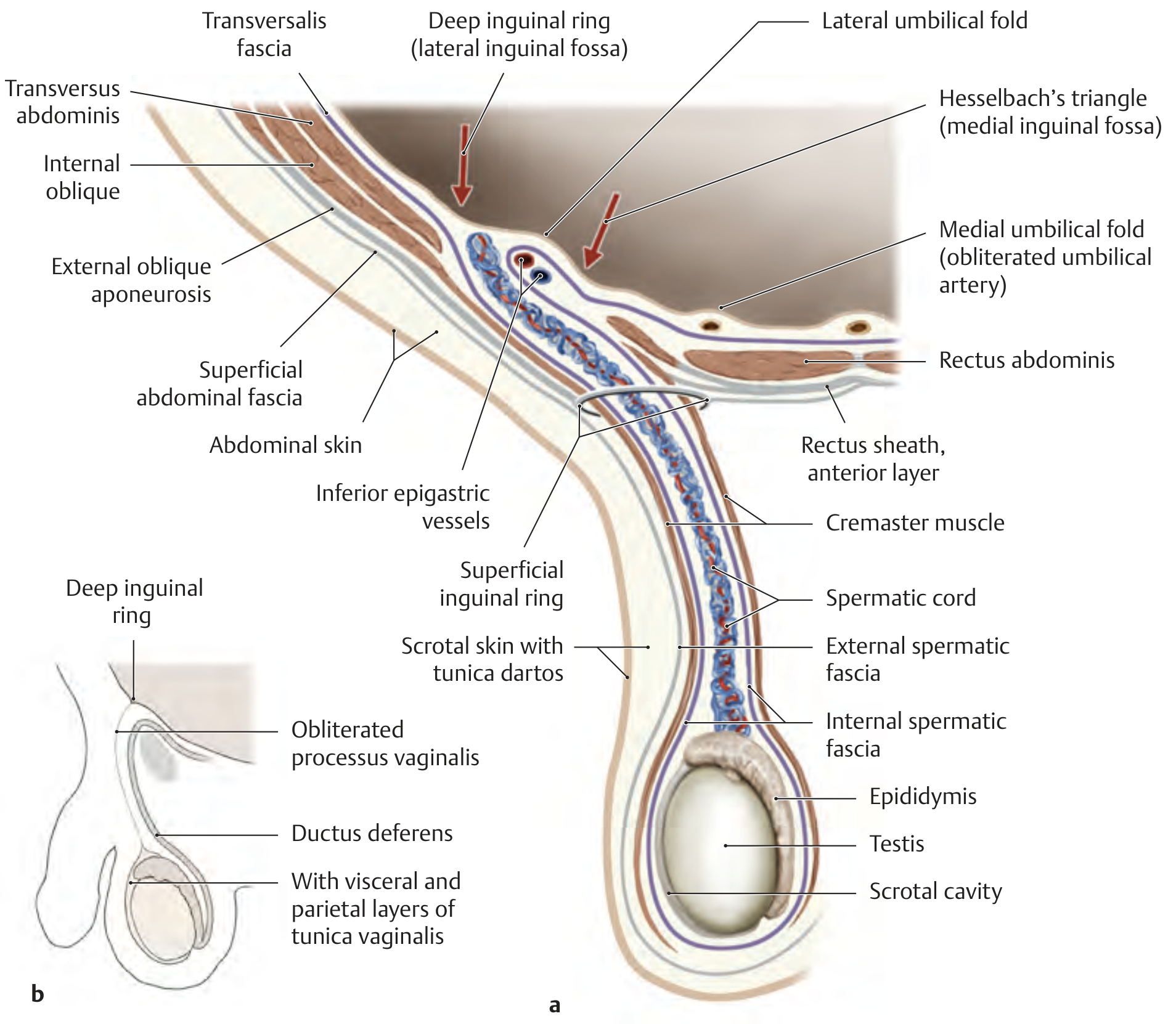
Relationship to Spermatic Cord (Clinically Important):
| Abdominal Wall Layer | Spermatic Cord/Testes Covering |
|---|---|
| Abdominal skin + membranous superficial fascia | Scrotal skin with tunica dartos |
| External oblique fascia | External spermatic fascia |
| Internal oblique muscle and fascia | Cremaster muscle with cremasteric fascia |
| Transversalis fascia | Internal spermatic fascia |
| Peritoneum | Tunica vaginalis (parietal and visceral layers) |
TOPIC 3: Blood Culture and Sensitivity (Blood C/S)
Source: Tietz Textbook of Laboratory Medicine, 7th Edition & Roberts and Hedges' Clinical Procedures in Emergency Medicine
What is Blood Culture and Sensitivity?
Types of Bloodstream Infections (BSI):
"BSIs occur for a variety of reasons. In general, they can be categorised as either intravascular or extravascular. Intravascular infections include infective endocarditis and intravenous catheter-related infection. Extravascular BSI results when organisms multiply at a body site, are drained by the lymphatic system, and gain access to the bloodstream. Common extravascular sites that lead to BSI are abscesses, wounds, surgical wounds, the respiratory tract, and the genitourinary tract."
- Tietz Textbook of Laboratory Medicine, 7th Edition
Specimen Collection - Critical Points:
"A critical aspect of blood culture draws is the disinfection of the skin (for peripheral blood draws). The CDC recommends disinfecting the site with 70% alcohol and then using povidone-iodine. Alternative methods include the use of chlorhexidine and chlorine peroxide."
- Tietz Textbook of Laboratory Medicine, 7th Edition
"The most critical factor influencing the sensitivity of blood cultures for the detection of BSI is the volume of blood drawn."
- Tietz Textbook of Laboratory Medicine, 7th Edition
| Patient | Recommended Volume | Notes |
|---|---|---|
| Adults | 40-60 mL total (20 mL per draw) | Each bottle (aerobic + anaerobic) gets 10 mL each |
| Children | 1 mL per 2.5 kg body weight (max 4-5% of total blood volume) | Bacteraemia levels are 10x higher in neonates than adults |
"In adults, a number of studies have assessed the blood volume required to maximize sensitivity and found that the ideal total volume for blood culture is 40 to 60 mL. Each draw should be 20 mL, with each bottle (aerobic and anaerobic) inoculated with 10 mL."
- Tietz Textbook of Laboratory Medicine, 7th Edition
"Most institutions encourage the order of two blood culture sets that include both aerobic and anaerobic bottles. These cultures should be collected from two distinct sites (i.e., a peripheral venous draw from the right and left arm or a central line and peripheral venous draw)."
- Tietz Textbook of Laboratory Medicine, 7th Edition
"These paired cultures help the provider differentiate between contamination and true infection. True bacteraemia will result in multiple positive cultures, whereas single positive cultures with skin or respiratory microbiota suggest contamination."
- Tietz Textbook of Laboratory Medicine, 7th Edition
- In typhoid fever: only 40-60% sensitive (Harrison's Principles of Internal Medicine, 22e)
- With proper technique and adequate volume: up to 80% per set, nearly 99% with two sets of 20 mL each
"An individual set is typically not more than 80% sensitive... sensitivity is typically at approximately 99% with two sets consisting of 20 mL of blood per set."
- Roberts and Hedges' Clinical Procedures in Emergency Medicine
Summary Diagram: Blood Culture Process
PATIENT (suspected BSI/sepsis)
↓
SKIN DISINFECTION
(70% alcohol → povidone-iodine OR chlorhexidine)
↓
BLOOD DRAW (2 separate sites)
SET 1 (Left arm) SET 2 (Right arm)
↓ ↓ ↓ ↓
Aerobic Anaerobic Aerobic Anaerobic
bottle bottle bottle bottle
(10 mL) (10 mL) (10 mL) (10 mL)
↓
INCUBATION (5 days, continuously monitored)
↓
ORGANISM IDENTIFIED
↓
ANTIBIOTIC SENSITIVITY TESTING
(MIC determined → report Sensitive / Intermediate / Resistant)
↓
TARGETED ANTIBIOTIC THERAPY
"Levels of bacteremia are typically 10-fold higher in neonates than in adults. The sicker the child, the greater the likelihood of a high level of bacteremia."
- Roberts and Hedges' Clinical Procedures in Emergency Medicine
Quick Summary Table for PBL Revision
| Topic | Key Point | Source |
|---|---|---|
| Zinc in diarrhoea | Reduces duration + severity; protective for 2-3 months post-episode | Park's PSM |
| Zinc dose (<6 months) | 10 mg/day for 10-14 days with ORS | Park's PSM / WHO-UNICEF |
| Zinc dose (>6 months) | 20 mg/day for 10-14 days with ORS | Park's PSM / WHO-UNICEF |
| Abdominal layers | Skin → Camper → Scarpa → EO → IO → TA → Transversalis fascia → Extraperitoneal fat → Peritoneum | Mulholland's Surgery 7e |
| Linea alba | Formed by midline decussation of all 3 aponeuroses | Mulholland's Surgery 7e |
| Blood C/S key factor | Volume of blood drawn | Tietz Lab Medicine 7e |
| Adult volume | 40-60 mL total, 2 sets from 2 sites | Tietz Lab Medicine 7e |
| Two sets reason | Distinguish true bacteraemia from contamination | Tietz Lab Medicine 7e |
tell me about the toxic dose of pcm in a normal and alcholic person and tell me the mechanism of pcm in flowchart and why n-acetyl is given here also define technical terms used
Paracetamol (PCM) Toxicity - Complete PBL Notes
PART 1: Therapeutic vs Toxic vs Lethal Doses
Source: The Essentials of Forensic Medicine and Toxicology, 36th Edition (2026) - Table 31.1 & Sleisenger and Fordtran's GI and Liver Disease
| Category | Adults | Children |
|---|---|---|
| Therapeutic dose | 500-1000 mg per dose (max 4 g/day) | 10-15 mg/kg/dose (max 60 mg/kg/day) |
| Toxic dose | ≥150 mg/kg (single ingestion) | 150-200 mg/kg |
| Lethal dose | >350 mg/kg | 250-350 mg/kg |
"When a large dose (>150 mg/kg or >10g in an adult) is taken, it causes serious toxicity. Fatality is common with doses >250 mg/kg."
- P.C. Dikshit Textbook of Forensic Medicine and Toxicology, p. 578
"Overdoses of 20 g or more are potentially lethal."
- P.C. Dikshit Textbook of Forensic Medicine and Toxicology
PART 2: Toxic Dose in an ALCOHOLIC Person - Why is it LOWER?
Source: Sleisenger and Fordtran's Gastrointestinal and Liver Disease & Goldman-Cecil Medicine
"Among heavy drinkers, daily acetaminophen doses of 2 to 6 g have been associated with fatal hepatotoxicity."
- Sleisenger and Fordtran's GI and Liver Disease, p. 1670
"In chronic alcoholics, even 5-6 g/day taken for a few days can cause hepatotoxicity."
- P.C. Dikshit Textbook of Forensic Medicine and Toxicology
- CYP2E1 induction: Alcohol induces (upregulates) the CYP2E1 enzyme. This pushes MORE paracetamol down the toxic pathway, producing MORE NAPQI than normal.
- Glutathione depletion: Chronic alcohol use depletes hepatic glutathione stores, so even normal NAPQI production cannot be neutralised.
"Excessive ethanol intake can induce CYP2E1, resulting in a greater fraction of the dose being converted to NAPQI. Patient-specific factors that can significantly reduce the dose threshold for acetaminophen toxicity include poor nutrition and fasting (depleting glucuronidation substrates) as well as excessive ethanol intake."
- Goldman-Cecil Medicine, International Edition
Risk Factors That Lower the Toxic Threshold (Table from Sleisenger & Fordtran)
| Risk Factor | Effect |
|---|---|
| Chronic alcohol ingestion | Toxic dose threshold lowered; worsens prognosis; nephrotoxicity common |
| Fasting / malnutrition | Toxic dose threshold lowered ("therapeutic misadventure") |
| Concomitant drugs (isoniazid, phenytoin, zidovudine) | Toxic threshold lowered |
| Cirrhosis | Recommendation: do NOT exceed 2 g in 24 hours |
| Late presentation / delayed treatment (>16 hr) | Predicts worse outcome |
PART 3: Mechanism of PCM Toxicity - FLOWCHART
Source: The Essentials of Forensic Medicine and Toxicology, 36th Ed. (Fig. 31.1) & Goldman-Cecil Medicine & Rosen's Emergency Medicine
PARACETAMOL (PCM) ingested
|
├──────────────────────────────────────┐
▼ ▼
~90% SAFE PATHWAY ~10% TOXIC PATHWAY
| |
Conjugation with CYP2E1 (P450 enzyme)
GLUCURONIC ACID oxidizes PCM
and SULFATE |
| ▼
NON-TOXIC metabolites NAPQI formed
excreted in urine (N-Acetyl-p-Benzoquinone Imine)
|
┌──────────┴───────────┐
▼ ▼
NORMAL DOSE OVERDOSE
(Glutathione (Glutathione
adequate) EXHAUSTED)
| |
▼ ▼
NAPQI neutralized NAPQI accumulates
by glutathione freely in liver cells
| |
▼ ▼
CYSTEINE + COVALENT BINDING
MERCAPTURIC ACID to liver cell proteins
(non-toxic, excreted) (especially mitochondria)
|
▼
OXIDATIVE STRESS
↓
CENTRILOBULAR HEPATIC NECROSIS
↓
RENAL TUBULAR NECROSIS
↓
ACUTE LIVER FAILURE → DEATH
"N-acetyl-benzoquinone-imine is a highly reactive arylating minor metabolite of paracetamol, which is detoxified by conjugation with glutathione. When a very large dose is taken, hepatic glutathione is depleted and this metabolite binds covalently to proteins in liver cells and renal tubules causing necrosis. Toxicity thus shows a threshold phenomenon, manifesting only when glutathione is depleted to a critical point."
- P.C. Dikshit Textbook of Forensic Medicine and Toxicology, p. 578
"The binding of acetaminophen to hepatocyte macromolecules leads to hepatocyte necrosis... Mitochondrial enzymes are a particular target of NAPQI."
- Harrison's Principles of Internal Medicine, 22E & Sleisenger and Fordtran's
PART 4: Clinical Stages of PCM Poisoning
Source: The Essentials of Forensic Medicine and Toxicology, 36th Ed. - Table 31.2
| Stage | Timeframe | Clinical Features |
|---|---|---|
| Stage I | 0-24 hours | Nausea, vomiting, anorexia, malaise, diaphoresis (sweating); LFTs usually normal |
| Stage II | 24-72 hours | Right upper quadrant pain; rising AST/ALT and bilirubin; prolonged PT; possible renal involvement |
| Stage III | 72-96 hours | Peak hepatotoxicity: jaundice, coagulopathy, hypoglycaemia, hepatic encephalopathy, renal failure, metabolic acidosis, multi-organ failure |
| Stage IV | 4-14 days | Recovery if survived - gradual normalisation of liver function (liver can regenerate) |
PART 5: Why is N-Acetylcysteine (NAC) Given?
Source: P.C. Dikshit Textbook of Forensic Medicine and Toxicology & The Essentials of Forensic Medicine, 36th Ed.
"N-acetylcysteine... replenishes the glutathione stores of the liver and prevents binding of the toxic metabolite to other cellular constituents."
- P.C. Dikshit Textbook of Forensic Medicine, p. 578
"Acetaminophen toxicity is due to the depletion of hepatic glutathione, resulting in the accumulation of toxic metabolic intermediates."
- Morgan and Mikhail's Clinical Anesthesiology, 7e
NAC Dosing (from Textbooks):
- Loading: 150 mg/kg IV over 1 hour
- Then: 50 mg/kg over next 4 hours
- Then: 100 mg/kg over 16 hours
- Total: 21-hour regimen (FDA-approved)
- Loading: 140 mg/kg, then 70 mg/kg every 4 hours
- Duration: 24-72 hours (6-18 doses)
"Treatment should be started as early as possible as it is practically ineffective if started 16 hours or more after paracetamol ingestion."
- P.C. Dikshit Textbook of Forensic Medicine, p. 578
"Most beneficial when administered within 8-10 hours of overdose."
- The Essentials of Forensic Medicine and Toxicology, 36th Ed.
PART 6: Glossary of Technical Terms Used
| Term | Definition |
|---|---|
| NAPQI | N-Acetyl-p-Benzoquinone Imine - the toxic intermediate metabolite of paracetamol produced by CYP2E1 |
| CYP2E1 | Cytochrome P450 2E1 - a liver enzyme responsible for the toxic oxidative metabolism of paracetamol; induced by chronic alcohol |
| Glutathione | A tripeptide (glutamate-cysteine-glycine) and the body's main antioxidant/detoxifier; neutralizes NAPQI under normal conditions |
| Hepatotoxicity | Toxic damage to liver cells (hepatocytes) |
| Centrilobular necrosis | Death of liver cells in the central zone of the liver lobule (zone 3) - the area richest in CYP enzymes and most vulnerable to NAPQI |
| Covalent binding | A chemical bond where NAPQI permanently attaches to liver cell proteins, destroying their function |
| Glucuronidation | The safe metabolic pathway where paracetamol is conjugated (joined) with glucuronic acid to form a harmless, water-soluble metabolite |
| Sulfation | Another safe metabolic pathway - conjugation of PCM with sulfate to form a harmless metabolite |
| N-Acetylcysteine (NAC) | The antidote for PCM poisoning; replenishes glutathione; most effective within 8-10 hours |
| Hepatic encephalopathy | Brain dysfunction caused by liver failure - confusion, drowsiness, coma |
| Coagulopathy | Inability of blood to clot properly, caused by liver failure (the liver makes clotting factors) |
| ALT/AST | Alanine aminotransferase / Aspartate aminotransferase - liver enzymes that rise when liver cells are damaged |
| Threshold phenomenon | Toxicity only occurs AFTER glutathione is depleted below a critical level; below that level, the body safely neutralizes NAPQI |
| Mercapturic acid | The harmless end-product formed when glutathione neutralizes NAPQI; excreted safely in urine |
| Oxidative stress | Cell damage caused by accumulation of reactive oxygen species (free radicals), triggered when NAPQI binds to mitochondria |
Quick Summary for PBL
NORMAL ADULT: Toxic >150 mg/kg (>10 g single dose); Lethal >350 mg/kg
ALCOHOLIC: Toxic at just 5-6 g/day (threshold massively reduced)
REASON: Alcohol → ↑CYP2E1 → ↑NAPQI + depleted glutathione
ANTIDOTE: NAC - replenishes glutathione; must give within 8-10 hours
- P.C. Dikshit Textbook of Forensic Medicine and Toxicology (p. 578)
- The Essentials of Forensic Medicine and Toxicology, 36th Edition (2026) (Table 31.1, 31.2, Fig. 31.1)
- Goldman-Cecil Medicine, International Edition
- Sleisenger and Fordtran's GI and Liver Disease (Table 88.5, p. 1670)
- Rosen's Emergency Medicine
- Morgan and Mikhail's Clinical Anesthesiology, 7e
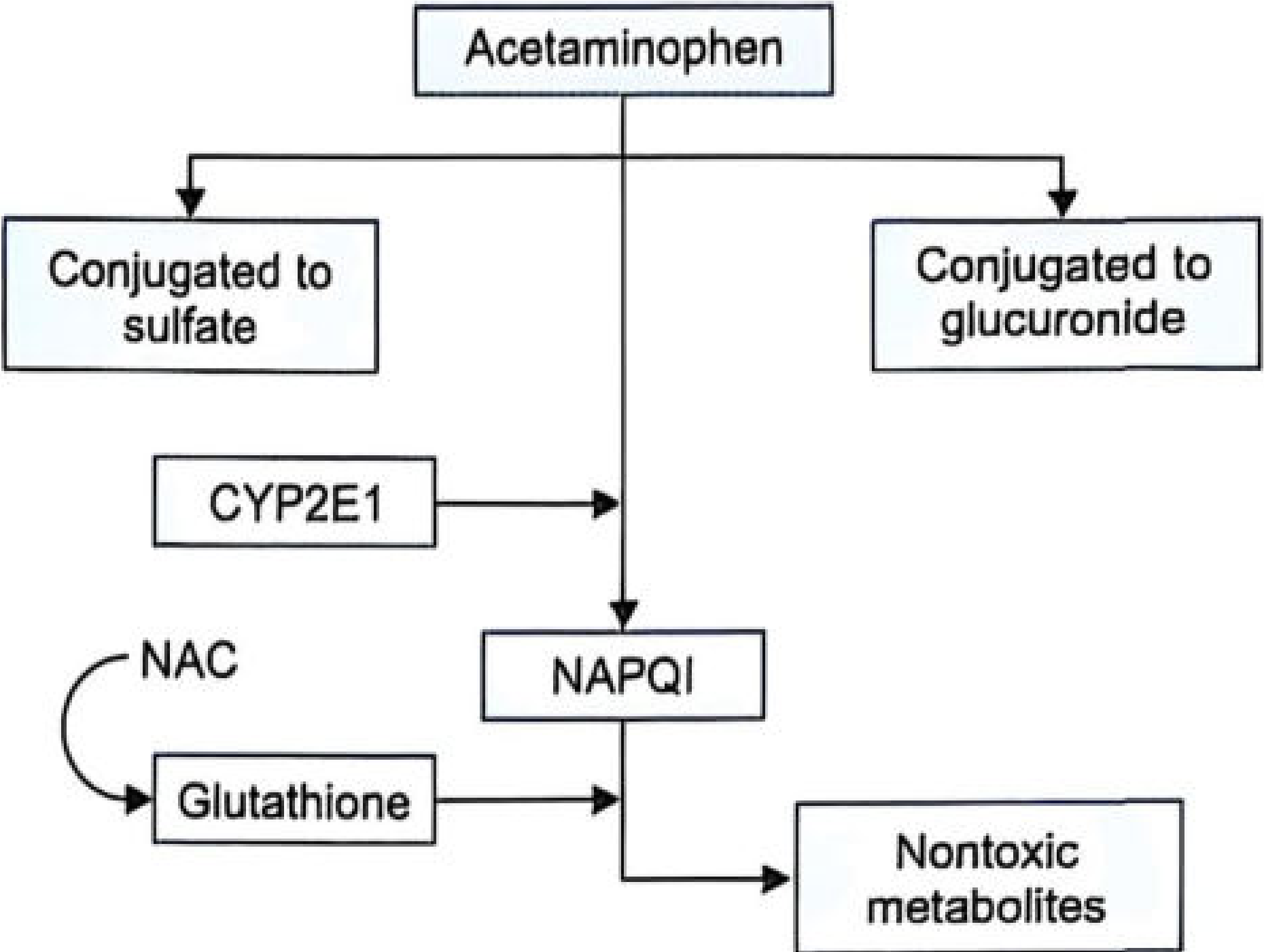
Tell me the metabolism chart of pcm
-
Acetaminophen (PCM) at the top splits into 3 paths:
- Left - Conjugated to sulfate (safe, non-toxic)
- Right - Conjugated to glucuronide (safe, non-toxic)
- Middle (the dangerous path) - Goes through CYP2E1 enzyme → produces NAPQI (the toxic metabolite)
-
NAPQI is then neutralized by Glutathione → forms non-toxic metabolites (safely excreted)
-
NAC (N-Acetylcysteine) shown on the left feeds into glutathione - this is exactly why NAC is the antidote. It replenishes glutathione so it can keep neutralizing NAPQI.
I am asking the metabolism pathway of PCM in a normal person.
Metabolism of Paracetamol (PCM) in a Normal Person
Textbook Diagram (Figure 43.2):
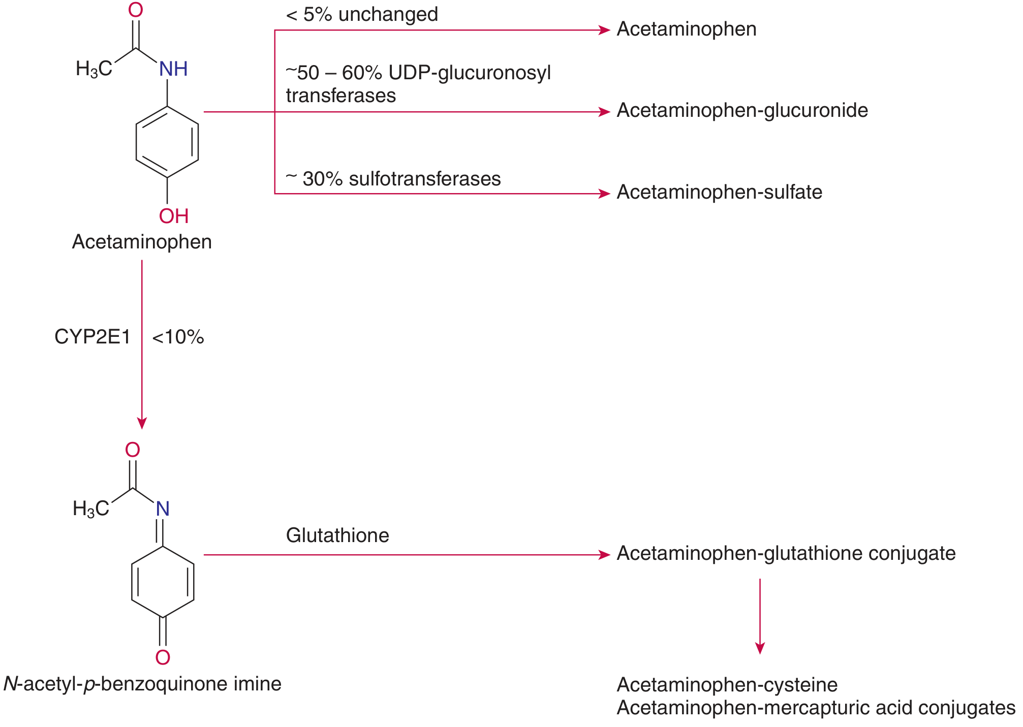
Reading the Chart Step by Step:
| Route | Enzyme Used | % of Dose | Product | Safe or Toxic? |
|---|---|---|---|---|
| Glucuronidation | UDP-glucuronosyltransferases | ~50-60% | Acetaminophen-glucuronide | ✅ Safe - excreted in urine |
| Sulfation | Sulfotransferases | ~30% | Acetaminophen-sulfate | ✅ Safe - excreted in urine |
| Unchanged excretion | None | <5% | Acetaminophen unchanged | ✅ Excreted in urine |
| CYP2E1 oxidation | CYP2E1 (P450 enzyme) | <10% | NAPQI (toxic intermediate) | ⚠️ Toxic BUT neutralized by glutathione |
"Acetaminophen is normally metabolized in the liver to glucuronide (50 to 60%) and sulfate (<30%) conjugates. A smaller amount (<10%) is metabolized by a cytochrome P450 mixed-function oxidase pathway... forming NAPQI. This intermediate normally undergoes electrophilic conjugation with glutathione and then subsequent transformation to cysteine and mercapturic acid conjugates of acetaminophen."
- Tietz Textbook of Laboratory Medicine, 7th Edition
The NAPQI pathway in a NORMAL person (safe outcome):
PCM → CYP2E1 → NAPQI
↓
+ Glutathione (abundant in normal liver)
↓
Acetaminophen-glutathione conjugate
↓
Acetaminophen-CYSTEINE conjugate
+
Acetaminophen-MERCAPTURIC ACID conjugate
↓
Safely excreted in URINE
Why does this go wrong in overdose?
"With acetaminophen overdose, the sulfation pathway becomes saturated; consequently, a greater portion is metabolized by the P450 mixed-function oxidase pathway. When the tissue stores of glutathione become depleted, arylation of cellular molecules by the benzoquinoneimine intermediate leads to hepatic necrosis."
- Tietz Textbook of Laboratory Medicine, 7th Edition
Simply teach me the metabolism pathway of pcm in normal human
PCM Metabolism in a Normal Person
The Diagram (from Tietz Textbook of Laboratory Medicine, 7th Ed.):
Simple Explanation:
PCM taken orally
|
Absorbed from gut
|
Reaches LIVER
|
┌───────────────┼─────────────────┐
▼ ▼ ▼
~50-60% ~30% ~10%
GLUCURONIDATION SULFATION CYP2E1 enzyme
↓ ↓ ↓
PCM-glucuronide PCM-sulfate NAPQI
(safe, harmless) (safe, harmless) ↓
↓ ↓ + Glutathione
Excreted in Excreted in ↓
URINE URINE PCM-mercapturic acid
(safe, harmless)
↓
Excreted in URINE
In Simple Words:
| Path | What happens | Safe? |
|---|---|---|
| 50-60% | Joins with glucuronic acid → flushed out in urine | ✅ Yes |
| ~30% | Joins with sulfate → flushed out in urine | ✅ Yes |
| ~10% | Liver enzyme CYP2E1 converts it to NAPQI → glutathione immediately destroys NAPQI → flushed out in urine | ✅ Yes (in normal dose) |
Source: Tietz Textbook of Laboratory Medicine, 7th Edition, Figure 43.2
explain the chart in simple words and also define the terms
Step-by-Step Simple Explanation
STEP 1 - You swallow a PCM tablet
STEP 2 - The liver breaks PCM down into 4 routes
🔵 STREAM 1 - The Biggest Safe Route (~50-60%)
- The liver sticks a molecule called glucuronic acid onto PCM
- This makes PCM water-soluble (dissolves in water)
- The kidney then flushes it out in your urine
- Completely harmless
Terms defined:
Glucuronidation - A process where the liver adds glucuronic acid to a drug to make it water-soluble so the kidneys can remove it. Think of it as "gift wrapping" the drug so the kidney can throw it away.
UDP-glucuronosyltransferase - The enzyme (a biological tool/machine) in the liver that performs glucuronidation. "UDP" is just the energy molecule that powers this reaction.
Conjugation - The general word for "joining/attaching" one molecule to another. Glucuronidation and sulfation are both types of conjugation.
🟡 STREAM 2 - Second Safe Route (~30%)
- The liver sticks a sulfate group (a sulfur-containing molecule) onto PCM
- Again becomes water-soluble → flushed out in urine
- Completely harmless
Terms defined:
Sulfation - Similar to glucuronidation, but instead of glucuronic acid, a sulfate molecule is attached to PCM. End result is the same - PCM is made harmless and removed in urine.
Sulfotransferase - The enzyme that performs sulfation in the liver.
🟢 STREAM 3 - Tiny Unchanged Route (<5%)
- A tiny bit of PCM escapes the liver without being changed at all
- Goes straight out in urine as PCM
- Harmless (too small an amount to matter)
🔴 STREAM 4 - The Important Dangerous Route (<10%)
PCM → CYP2E1 → NAPQI (toxic!)
↓
Glutathione grabs it
↓
PCM-Glutathione conjugate
↓
PCM-Cysteine + PCM-Mercapturic acid
↓
Out in URINE (safe ✅)
All Technical Terms - Simple Definitions
| Term | Simple Definition |
|---|---|
| Metabolism | How the body chemically breaks down and processes a drug |
| Liver | The body's main "drug-processing factory" - handles almost all medications |
| Enzyme | A biological tool/machine made of protein that speeds up a chemical reaction in the body |
| Glucuronidation | Liver sticks glucuronic acid onto PCM → makes it water-soluble → kidney removes it in urine. The BIGGEST safe pathway (~50-60%) |
| Glucuronic acid | A sugar molecule that the liver attaches to drugs to make them easier to remove |
| UDP-glucuronosyltransferase | The enzyme that performs glucuronidation. "UDP" = uridine diphosphate, the energy source |
| Sulfation | Liver sticks a sulfate group onto PCM → also water-soluble → removed in urine. Second safe pathway (~30%) |
| Sulfotransferase | The enzyme that performs sulfation |
| Conjugation | "Joining" - the general term for attaching one molecule to another to make it harmless |
| CYP2E1 | Cytochrome P450 2E1 - a liver enzyme that converts PCM into the dangerous NAPQI. Works on about 10% of PCM in a normal person. Alcohol INCREASES this enzyme |
| Cytochrome P450 | A family of liver enzymes responsible for metabolising most drugs. They use oxygen to chemically modify drug molecules |
| NAPQI | N-Acetyl-p-Benzoquinone Imine - the TOXIC chemical produced from PCM by CYP2E1. The "villain" in PCM toxicity |
| Glutathione | The body's natural "fire extinguisher" - a protective molecule in the liver that grabs and neutralizes NAPQI. Made of 3 amino acids (glutamate + cysteine + glycine) |
| Glutathione conjugate | The safe product formed when glutathione grabs NAPQI - completely harmless |
| Cysteine conjugate | A further breakdown product of the glutathione conjugate - harmless, excreted in urine |
| Mercapturic acid | The final harmless end-product - excreted in urine. Seeing it in urine is proof that PCM was processed safely |
| Water-soluble | Can dissolve in water - important because kidneys can only remove water-soluble substances in urine |
| Excretion | Removing waste products from the body - mainly through urine (kidneys) |
The One-Line Summary
In a normal person, 90% of PCM is safely converted to harmless products (glucuronide + sulfate) and flushed out in urine. The remaining 10% becomes toxic NAPQI, but the liver's glutathione immediately destroys it before it can cause any harm.
what is cpk, alp,tlc what is its normal range ,significance and when does it increas
CPK, ALP, and TLC - Complete Notes for PBL
1. CPK (Creatine Phosphokinase) = Also called CK (Creatine Kinase)
What is it?
"Creatine kinase (CK) catalyzes the reversible phosphorylation of creatine by ATP... Physiologically, when muscle contracts, ATP is converted to ADP, and CK catalyzes the re-phosphorylation of ADP to ATP using creatine phosphate as the phosphorylation reservoir."
- Tietz Textbook of Laboratory Medicine, 7th Edition
Where is it found in the body?
| Source | CPK content |
|---|---|
| Skeletal muscle | Highest - 2500 U/g protein |
| Heart muscle | High - 550 U/g protein |
| Brain | Some |
| Liver & red blood cells | Almost NONE |
Normal Range
Males: 46-171 U/L | Females: 34-145 U/L (Tietz Textbook of Laboratory Medicine, 7th Edition)
CPK Isoforms (Sub-types) - Very Important!
| Isoform | Full Name | Found in | Clinical use |
|---|---|---|---|
| CK-MM (CK-3) | Muscle-Muscle | Skeletal muscle | 97-100% of normal serum CK |
| CK-MB (CK-2) | Muscle-Brain | Heart muscle | Marker of heart attack (MI) |
| CK-BB (CK-1) | Brain-Brain | Brain | Brain injury (rarely measured) |
When does CPK INCREASE?
| Cause | Why |
|---|---|
| Myocardial Infarction (heart attack) | Heart muscle cells die → CK-MB released into blood |
| Muscular Dystrophy | Muscle fibres breaking down |
| Rhabdomyolysis | Massive muscle destruction (crush injury, extreme exercise) |
| Polymyositis / Dermatomyositis | Inflammatory muscle disease |
| After surgery / IM injection | Muscle trauma |
| Hypothyroidism | Slow metabolism affects muscle |
| Strenuous exercise | Temporary rise - normal |
| Epileptic seizures | Muscle overactivity |
| Blunt chest trauma | Heart/muscle injury |
| Brain injury / stroke | CK-BB released (rarely elevated in serum) |
"An elevated serum creatine kinase (CK) concentration can result from recent exercise, electromyography (EMG), intramuscular injection, liver disease, or myocardial infarction (MI) as well as from a primary muscle disease."
- Bradley and Daroff's Neurology in Clinical Practice
"CK-MB is no longer a recommended marker for the initial diagnosis of NSTEMI. It lacks specificity because it is present in both skeletal and cardiac muscle cells... Troponin is the recommended biomarker for assessment of myocardial necrosis."
- The Washington Manual of Medical Therapeutics
2. ALP (Alkaline Phosphatase)
What is it?
Where is it found?
"Alkaline phosphatase (ALP) is expressed in liver, bile ducts, bone, intestine, placenta, kidney, and leukocytes."
- Sabiston Textbook of Surgery, The Biological Basis of Modern Surgical Practice
Normal Range
30-140 IU/L
- Bailey and Love's Short Practice of Surgery, 28th Edition - Table 69.1
ALP Isoforms by source:
| Source | When elevated |
|---|---|
| Liver ALP | Liver disease, biliary obstruction |
| Bone ALP | Bone disorders, fractures, growing children |
| Placental ALP | Normal in pregnancy |
| Intestinal ALP | Intestinal disease |
When does ALP INCREASE?
| Cause | Explanation |
|---|---|
| Cholestasis (blocked bile flow) | ALP is most elevated here - bile duct obstruction causes ALP to be produced in huge amounts |
| Biliary obstruction (gallstones, tumour) | Bile backs up → ALP rises dramatically |
| Hepatitis / Liver disease | Liver cell damage |
| Liver cancer / metastasis | Tumour stimulates ALP production |
| Primary Biliary Cirrhosis | Autoimmune bile duct destruction |
"The serum ALP is particularly elevated with cholestatic liver disease or biliary obstruction."
- Bailey and Love's Short Practice of Surgery, 28th Edition
| Cause | Explanation |
|---|---|
| Paget's Disease of bone | Overactive bone remodelling |
| Bone fractures (healing) | Bone formation requires ALP |
| Bone cancer / metastasis | Tumour invades bone |
| Osteomalacia / Rickets | Vitamin D deficiency → abnormal bone |
| Hyperparathyroidism | Excess PTH stimulates bone breakdown |
| Growing children | Normally higher due to bone growth |
- Pregnancy (placental ALP - normal)
- Prostate cancer with bone metastasis
"It is important to note that routine laboratory analysis of ALP is not isoform specific and so ALP from a skeletal source may also lead to elevation, particularly Paget's disease and prostate cancer."
- Bailey and Love's Short Practice of Surgery, 28th Edition
- If GGT is also raised → ALP is from the liver
- If GGT is normal → ALP is from bone
3. TLC (Total Leukocyte Count) = WBC Count (White Blood Cell Count)
What is it?
Normal Range
4,500 - 10,000 cells/μL (mean = 7,500/μL)
- Goldman-Cecil Medicine, International Edition
WBC Differential (breakdown of types):
| Cell Type | % of WBC | Function |
|---|---|---|
| Neutrophils | ~60% | Fight bacteria (most important) |
| Lymphocytes | ~30% | Fight viruses, produce antibodies |
| Monocytes | ~5% | Engulf and destroy pathogens |
| Eosinophils | ~2-4% | Fight parasites, allergic reactions |
| Basophils | ~0.5-1% | Allergic reactions |
"The normal peripheral white blood cell count ranges between 4500/μL and 10,000/μL, with a mean of 7500/μL, and is composed of neutrophils, lymphocytes, monocytes, basophils, and eosinophils. Because neutrophils usually represent about 60% of the peripheral WBC, derangement in the WBC usually reflects elevation or reduction in the absolute neutrophil count."
- Goldman-Cecil Medicine, International Edition
When TLC INCREASES (Leukocytosis = WBC > 10,000/μL):
| Cause | Type of WBC elevated |
|---|---|
| Bacterial infection | Neutrophilia (↑ neutrophils) |
| Viral infection | Lymphocytosis (↑ lymphocytes) |
| Parasitic infection / Allergy | Eosinophilia (↑ eosinophils) |
| Sepsis / severe infection | Very high neutrophils + "left shift" |
| Leukaemia (blood cancer) | All types - massive increase |
| Myocardial Infarction | Reactive neutrophilia |
| Chronic inflammation | Neutrophilia (e.g., TB, abscess, Crohn's) |
| Corticosteroid drugs | Neutrophilia |
| Cigarette smoking | Neutrophilia |
| Post-splenectomy | Lymphocytosis |
| Physical/emotional stress | Neutrophilia (mobilized from marginated pool) |
"Many acute bacterial infections can present with leukocytosis with a 'left shift', referring to the circulation of more immature myeloid cells."
- Goldman-Cecil Medicine, International Edition
When TLC DECREASES (Leukopenia = WBC < 4,000/μL):
| Cause | Explanation |
|---|---|
| Viral infections (HIV, dengue, typhoid) | Virus suppresses marrow |
| Bone marrow failure | Not producing enough cells |
| Chemotherapy / radiation | Destroys marrow cells |
| Autoimmune disease (SLE) | Antibodies destroy WBCs |
| Severe sepsis / early sepsis | WBCs consumed rapidly |
| Nutritional deficiency (B12, folate) | Marrow needs these to make cells |
Quick Summary Table
| Test | Full Form | Normal Range | Main Organ | Increases When |
|---|---|---|---|---|
| CPK/CK | Creatine Phosphokinase | 46-171 U/L (M), 34-145 U/L (F) | Skeletal muscle, Heart | MI, muscle disease, trauma, rhabdomyolysis |
| ALP | Alkaline Phosphatase | 30-140 IU/L | Liver, Bone | Biliary obstruction, cholestasis, bone disease |
| TLC | Total Leukocyte Count | 4,500-10,000/μL | Blood (bone marrow) | Infection, inflammation, leukaemia, drugs |
- Tietz Textbook of Laboratory Medicine, 7th Edition (CPK normal range, isoenzymes)
- Bailey and Love's Short Practice of Surgery, 28th Edition (ALP normal range, Table 69.1)
- Goldman-Cecil Medicine, International Edition (TLC normal range, leukocytosis)
- Sabiston Textbook of Surgery (ALP sources)
- Bradley and Daroff's Neurology in Clinical Practice (CPK causes)