
Mcqs on brain stem having
I need a bit more information before generating the MCQs. Let me ask quickly:
Asking for Preferences
Which aspects of the brainstem do you want the MCQs to cover? · How many MCQs do you need? · What difficulty level?
Searching sources...
Reading File
Finding Sources
Finding Sources
Reading File
Reading File
Finding Sources
I now have rich textbook content from Neuroanatomy through Clinical Cases and Principles of Neural Science. I have enough to construct 20 high-quality UG-level MCQs covering brainstem anatomy/structure and cranial nerve nuclei. Let me compile them now.
20 MCQs on Brainstem - Anatomy & Cranial Nerve Nuclei (UG / MBBS Level)
SECTION A: Brainstem Structure & Anatomy (Q 1-10)
Q1. The brainstem, in rostrocaudal order, consists of:
- A) Medulla - Pons - Midbrain
- B) Midbrain - Medulla - Pons
- C) Pons - Midbrain - Medulla
- D) Midbrain - Pons - Medulla ✅
The brainstem consists of three subdivisions in rostrocaudal order: midbrain, pons, and medulla oblongata. (Neuroanatomy through Clinical Cases, 3rd Ed.)
Q2. In the brainstem, which of the following is the dividing line between motor and sensory nuclei?
- A) Median sulcus
- B) Sulcus limitans ✅
- C) Lateral sulcus
- D) Central canal
Motor cranial nerve nuclei lie ventral to the sulcus limitans; sensory nuclei lie dorsal to it - an arrangement analogous to the spinal cord. (Neuroanatomy through Clinical Cases, 3rd Ed.)
Q3. The "floor of the fourth ventricle" forms the dorsal surface of which brainstem structure?
- A) Midbrain
- B) Medulla only
- C) Pons and medulla ✅
- D) Midbrain and pons
The floor of the fourth ventricle is contributed by both the pons (upper part) and the medulla (lower part). The facial colliculus and hypoglossal trigone are landmarks on this floor.
Q4. The medial longitudinal fasciculus (MLF) connects which of the following nuclei?
- A) Facial, trigeminal, and hypoglossal
- B) Oculomotor, trochlear, abducens, and vestibular nuclei ✅
- C) Dorsal vagal, ambiguus, and hypoglossal
- D) Edinger-Westphal, ciliary, and superior cervical ganglia
The MLF interconnects the oculomotor, trochlear, and abducens nuclei with vestibular nuclei, coordinating conjugate eye movements. (Neuroanatomy through Clinical Cases, 3rd Ed.)
Q5. The periaqueductal gray (PAG) surrounds which structure in the midbrain?
- A) Cerebral peduncle
- B) Substantia nigra
- C) Cerebral aqueduct (aqueduct of Sylvius) ✅
- D) Red nucleus
The oculomotor (CN III) and trochlear (CN IV) nuclei lie just ventral to the periaqueductal gray in the midbrain.
Q6. The "facial colliculus" seen on the floor of the fourth ventricle is formed by:
- A) Facial nucleus itself
- B) Fibers of CN VII looping over the abducens nucleus ✅
- C) Abducens nucleus alone
- D) MLF fibers crossing the pons
The abducens nucleus helps form the facial colliculus in the mid-to-lower pons, where CN VII fibers loop over it before exiting the brainstem. (Neuroanatomy through Clinical Cases, 3rd Ed.)
Q7. The "hypoglossal trigone" is a surface landmark on the floor of the fourth ventricle overlying which nucleus?
- A) Dorsal motor nucleus of vagus
- B) Nucleus ambiguus
- C) Hypoglossal nucleus (CN XII) ✅
- D) Solitary nucleus
The hypoglossal nuclei form the hypoglossal trigones on the floor of the fourth ventricle in the medulla. (Neuroanatomy through Clinical Cases, 3rd Ed.)
Q8. From medial to lateral, three longitudinal nuclei run along the floor/central canal of the medulla in a constant relationship. What is the correct medial-to-lateral order?
- A) Solitary - Dorsal motor X - Hypoglossal
- B) Dorsal motor X - Hypoglossal - Solitary
- C) Hypoglossal - Dorsal motor X - Solitary ✅
- D) Solitary - Hypoglossal - Dorsal motor X
From medial to lateral: hypoglossal nucleus, dorsal motor nucleus of X, and solitary nucleus. (Neuroanatomy through Clinical Cases, 3rd Ed.)
Q9. The brainstem tegmentum is distinguished from the basis (base) by its position:
- A) Ventral to the tectum
- B) Dorsal to the cerebral aqueduct
- C) Dorsal to the basis, ventral to the tectum ✅
- D) Only present in the midbrain
Any cross-section of the brainstem shows three laminae rostrocaudally: tectum (dorsal), tegmentum (middle), and basis (ventral). (Localization in Clinical Neurology, 8e)
Q10. During development, which group of cranial nerve motor nuclei migrates ventrolaterally away from the midline into the tegmentum?
- A) Somatic motor (GSE) nuclei
- B) Parasympathetic (GVE) nuclei
- C) Branchial motor (SVE) nuclei ✅
- D) Special visceral afferent nuclei
Branchial motor nuclei initially lie lateral to the somatic motor nuclei but gradually migrate ventrolaterally. Somatic motor nuclei stay adjacent to the midline. (Neuroanatomy through Clinical Cases, 3rd Ed.)
SECTION B: Cranial Nerve Nuclei (Q 11-20)
Q11. Which cranial nerve nuclei are classified as "somatic motor (GSE)"?
- A) CN V, VII, IX, X
- B) CN III, VII, IX, XI
- C) CN III, IV, VI, XII ✅
- D) CN IX, X, XI, XII
The somatic motor (GSE) nuclei are the oculomotor (III), trochlear (IV), abducens (VI), and hypoglossal (XII) - all innervating muscles derived from occipital somites. (Neuroanatomy through Clinical Cases, 3rd Ed.)
Q12. The nucleus ambiguus is the motor nucleus for which cranial nerves?
- A) CN V, VII
- B) CN VII, VIII
- C) CN IX, X (and part of XI) ✅
- D) CN XI, XII
The nucleus ambiguus contains the branchial motor (SVE) neurons for CN IX and X, supplying pharyngeal and laryngeal muscles. It runs longitudinally through the medulla.
Q13. Which parasympathetic (GVE) nucleus is associated with CN III and controls pupillary constriction?
- A) Superior salivatory nucleus
- B) Inferior salivatory nucleus
- C) Dorsal motor nucleus of X
- D) Edinger-Westphal nucleus ✅
The Edinger-Westphal nucleus is the parasympathetic nucleus of CN III. It forms a V-shaped cap over the oculomotor nuclei in the rostral midbrain and is responsible for pupillary constriction and accommodation. (Neuroanatomy through Clinical Cases, 3rd Ed.)
Q14. Taste sensation (special visceral afferent, SVA) from CN VII, IX, and X is relayed to which brainstem nucleus?
- A) Trigeminal nuclei
- B) Nucleus solitarius (rostral/gustatory portion) ✅
- C) Cochlear nuclei
- D) Vestibular nuclei
The rostral portion of the nucleus solitarius is the gustatory nucleus, receiving taste input from CN VII, IX, and X. The caudal portion handles cardiorespiratory and visceral inputs. (Neuroanatomy through Clinical Cases, 3rd Ed.)
Q15. Touch, pain, temperature, and proprioception from the face are processed by which brainstem nuclei?
- A) Cochlear and vestibular nuclei
- B) Nucleus solitarius
- C) Trigeminal nuclei ✅
- D) Dorsal motor nucleus of X
The trigeminal nuclei (principal/chief sensory, spinal, and mesencephalic) process general somatic afferent (GSA) inputs - touch, pain, temperature, and vibration from the face, sinuses, and meninges (CN V, VII, IX, X). (Neuroanatomy through Clinical Cases, 3rd Ed.)
Q16. The trigeminal motor nucleus (CN V) is located in which part of the brainstem?
- A) Midbrain
- B) Upper to mid pons ✅
- C) Lower pons and upper medulla
- D) Medulla
The trigeminal motor nucleus lies in the upper to mid pons, just medial to the chief trigeminal sensory nucleus, near the level where CN V exits the brainstem. (Neuroanatomy through Clinical Cases, 3rd Ed.)
Q17. The spinal accessory nucleus (CN XI) is anatomically located in:
- A) Medulla oblongata
- B) Lower pons
- C) Midbrain
- D) Upper five segments of the cervical spinal cord ✅
Unlike other cranial nerve nuclei, the spinal accessory nucleus (CN XI) is located in the upper five cervical spinal cord segments, protruding laterally between the dorsal and ventral horns. (Neuroanatomy through Clinical Cases, 3rd Ed.)
Q18. Hearing (cochlear) and vestibular sensation are relayed by CN VIII to which category of brainstem nuclei?
- A) General visceral afferent
- B) General somatic afferent
- C) Special visceral afferent
- D) Special somatic afferent (SSA) ✅
Cochlear and vestibular nuclei receive special somatic afferent (SSA) input via CN VIII. Note: olfaction and vision are also SSA but do NOT have nuclei in the brainstem.
Q19. The superior salivatory nucleus is the parasympathetic nucleus associated with which cranial nerve?
- A) CN IX
- B) CN X
- C) CN III
- D) CN VII ✅
Parasympathetic nuclei: Edinger-Westphal (CN III), superior salivatory nucleus (CN VII), inferior salivatory nucleus (CN IX), dorsal motor nucleus of X (CN X). The superior salivatory nucleus controls submandibular and sublingual glands via CN VII.
Q20. The oculomotor nucleus (CN III) and trochlear nucleus (CN IV) are located in which part of the brainstem, and what is their relationship to each other?
- A) Both in pons; IV is rostral to III
- B) Both in midbrain; IV is rostral to III
- C) III in medulla, IV in pons
- D) III in rostral midbrain, IV in caudal midbrain; both just ventral to the PAG ✅
The oculomotor nuclei (CN III) are in the rostral midbrain and the trochlear nuclei (CN IV) are in the caudal midbrain. Both lie just ventral to the periaqueductal gray (PAG). (Neuroanatomy through Clinical Cases, 3rd Ed.)
Quick Reference Summary Table
| CN | Nucleus | Type | Location |
|---|---|---|---|
| III | Oculomotor | Somatic motor (GSE) | Rostral midbrain |
| III | Edinger-Westphal | Parasympathetic (GVE) | Rostral midbrain |
| IV | Trochlear | Somatic motor (GSE) | Caudal midbrain |
| V | Trigeminal motor | Branchial motor (SVE) | Upper-mid pons |
| V | Trigeminal sensory | Somatic sensory (GSA) | Pons to medulla |
| VI | Abducens | Somatic motor (GSE) | Mid-lower pons |
| VII | Facial nucleus | Branchial motor (SVE) | Lower pons |
| VII | Superior salivatory | Parasympathetic (GVE) | Pons |
| VIII | Cochlear/Vestibular | Special somatic sensory (SSA) | Pontomedullary junction |
| IX/X | Nucleus ambiguus | Branchial motor (SVE) | Medulla |
| IX/X | Nucleus solitarius | Visceral sensory (SVA/GVA) | Medulla |
| X | Dorsal motor nucleus | Parasympathetic (GVE) | Medulla |
| XII | Hypoglossal | Somatic motor (GSE) | Medulla |
All answers are grounded in Neuroanatomy through Clinical Cases, 3rd Ed. and Principles of Neural Science, 6th Ed. (Kandel). These 20 questions cover the key UG examination points for brainstem anatomy and cranial nerve nuclei.
Explain all the nucleus of thalamus withb there functions
Searching sources...
Finding Sources
Finding Sources
Reading File
Reading File
Reading File
Reading File
Finding Sources
Finding Sources
Reading File
Finding Sources
Finding Sources
Reading File
Loading Image
I now have comprehensive textbook content from multiple authoritative sources. Let me compile the full explanation.
Nuclei of the Thalamus - Complete Guide with Functions
The thalamus is the "gateway to the cerebral cortex" - it processes virtually all information that reaches the cortex. It sits within the diencephalon and is divided into groups of nuclei by a Y-shaped sheet of white matter called the internal medullary lamina.
Here is the thalamic diagram showing the major nuclei and their inputs:
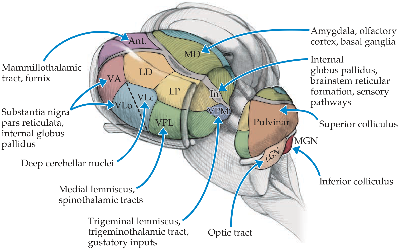
Classification Overview
Thalamic nuclei are divided into three broad categories:
| Category | Cortical Projection | Examples |
|---|---|---|
| Specific relay nuclei | Focal, discrete cortical areas | VPL, VPM, LGN, MGN, VL, VA |
| Association nuclei | Widespread, association areas | Pulvinar, MD, LD |
| Non-specific nuclei | Diffuse, wide cortex + striatum | Intralaminar, Midline, Reticular |
GROUP 1: SPECIFIC (RELAY) NUCLEI
These relay specific sensory or motor information to discrete cortical areas.
1. Ventral Posterior Lateral Nucleus (VPL)
| Inputs | Medial lemniscus (posterior column pathway) + Spinothalamic tract |
| Outputs | Primary somatosensory cortex (postcentral gyrus) |
| Function | Relays touch, pain, temperature, proprioception from the trunk and limbs to cortex |
| Clinical note | Lesion causes contralateral sensory loss; thalamic pain syndrome (Dejerine-Roussy syndrome) |
2. Ventral Posterior Medial Nucleus (VPM)
| Inputs | Trigeminal lemniscus, trigeminothalamic tract, taste fibers (CN VII, IX, X) |
| Outputs | Primary somatosensory cortex + gustatory (taste) cortex |
| Function | Relays somatosensory information from the face and head; also relays taste |
| Mnemonic | VPL = limbs, VPM = medial (face/head) |
3. Lateral Geniculate Nucleus (LGN)
| Inputs | Optic tract (from retina) |
| Outputs | Primary visual cortex (V1, striate cortex, occipital lobe - area 17) |
| Function | Relays visual information from retina to visual cortex |
| Mnemonic | "Lateral = Light" (vision) |
| Clinical note | Lesion causes contralateral homonymous hemianopia |
4. Medial Geniculate Nucleus (MGN)
| Inputs | Inferior colliculus (auditory pathway) |
| Outputs | Primary auditory cortex (Heschl's gyrus, temporal lobe) |
| Function | Relays auditory (hearing) information to auditory cortex |
| Mnemonic | "Medial = Music" (audition) |
| Clinical note | Lesion causes cortical deafness |
5. Ventral Lateral Nucleus (VL)
| Inputs | Deep cerebellar nuclei (dentate, interposed) + Internal globus pallidus + Substantia nigra pars reticulata |
| Outputs | Motor cortex (M1), premotor cortex, supplementary motor area |
| Function | Relays cerebellar and basal ganglia signals to motor cortex for coordinating movement |
| Sub-divisions | VL anterior part: receives from globus pallidus → SMA; VL posterior part: receives from cerebellum → motor/premotor cortex |
| Clinical note | Target for deep brain stimulation (DBS) in Parkinson's disease and essential tremor |
6. Ventral Anterior Nucleus (VA)
| Inputs | Substantia nigra pars reticulata + Internal globus pallidus + Deep cerebellar nuclei |
| Outputs | Widespread frontal lobe - prefrontal, premotor, motor, supplementary motor cortex |
| Function | Relays basal ganglia output to frontal cortex; broader projections than VL |
| Note | Works with VL as the thalamic motor relay for basal ganglia output |
GROUP 2: ASSOCIATION NUCLEI
These connect with association cortices and integrate information from multiple sources.
7. Pulvinar
| Inputs | Superior colliculus (extrageniculate visual pathway), primary visual/auditory/somatosensory cortex |
| Outputs | Parietotemporo-occipital association cortex (large areas) |
| Function | Behavioral orientation toward relevant visual and other stimuli; regulates information transmission across the visual system during attention; integrates multisensory inputs |
| Anatomy | Largest thalamic nucleus; "pillow-shaped" (pulvinar = Latin for cushion/couch); occupies most of posterior thalamus |
| Clinical note | Involved in attention and visual neglect |
8. Mediodorsal Nucleus (MD) / Dorsomedial Nucleus (DM)
| Inputs | Amygdala, olfactory cortex, limbic basal ganglia, prefrontal cortex |
| Outputs | Frontal association (prefrontal) cortex |
| Function | Major thalamic relay for the frontal lobe; limbic pathways, emotion, cognition, working memory, executive function |
| Clinical note | Damaged in Korsakoff syndrome (along with mammillary bodies) → anterograde amnesia; implicated in schizophrenia (dorsolateral prefrontal loop) |
9. Anterior Nucleus (AN)
| Inputs | Mammillary bodies (via mammillothalamic tract), hippocampal formation |
| Outputs | Cingulate gyrus (limbic cortex) |
| Function | Part of the Papez circuit (hippocampus → fornix → mammillary bodies → mammillothalamic tract → anterior nucleus → cingulate gyrus → hippocampus); involved in memory and emotion |
| Clinical note | Damaged in bilateral lesions (e.g., Korsakoff syndrome) → memory loss; target for DBS in epilepsy |
10. Lateral Dorsal Nucleus (LD)
| Inputs | Hippocampus (similar connections to anterior nucleus) |
| Outputs | Cingulate cortex |
| Function | Functions closely with the anterior nucleus; part of limbic circuitry |
11. Lateral Posterior Nucleus (LP)
| Inputs | Superior colliculus, primary visual/auditory/somatosensory cortex |
| Outputs | Posterior parietal and lateral temporal association cortex |
| Function | Functions closely with the pulvinar; involved in multisensory association and spatial processing |
GROUP 3: NON-SPECIFIC NUCLEI
These have widespread, diffuse projections to many cortical and subcortical areas.
12. Intralaminar Nuclei
Located within the internal medullary lamina. Divided into two functional groups:
A. Rostral Intralaminar Nuclei (Central medial, Paracentral, Central lateral)
| Inputs | Ascending reticular activating system (ARAS), deep cerebellar nuclei, globus pallidus |
| Outputs | Widespread cerebral cortex + striatum |
| Function | Maintain alert, conscious state by relaying ARAS inputs to cortex; also motor relay for basal ganglia and cerebellum |
B. Caudal Intralaminar Nuclei (Centromedian, Parafascicular)
| Inputs | Globus pallidus (internal), ARAS, sensory pathways |
| Outputs | Striatum (putamen, caudate), cerebral cortex |
| Function | Mainly motor relay for basal ganglia circuitry; the centromedian nucleus is the largest of these |
| Clinical note | Centromedian nucleus targeted in DBS for Tourette syndrome; involved in seizure genesis |
13. Midline Nuclei (Paraventricular, Parataenial, Reuniens/Medial Ventral, etc.)
| Inputs | Hypothalamus, basal forebrain, amygdala, hippocampus |
| Outputs | Amygdala, hippocampus, limbic cortex |
| Function | Limbic pathways; visceral and arousal functions; connection between hypothalamus and limbic system |
14. Reticular Nucleus (TRN)
| Inputs | Cerebral cortex, other thalamic relay and intralaminar nuclei, ARAS, basal forebrain |
| Outputs | Back to thalamic relay and intralaminar nuclei (does NOT project to cortex) |
| Neurotransmitter | Pure GABAergic (inhibitory) - the only inhibitory neurons in the thalamus |
| Anatomy | Thin sheet just lateral to the rest of the thalamus, medial to the internal capsule |
| Function | Acts as a "gatekeeper" - regulates and modulates activity of all other thalamic nuclei; role in sleep spindles, attention, and alertness |
| Key fact | The ONLY thalamic nucleus that does NOT project to the cortex |
Master Summary Table
| Nucleus | Key Input | Key Output | Main Function |
|---|---|---|---|
| VPL | Medial lemniscus, spinothalamic tract | Somatosensory cortex | Body sensation (limbs/trunk) |
| VPM | Trigeminal lemniscus, taste | Somatosensory + taste cortex | Face sensation + taste |
| LGN | Optic tract (retina) | Primary visual cortex | Vision |
| MGN | Inferior colliculus | Primary auditory cortex | Hearing |
| VL | Cerebellum, globus pallidus | Motor cortex | Motor coordination relay |
| VA | Basal ganglia, cerebellum | Frontal/premotor cortex | Motor planning relay |
| Pulvinar | Superior colliculus | Parieto-temporo-occipital cortex | Visual attention, orientation |
| MD | Amygdala, olfactory, limbic BG | Prefrontal cortex | Emotion, cognition, executive function |
| Anterior | Mammillary bodies, hippocampus | Cingulate gyrus | Memory, emotion (Papez circuit) |
| LD | Hippocampus | Cingulate cortex | Limbic (with anterior nucleus) |
| LP | Superior colliculus | Posterior parietal cortex | Multisensory association (with pulvinar) |
| Intralaminar (rostral) | ARAS, cerebellum, BG | Cortex, striatum | Alertness/consciousness |
| Intralaminar (caudal - CM/Pf) | Globus pallidus, sensory | Striatum, cortex | Basal ganglia motor relay |
| Midline | Hypothalamus, amygdala | Limbic cortex, hippocampus | Visceral/limbic functions |
| Reticular | Cortex + thalamic nuclei | Thalamus only (no cortex!) | Regulates all thalamic activity; GABAergic |
Key Clinical Correlates
| Condition | Nucleus Involved |
|---|---|
| Thalamic pain syndrome (Dejerine-Roussy) | VPL/VPM infarct (posterior thalamus) |
| Korsakoff's amnesia | Anterior nucleus + MD (mammillothalamic tract) |
| Homonymous hemianopia | LGN lesion |
| DBS for Parkinson's / essential tremor | VL nucleus |
| DBS for epilepsy | Anterior nucleus |
| DBS for Tourette syndrome | Centromedian nucleus |
| Impaired consciousness (bilateral) | Intralaminar nuclei |
Sources: Neuroanatomy through Clinical Cases, 3rd Ed. (Blumenfeld) | Ganong's Review of Medical Physiology, 26th Ed. | Gray's Anatomy for Students | Principles of Neural Science, 6th Ed. (Kandel)
Mcqs on thalamus
Based on our detailed discussion of thalamic nuclei, I have everything needed to generate strong MCQs right away.
20 MCQs on Thalamus (UG / MBBS Level)
SECTION A: General Anatomy & Organisation (Q 1-5)
Q1. The thalamus is described as the "gateway to the cerebral cortex" because:
- A) It is the largest structure in the diencephalon
- B) It generates the cerebral cortex during development
- C) It processes virtually all information that reaches the cortex ✅
- D) It connects the two cerebral hemispheres
The thalamus is called the gateway to the cortex because it processes virtually all information reaching the cortex, and also receives input back from the cortex. (Ganong's Review of Medical Physiology, 26th Ed.)
Q2. The thalamus is divided internally into groups of nuclei by a Y-shaped sheet of white matter called the:
- A) External medullary lamina
- B) Zona incerta
- C) Internal medullary lamina ✅
- D) Internal capsule
The internal medullary lamina is a Y-shaped sheet of white matter that divides the thalamus into its major nuclear groups. The intralaminar nuclei lie within it.
Q3. Which of the following is the ONLY thalamic nucleus that does NOT project to the cerebral cortex?
- A) Intralaminar nuclei
- B) Mediodorsal nucleus
- C) Anterior nucleus
- D) Reticular nucleus ✅
The reticular nucleus is the only thalamic nucleus whose axons do not project to the cortex. It receives input from thalamic and cortical neurons and projects back only to other thalamic nuclei. (Neuroanatomy through Clinical Cases, 3rd Ed.)
Q4. The neurotransmitter used by the thalamic reticular nucleus is:
- A) Glutamate
- B) Dopamine
- C) Acetylcholine
- D) GABA ✅
Unlike most thalamic neurons (which are excitatory and release glutamate), the reticular nucleus consists of an almost pure population of inhibitory GABAergic neurons. (Neuroanatomy through Clinical Cases, 3rd Ed.)
Q5. Specific thalamic relay nuclei project to which layer(s) of the neocortex primarily?
- A) Layer I only
- B) Layers I-IV (nonspecific pattern)
- C) Layer IV primarily ✅
- D) Layers V and VI only
Afferents from specific thalamic nuclei terminate primarily in layer IV of the neocortex, while nonspecific nuclei project to layers I-IV. (Ganong's Review of Medical Physiology, 26th Ed.)
SECTION B: Sensory Relay Nuclei (Q 6-10)
Q6. The ventral posterior lateral (VPL) nucleus receives inputs from which pathways?
- A) Optic tract and trigeminal lemniscus
- B) Inferior colliculus and medial lemniscus
- C) Medial lemniscus and spinothalamic tract ✅
- D) Fornix and mammillothalamic tract
The VPL receives inputs from the medial lemniscus (carrying posterior column information) and the spinothalamic tract (pain, temperature), relaying somatosensory information from the trunk and limbs to the somatosensory cortex. (Neuroanatomy through Clinical Cases, 3rd Ed.)
Q7. Touch, pain, and temperature sensation from the FACE is relayed through which thalamic nucleus?
- A) VPL
- B) VPM ✅
- C) LGN
- D) Anterior nucleus
The VPM (ventral posterior medial) receives inputs from the trigeminal lemniscus and trigeminothalamic tract, relaying somatosensory information from the face and head. The VPL handles the trunk and limbs. (Neuroanatomy through Clinical Cases, 3rd Ed.)
Q8. Which thalamic nucleus also relays taste sensation to the gustatory cortex?
- A) VPL
- B) Mediodorsal nucleus
- C) VPM ✅
- D) Pulvinar
In addition to face sensation, the VPM receives taste inputs via CN VII, IX, and X and relays them to the gustatory cortex. (Neuroanatomy through Clinical Cases, 3rd Ed.)
Q9. A useful mnemonic for the two geniculate bodies is "lateral light, medial music." This means:
- A) LGN relays auditory input; MGN relays visual input
- B) LGN relays visual input; MGN relays auditory input ✅
- C) LGN relays pain; MGN relays touch
- D) Both relay somatosensory information
Lateral geniculate nucleus (LGN) receives input from the optic tract and projects to the primary visual cortex. Medial geniculate nucleus (MGN) receives input from the inferior colliculus and projects to the primary auditory cortex. (Neuroanatomy through Clinical Cases, 3rd Ed.)
Q10. The lateral geniculate nucleus (LGN) receives its primary input from:
- A) Inferior colliculus
- B) Superior colliculus
- C) Visual association cortex
- D) Optic tract (retina) ✅
The LGN receives direct input from the retina via the optic tract and projects to the primary visual cortex (area 17, striate cortex). (Neuroanatomy through Clinical Cases, 3rd Ed.)
SECTION C: Motor & Association Nuclei (Q 11-15)
Q11. The ventral lateral (VL) nucleus of the thalamus receives inputs from which two major sources?
- A) Retina and inferior colliculus
- B) Hippocampus and mammillary bodies
- C) Deep cerebellar nuclei and internal globus pallidus ✅
- D) Amygdala and olfactory cortex
The VL receives inputs from the deep cerebellar nuclei (dentate, interposed) and the internal globus pallidus, relaying this motor information to the motor, premotor, and supplementary motor cortex. (Neuroanatomy through Clinical Cases, 3rd Ed.)
Q12. Deep brain stimulation (DBS) targeting the ventral lateral (VL) nucleus of the thalamus is used to treat:
- A) Depression and OCD
- B) Alzheimer's disease
- C) Parkinson's disease and essential tremor ✅
- D) Absence epilepsy
The VL nucleus is the classic DBS target for tremor control in Parkinson's disease and essential tremor, as it is the relay point for both cerebellar and basal ganglia motor outputs to cortex.
Q13. The LARGEST nucleus in the posterior thalamus, described as "pillow-shaped" (from its Latin name meaning cushion), is the:
- A) Mediodorsal nucleus
- B) Centromedian nucleus
- C) Lateral geniculate nucleus
- D) Pulvinar ✅
The pulvinar (Latin for "cushion/couch") is a large, pillow-shaped nucleus occupying most of the posterior thalamus. It receives inputs from the superior colliculus and projects to large parieto-temporo-occipital association areas. (Neuroanatomy through Clinical Cases, 3rd Ed.)
Q14. The pulvinar nucleus projects mainly to which cortical region?
- A) Primary visual cortex
- B) Primary somatosensory cortex
- C) Prefrontal cortex
- D) Parietotemporo-occipital association cortex ✅
The pulvinar receives extrageniculate visual and multisensory inputs and projects to wide areas of parietal, temporal, and occipital association cortex, involved in behavioral orientation toward relevant stimuli. (Neuroanatomy through Clinical Cases, 3rd Ed.)
Q15. The mediodorsal (dorsomedial) nucleus of the thalamus serves as the major thalamic relay to which cortical area?
- A) Primary visual cortex
- B) Somatosensory cortex
- C) Cingulate gyrus
- D) Prefrontal (frontal association) cortex ✅
The mediodorsal (MD) nucleus receives inputs from the amygdala, olfactory cortex, and limbic basal ganglia, and projects to the frontal association/prefrontal cortex, relaying limbic information and supporting cognition and executive function. (Neuroanatomy through Clinical Cases, 3rd Ed.)
SECTION D: Limbic & Non-Specific Nuclei + Clinical (Q 16-20)
Q16. The anterior nucleus of the thalamus is a crucial relay in which memory circuit?
- A) Basal ganglia-thalamocortical circuit
- B) Visual Geniculocalcarine pathway
- C) Papez circuit ✅
- D) Dorsal column-medial lemniscal pathway
The Papez circuit runs: Hippocampus → Fornix → Mammillary bodies → Mammillothalamic tract → Anterior nucleus of thalamus → Cingulate gyrus → back to Hippocampus. It is essential for memory and emotion. (Kaplan & Sadock's Synopsis of Psychiatry)
Q17. The anterior nucleus of the thalamus receives its main input via the mammillothalamic tract from the:
- A) Amygdala
- B) Olfactory cortex
- C) Prefrontal cortex
- D) Mammillary bodies ✅
The mammillothalamic tract connects the mammillary bodies to the anterior thalamic nucleus, the key link in the Papez circuit. Bilateral damage (as in Korsakoff syndrome) causes anterograde amnesia. (Neuroanatomy through Clinical Cases, 3rd Ed.)
Q18. The intralaminar nuclei of the thalamus are involved in maintaining the alert, conscious state by receiving inputs from which system?
- A) Visual system (optic radiations)
- B) Somatosensory pathways only
- C) Limbic system and hippocampus
- D) Ascending reticular activating system (ARAS) ✅
The rostral intralaminar nuclei receive inputs from the ARAS and project widely to the cerebral cortex, maintaining alertness and consciousness. Bilateral intralaminar lesions can cause coma. (Neuroanatomy through Clinical Cases, 3rd Ed.)
Q19. Thalamic pain syndrome (Dejerine-Roussy syndrome) results from a lesion of:
- A) Anterior nucleus
- B) Mediodorsal nucleus
- C) Intralaminar nuclei
- D) VPL/VPM nucleus (posterior thalamus) ✅
Lesions of the VPL/VPM (posterior thalamus) can cause Dejerine-Roussy syndrome - severe contralateral pain, dysesthesia, and sensory loss. It is most commonly due to a posterior thalamic infarct (thalamogeniculate artery territory). (Neuroanatomy through Clinical Cases, 3rd Ed.)
Q20. A patient with Korsakoff syndrome develops severe anterograde amnesia. Which TWO thalamic structures are classically damaged in this condition?
- A) VPL and VPM
- B) LGN and MGN
- C) Reticular nucleus and pulvinar
- D) Anterior nucleus and mediodorsal nucleus ✅
Korsakoff syndrome (from thiamine deficiency/chronic alcoholism) causes bilateral damage to the mammillary bodies, the mammillothalamic tract, the anterior thalamic nucleus, and the mediodorsal nucleus - all disrupting the Papez circuit and frontal connections, resulting in profound memory failure. (Neuroanatomy through Clinical Cases, 3rd Ed.; Kaplan & Sadock's)
Quick Revision Cheat Sheet
| Nucleus | Remember as... |
|---|---|
| VPL | Body sensation (Limbs) |
| VPM | Face sensation + taste (Medial = face) |
| LGN | Lateral = Light (vision) |
| MGN | Medial = Music (hearing) |
| VL / VA | Motor relay (basal ganglia + cerebellum → cortex) |
| Pulvinar | Largest posterior nucleus; visual attention |
| MD | Frontal/prefrontal relay; emotion + cognition |
| Anterior | Papez circuit; memory |
| Intralaminar | ARAS relay; consciousness |
| Reticular | GABAergic; regulates thalamus; NO cortical projection |
All answers based on: Neuroanatomy through Clinical Cases 3rd Ed. (Blumenfeld), Ganong's Review of Medical Physiology 26th Ed., Gray's Anatomy for Students, and Principles of Neural Science 6th Ed. (Kandel).
This is a shared conversation. Sign in to Orris to start your own chat.