
What is cor pulmonale
Reading File
Now let me search the medical library and PubMed in parallel.
Finding Sources
Searching PubMed
"cor pulmonale" AND pulmonary hypertension
Good - found excellent sources. Let me read the key sections in parallel.
Reading File
Reading File
Reading File
Excellent content retrieved. Let me also read the detailed pathophysiology and management sections from Fuster and Hurst's and fetch the key image.
Reading File
Loading Image
I now have comprehensive content from three major textbooks. Let me compile the full answer.
Cor Pulmonale
Definition
Cor pulmonale is right ventricular (RV) hypertrophy and/or dilation - often progressing to right-sided heart failure - caused by pulmonary hypertension (PH) that arises from primary disorders of the lungs or pulmonary vasculature. The term was coined by Paul Dudley White nearly a century ago.
Two critical exclusions define it:
- RV enlargement from left-sided heart failure does NOT qualify
- RV enlargement from congenital heart disease does NOT qualify
Pulmonary arterial hypertension (PAH) - defined as mean pulmonary artery pressure >25 mmHg at rest by right heart catheterization - is the sine qua non of cor pulmonale. - Fuster and Hurst's The Heart, 15th Ed., p. 1798
Types
| Acute | Chronic | |
|---|---|---|
| Onset | Sudden | Insidious |
| Cause | Massive pulmonary embolism | COPD, interstitial fibrosis, pulmonary vascular disease |
| RV finding | Dilation only (may be normal size if sudden death) | Hypertrophy + dilation |
| Prognosis | Often fatal acutely | Progressive; 50% 5-year survival if mPAP >25 mmHg |
Causes / Etiology
WHO Group 3 (most common) - Lung disease and/or hypoxia:
- COPD (most common cause overall)
- Interstitial lung disease / pulmonary fibrosis
- Sleep-disordered breathing (OSA, obesity-hypoventilation)
- High altitude exposure
WHO Group 4:
- Chronic thromboembolic pulmonary hypertension (CTEPH)
WHO Groups 1 & 5 (selected cases):
- Idiopathic PAH, connective tissue disease-related PAH
- Various miscellaneous causes
Excluded: WHO Group 2 (left-heart disease)
Pathophysiology
The pathogenic sequence follows this chain:
- Airway obstruction / lung fibrosis → loss of pulmonary vascular surface area + impaired gas exchange
- Hypoxemia (±hypercapnia) → potent stimulus for hypoxic pulmonary vasoconstriction and vascular remodeling
- Polycythemia from hypoxia-driven erythropoiesis → increased blood viscosity → higher pulmonary vascular resistance (PVR)
- Endothelial dysfunction → further vasoconstriction and structural remodeling of pulmonary vessels
- Elevated PVR → sustained increase in pulmonary artery pressure
- RV pressure overload → RV hypertrophy (compensated)
- With progressive disease: RV dilation, tricuspid regurgitation, RV failure
The normal pulmonary circulation is a high-flow, low-resistance system - even the loss of 50% of the vascular surface area (e.g., pneumonectomy) causes little change in resting pressures. PH only develops when this reserve is exhausted. - Fuster and Hurst's The Heart, 15th Ed., p. 1798
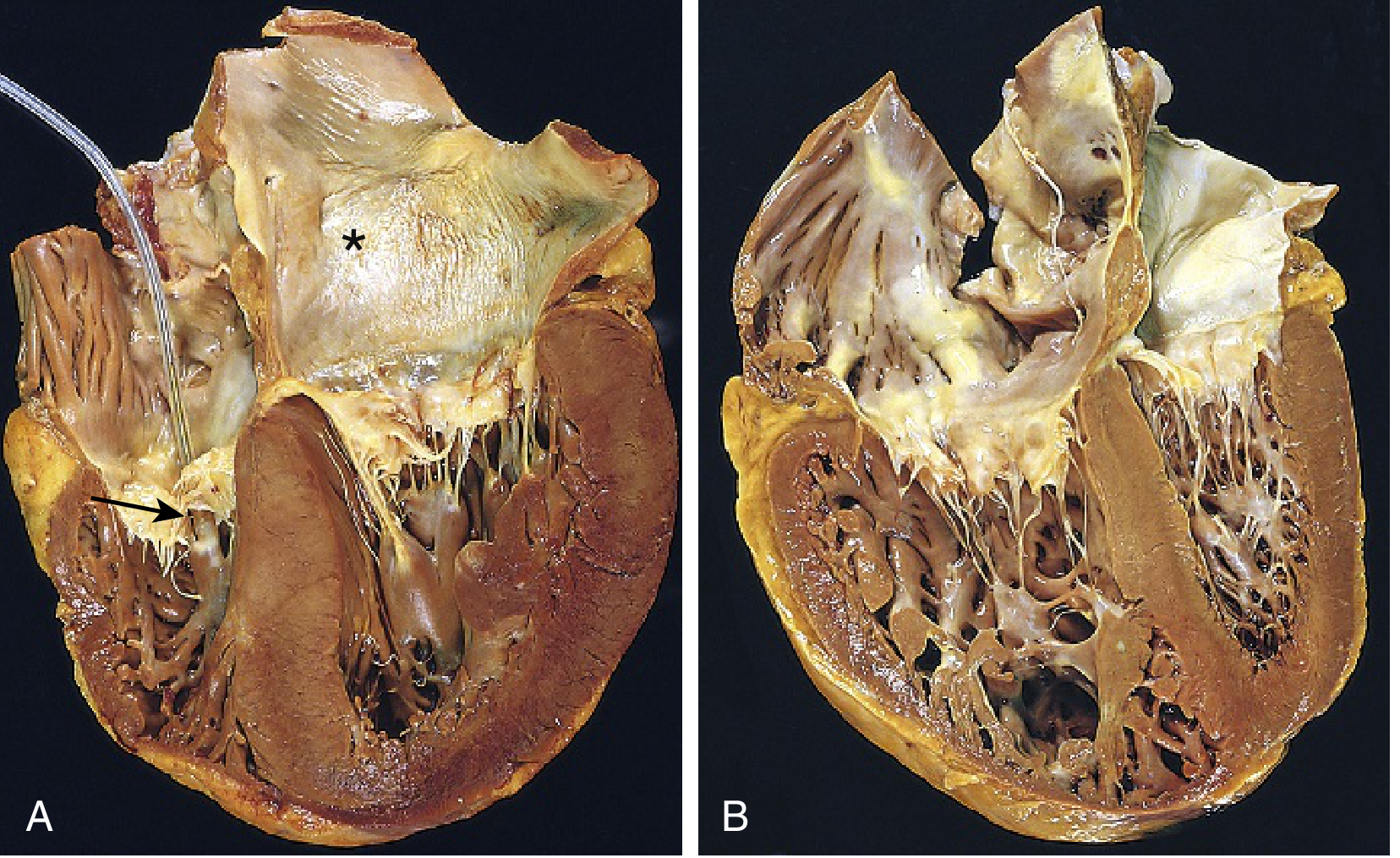
Fig. 9.16B (Robbins Pathology): Chronic cor pulmonale - the right ventricle (left) is markedly dilated and hypertrophied, with a thickened free wall and hypertrophied trabeculae. The right ventricle distorts the shape and volume of the adjacent left ventricle.
Morphology (Pathology)
- Acute cor pulmonale: RV shows only dilation; if death is sudden (e.g., massive PE), the heart may appear near-normal in size
- Chronic cor pulmonale: RV (and often RA) hypertrophy; in severe cases, RV wall thickness may equal or exceed that of the LV; when failure develops, both RV and RA dilate; pulmonary arteries may show abnormal intimal thickening from chronic PH - Robbins & Kumar Basic Pathology, p. 363
Clinical Features
- Dyspnea (often from underlying lung disease)
- Signs of right heart failure: peripheral edema, raised JVP, hepatomegaly, ascites
- Note: Peripheral edema correlates poorly with resting right atrial pressure - it may reflect activation of the renin-angiotensin-aldosterone system rather than RV failure alone
- Cyanosis (central, from hypoxemia)
- Parasternal heave, loud P2, RV S3 gallop, tricuspid regurgitation murmur
Investigations
| Investigation | Findings |
|---|---|
| ECG | Right axis deviation, P pulmonale (tall P in II), RBBB, RV strain pattern (V1-V3) |
| CXR | Cardiomegaly (RV), prominent pulmonary arteries, signs of underlying lung disease |
| Echocardiogram | RV dilation/hypertrophy, elevated RVSP; note - Doppler-estimated PASP correlates poorly with right heart catheterization values |
| CT/MRI | Pulmonary artery dilation, RV changes; cardiac MRI is more reliable for RV function |
| Right Heart Catheterization | Gold standard for confirming PH (mPAP >25 mmHg) and measuring hemodynamics |
| PFTs / ABG | Hypoxemia, hypercapnia; FEV1/FVC ratio reduction in COPD |
Physical findings of venous engorgement and RV hypertrophy are late signs and can be difficult to detect clinically. - Fishman's Pulmonary Diseases & Disorders, p. 734
Management
The cornerstone is treating the underlying lung disease and correcting hypoxemia:
- Supplemental oxygen (continuous) - the most evidence-based intervention; corrects hypoxic vasoconstriction and reduces erythropoiesis; in patients sensitive to O2, Venturi masks are preferred over nasal cannula
- Diuretics - to optimize volume status and reduce RV preload
- Treat the underlying condition - bronchodilators for COPD, antifibrotics for ILD, anticoagulation + pulmonary endarterectomy for CTEPH
- Vasodilators (calcium channel blockers, PDE-5 inhibitors, prostacyclins): may dilate the pulmonary circulation, but calcium channel blockers can worsen hypoxemia, and their benefit in cor pulmonale from COPD is not well established
- Noninvasive ventilation (NIV/CPAP) - for hypoventilation syndromes and OSA overlap
- Phlebotomy for severe polycythemia (though recent data suggest polycythemia is less common and has uncertain outcome impact)
Prognosis
- Once cor pulmonale develops, survival is significantly reduced
- If mPAP exceeds 25 mmHg, average 5-year survival is reduced by ~50%
- Prognosis is largely tied to the severity of the underlying lung disease and the degree of RV dysfunction
Sources:
- Robbins & Kumar Basic Pathology, p. 363 (Elsevier, 2023)
- Fuster and Hurst's The Heart, 15th Ed., Chapter 59, pp. 1773-1778
- Fishman's Pulmonary Diseases and Disorders, p. 734
Investigations for copd
Finding Sources
Searching PubMed
"COPD" AND "diagnosis" AND "spirometry"
Finding Sources
Finding Sources
Reading File
That block is about sleep. Let me search specifically for COPD investigations/diagnosis content.
Finding Sources
Reading File
Reading File
Finding Sources
Reading File
Finding Sources
Finding Sources
Reading File
Reading File
Reading File
Good - I have enough content. Let me also fetch the CXR image to show the user. Let me now compile a comprehensive answer from all the gathered textbook content.
Loading Image
Loading Image
I now have comprehensive textbook content and images. Here is the full answer:
Investigations for COPD
1. Spirometry (Gold Standard / Mandatory)
Spirometry is the definitive investigation for diagnosing COPD. It is simple, noninvasive, and inexpensive - it is used to both diagnose and monitor disease progression. - Fishman's Pulmonary Diseases & Disorders, p. 726
Key diagnostic criterion:
- Post-bronchodilator FEV1/FVC < 0.70 confirms fixed (irreversible) airflow obstruction
Severity classification (GOLD criteria):
| Stage | Label | FEV1 (% predicted) |
|---|---|---|
| I | Mild | ≥ 80% |
| II | Moderate | 50-79% |
| III | Severe | 30-49% |
| IV | Very Severe | < 30% (or <50% with PaO2 < 60 mmHg) |
Note: A 2025 GOLD Science Committee guideline (PMID 39638416) recommends post-bronchodilator spirometry as the standard for COPD diagnosis, reinforcing the established threshold.
2. Lung Volume Measurements
- Plethysmography, helium dilution, nitrogen washout, or single-breath methods
- Show hyperinflation (elevated Total Lung Capacity, TLC) and air trapping (elevated Residual Volume, RV)
- Useful to exclude restrictive lung diseases, which show reduced TLC
3. Diffusing Capacity (DLCO)
- Carbon monoxide diffusing capacity (DLCO) is reduced in emphysema
- Acts as an independent predictor of mortality in COPD
- Helps distinguish COPD from asthma (asthma typically has normal DLCO)
4. Arterial Blood Gas (ABG)
| Finding | Significance |
|---|---|
| PaO2 < 60 mmHg | Hypoxemia; criterion for very severe COPD; indicates need for LTOT |
| PaCO2 > 45 mmHg | Type II respiratory failure / chronic hypercapnic failure |
| pH, HCO3- | Assess for compensated vs. acute-on-chronic respiratory acidosis |
5. Chest Radiograph (CXR)
CXR is insensitive for early disease but shows classic features in advanced COPD:
- Hyperinflation: flattened, low-lying diaphragm; barrel chest
- Increased anteroposterior diameter (lateral view)
- Widened retrosternal airspace (lateral view)
- Reduced peripheral vascular markings (oligemia)
- Bullae (avascular transradiant areas)
- Narrow transverse cardiac silhouette
- Irregular diaphragm contour (insertions visible on ribs)
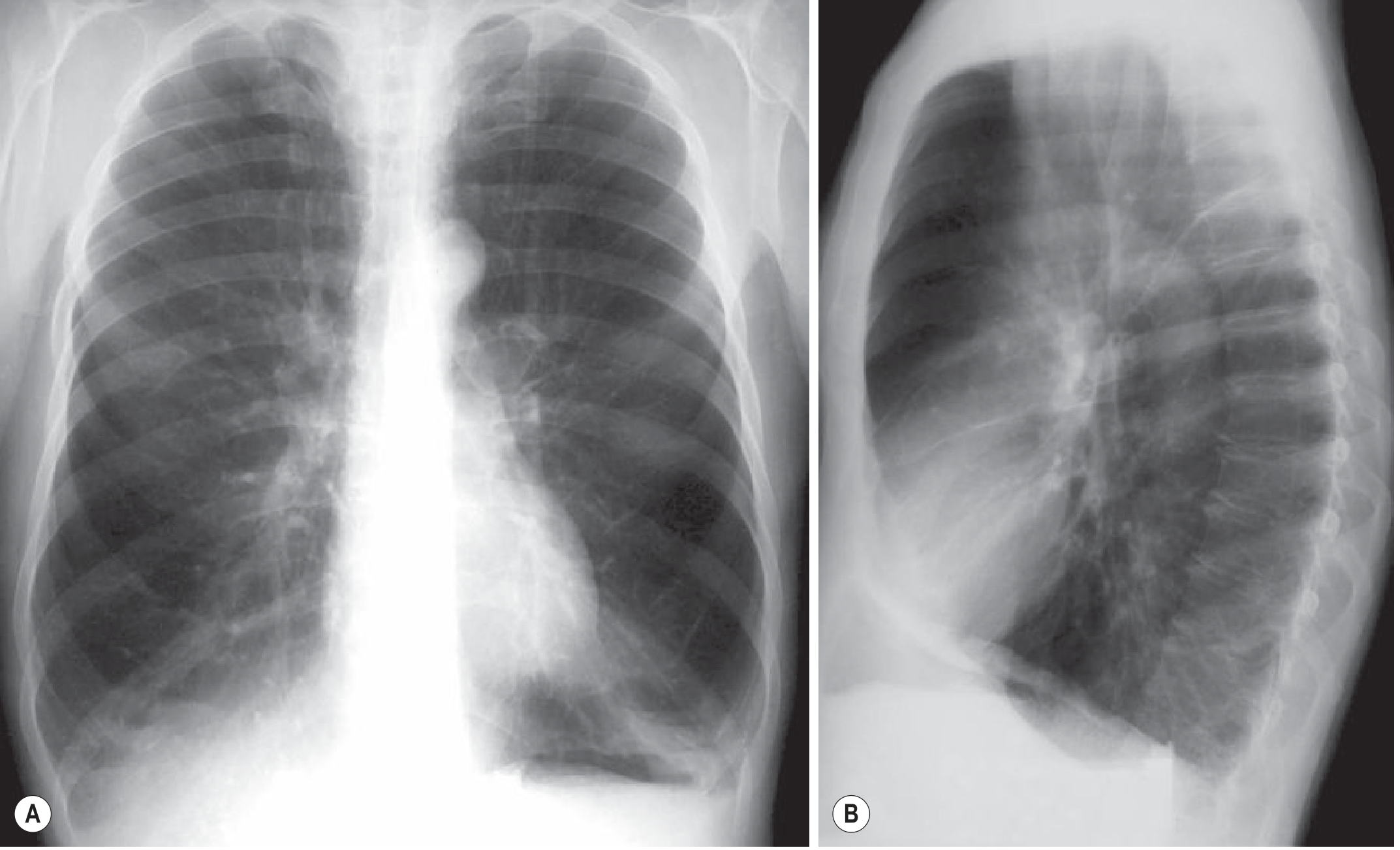
Fig. 6.27 - Severe Diffuse Emphysema, PA and lateral CXR (Grainger & Allison's Diagnostic Radiology)
6. High-Resolution CT (HRCT) Chest
HRCT is the best imaging modality for COPD. It is particularly useful for:
- Confirming and characterizing emphysema (type and extent)
- Phenotyping: distinguishing emphysema-predominant vs. airway-predominant disease
- Detecting bullae, air trapping, and bronchiectasis
- Early detection of emphysema before spirometric changes are apparent
- Guiding surgical decisions (bullectomy, lung volume reduction)
HRCT features of emphysema:
- Centrilobular (most common, upper lobe, smoking-related): small round low-attenuation areas around centrilobular arteries
- Panlobular (lower lobe, alpha-1 antitrypsin deficiency): diffuse destruction of entire acinus
- Paraseptal (near pleura/septa): associated with spontaneous pneumothorax
- Vessels in emphysematous areas appear straightened and splayed with decreased branching
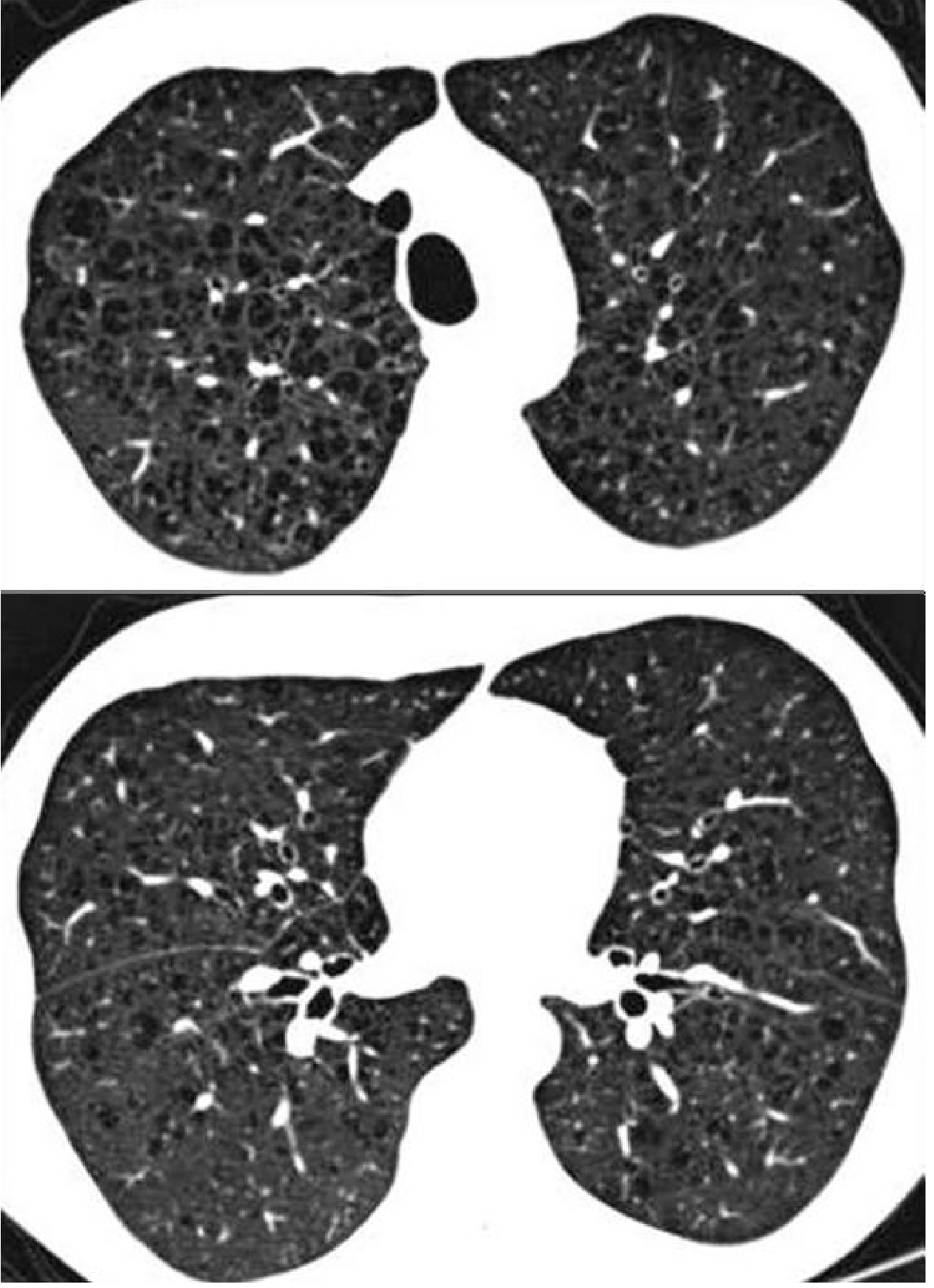
Fig. 6.30 - Moderate Centrilobular Emphysema on HRCT (Grainger & Allison's Diagnostic Radiology)
Quantitative CT analysis is a validated research tool for early emphysema detection, but is not yet routine in clinical practice. - Fishman's Pulmonary Diseases & Disorders, p. 726
7. Pulse Oximetry / Overnight Oximetry
- Screening tool for resting hypoxemia (SpO2 < 88% at rest = indication for LTOT)
- Overnight oximetry can detect nocturnal desaturation and screen for OSA-COPD overlap syndrome (though it cannot differentiate the relative contribution of each)
8. Alpha-1 Antitrypsin (AAT) Level
- Should be measured when features suggest AAT deficiency (see below)
- Some experts recommend testing all patients with emphysema or fixed airflow obstruction, as treatments exist for severe deficiency
Indications to test:
| Feature |
|---|
| Emphysema onset < 45 years |
| Emphysema in a non-smoker |
| Predominantly basal emphysema (panacinar pattern) |
| Family history of early-onset emphysema |
| Family history of cirrhosis |
| Bronchiectasis without clear etiology |
| c-ANCA positive vasculitis |
9. ECG and Echocardiogram
- ECG: look for P pulmonale, right axis deviation, RV hypertrophy (cor pulmonale), atrial arrhythmias (common in COPD due to hypoxemia and RAE)
- Echocardiogram: assess RV function, estimate pulmonary artery pressure, rule out left heart disease as a contributor
10. Full Blood Count (FBC)
- Polycythemia (raised Hct/Hb): secondary to chronic hypoxemia
- Anaemia: a poor prognostic marker in COPD; also worsens dyspnea
- Eosinophilia: may identify patients likely to respond to inhaled corticosteroids
11. Sputum Analysis
- Routine culture during exacerbations to identify bacterial pathogens (H. influenzae, S. pneumoniae, M. catarrhalis, P. aeruginosa in severe disease)
- Sputum eosinophilia: identifies a treatable trait
12. Exercise Testing / 6-Minute Walk Test (6MWT)
- 6MWT is simple and widely used; distance <350 m indicates poor prognosis
- Formal cardiopulmonary exercise testing (CPET): measures VO2 max, useful pre-operatively and in complex cases
- Exercise desaturation identifies patients who may need ambulatory O2
13. Sleep Study (Polysomnography / HSAT)
- Indicated when OSA-COPD overlap syndrome is suspected
- PSG with CO2 monitoring preferred in moderate-severe COPD or when hypercapnia is anticipated
- HSAT may be used in mild COPD without hypoxemia - Murray & Nadel's Textbook of Respiratory Medicine
Prognostic Markers (Additional Assessments)
| Marker | Significance |
|---|---|
| Low FEV1 | Most important prognostic spirometric marker |
| Low DLCO | Independent mortality predictor |
| Low 6MWT distance | Poor functional status |
| Hypoxemia (PaO2 < 60) | Cor pulmonale risk, LTOT indication |
| mMRC dyspnea scale ≥ 2 | Symptom burden; used in GOLD ABCD assessment |
| Frequent exacerbations (≥2/year) | Accelerated decline, mortality |
| Low BMI / malnutrition | Poor prognosis |
| CAT score (COPD Assessment Test) | Health status; used in GOLD staging |
Sources:
- Fishman's Pulmonary Diseases and Disorders, 2-Volume Set, pp. 726-727
- Goldman-Cecil Medicine International Edition, p. 890
- Grainger & Allison's Diagnostic Radiology, pp. 165-167
- Murray & Nadel's Textbook of Respiratory Medicine
- GOLD 2025 Science Committee - pre/post-bronchodilator spirometry guideline (PMID 39638416, Eur Respir J 2025)
Lung volumes and capacities
Finding Sources
Reading File
Reading File
Reading File
Excellent - rich content from two physiology textbooks. Let me fetch the spirogram diagram and read the remaining sections on FRC measurement and changes in disease.
Loading Image
Loading Image
Reading File
Reading File
Now let me get the section on changes in lung volumes in disease and FRC measurement methods.
Reading File
Finding Sources
Finding Sources
Reading File
I now have everything needed for a comprehensive, well-illustrated answer. Here it is:
Lung Volumes and Capacities
Key Terminology
- Volume: a primary, non-overlapping compartment of air in the lung (4 volumes)
- Capacity: a combination of two or more volumes (4 capacities)
The total maximal volume of all airways in an adult is typically 5-6 litres.
The Four Primary Lung Volumes
| Volume | Symbol | Definition | Typical Value |
|---|---|---|---|
| Tidal Volume | TV (Vt) | Air inspired or expired with each normal quiet breath | ~500 mL |
| Inspiratory Reserve Volume | IRV | Additional air that can be inspired above tidal volume with maximal effort | ~1900-3000 mL |
| Expiratory Reserve Volume | ERV | Additional air that can be expired below tidal volume with maximal effort | ~1100-1200 mL |
| Residual Volume | RV | Air remaining in lungs after maximal forced expiration - cannot be emptied | ~1200-1900 mL |
Important: RV cannot be measured by spirometry alone, because no air flows out of the spirometer once it is reached. It requires special techniques (see below).
Why does RV exist?
- Prevents complete airway collapse - reinflating collapsed airways requires disproportionately high pressure
- Maintains continuous gas exchange between episodic breaths; prevents wild swings in blood PO2
The Four Lung Capacities
Each capacity is a sum of two or more volumes:
| Capacity | Symbol | Components | Typical Value | Measurable by Spirometer? |
|---|---|---|---|---|
| Inspiratory Capacity | IC | IRV + TV | ~3500 mL | Yes |
| Functional Residual Capacity | FRC | ERV + RV | ~2400 mL | No (contains RV) |
| Vital Capacity | VC | IRV + TV + ERV | ~4700 mL | Yes |
| Total Lung Capacity | TLC | All 4 volumes (VC + RV) | ~6000 mL | No (contains RV) |
FRC is the resting/equilibrium volume of the lung - the volume remaining after a normal tidal expiration when the inward recoil of the lungs exactly balances the outward recoil of the chest wall.
Spirogram
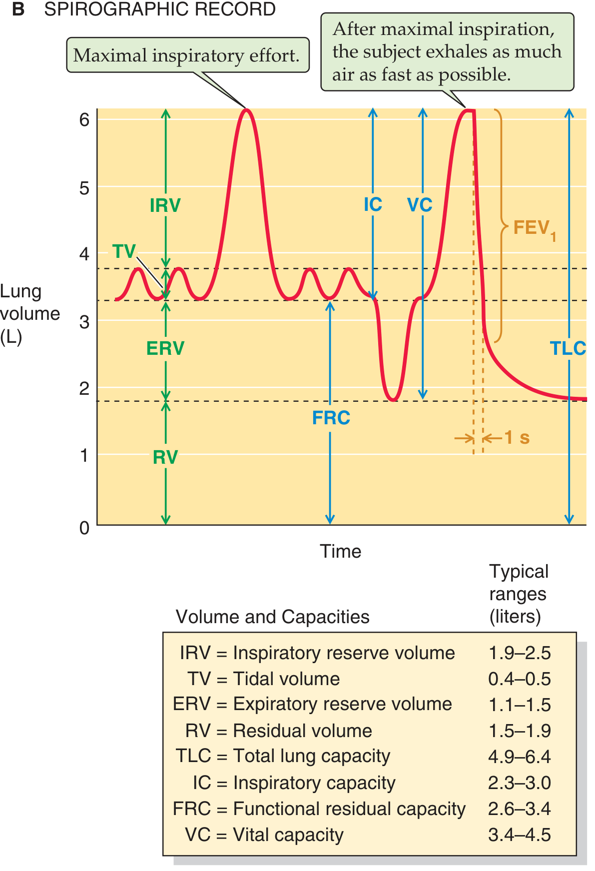
Spirographic record showing all four volumes and four capacities, with typical ranges. Note that RV, FRC, and TLC cannot be read from the spirogram since the tracing never reaches zero. (Medical Physiology, Boron & Boulpaep)
Memory Aid (Volumes from bottom to top)
TLC ──────────────────────  ← Maximal inspiration
      ↑ IRV (~3000 mL)
End of normal inspiration ──
      ↑ TV (~500 mL)
End of normal expiration ─── ← FRC level (= ERV + RV)
      ↑ ERV (~1200 mL)
      ↑ RV (~1200 mL)
0 ────────────────────────  ← Cannot reach this
How to Measure RV, FRC, and TLC
Since RV is not spirometrically measurable, three techniques are used:
1. Helium Dilution Method
- Subject breathes from a spirometer containing a known amount of He (insoluble in blood)
- He equilibrates between spirometer and lungs
- Using mass conservation:
[He]initial × V_spirometer = [He]final × (V_spirometer + V_lung) - Solves for V_lung = FRC (if measured after normal expiration)
- Limitation: underestimates FRC in COPD - He doesn't penetrate poorly ventilated (trapped) areas
2. Nitrogen Washout
- Subject breathes 100% O2; N2 is gradually washed out of lungs
- Total N2 exhaled is collected; since N2 starts at ~80% in alveolar gas, FRC can be calculated
- Same limitation as He dilution in obstructed lungs
3. Body Plethysmography (most accurate)
- Subject sits in an airtight box and attempts to breathe against a closed mouthpiece
- Applies Boyle's Law (P × V = constant)
- When the subject tries to inspire, chest expands, compressing the box gas, allowing calculation of lung volume
- Advantage: measures ALL gas in the lungs, including trapped air - gives the highest (most accurate) FRC in COPD - Costanzo Physiology, p. 197
Changes in Lung Volumes in Disease
| Parameter | Obstructive (COPD, Asthma) | Restrictive (Fibrosis, Scoliosis, Neuromuscular) |
|---|---|---|
| FVC | Normal or reduced | Reduced |
| FEV1 | Reduced | Reduced |
| FEV1/FVC | Reduced (<0.70) | Normal (>0.70) |
| TLC | Normal or increased (hyperinflation) | Reduced |
| RV | Increased (air trapping) | Reduced |
| RV/TLC ratio | Increased | Unchanged |
| FRC | Increased | Reduced |
| FEF 25-75% | Reduced | Normal or reduced |
Harriet Lane Handbook, 23rd Ed.
Key distinctions:
- In obstructive disease: air trapping raises RV and FRC; TLC may be elevated (hyperinflation); FEV1/FVC falls
- In restrictive disease: all volumes shrink proportionally; FEV1/FVC ratio is preserved or even elevated
- Plethysmography gives higher FRC than helium dilution in COPD, because it captures trapped air
Factors Affecting Lung Volumes
| Factor | Effect |
|---|---|
| Taller/larger body | Higher volumes |
| Male sex | Higher VC than females |
| Physical conditioning | Higher VC |
| Age | TLC and VC decrease with age |
| Recumbent position | IRV and FRC decrease (diaphragm pushed up) |
| Reduced compliance (fibrosis) | All volumes decrease |
| Muscle weakness | IRV, ERV, VC decrease |
| Obesity | FRC markedly decreased |
| Pregnancy | FRC decreases (elevated diaphragm) |
Additional Derived Values
| Measurement | Definition | Clinical Use |
|---|---|---|
| FEV1 | Volume exhaled forcefully in the first second of VC maneuver | Diagnose/grade airflow obstruction |
| FEV1/FVC | Ratio (normal ~80%) | < 0.70 = obstructive defect |
| FEF 25-75% | Mean expiratory flow in the middle half of FVC | Sensitive marker of small airway disease |
| PEFR | Peak expiratory flow rate | Monitoring asthma |
| Alveolar ventilation (VA) | (TV - Dead space) × RR | Effective ventilation reaching alveoli |
| Dead space (VD) | ~150 mL (anatomic); air ventilated but not perfused | Wasted ventilation |
Sources:
- Medical Physiology (Boron & Boulpaep), pp. 880-882
- Costanzo Physiology 7th Edition, pp. 196-197
- Fishman's Pulmonary Diseases & Disorders - Glossary Table 31-1
- Harriet Lane Handbook 23rd Ed., p. 839
This is a shared conversation. Sign in to Orris to start your own chat.