
Gall bladder stone
gallstones cholelithiasis gallbladder ultrasound
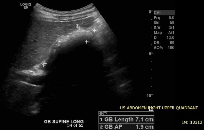
Diagnostic Image: Abdominal Ultrasound (US). Modality: B-mode sonography of the Right Upper Quadrant (RUQ) in the supine longitudinal plane. Anatomical Region: Gallbladder and adjacent liver parenchyma. Findings: The gallbladder lumen contains multiple well-defined, hyperechoic structures consistent with cholelithiasis (gallstones). These structures exhibit prominent posterior acoustic shadowing, a characteristic feature of mineralized calculi. Calipers measure the gallbladder length at 7.1 cm and the anteroposterior (AP) diameter at 1.9 cm, both of which fall within normal physiological limits. There is no visible evidence of gallbladder wall thickening or pericholecystic fluid in this view. Clinical Significance: The image demonstrates uncomplicated cholelithiasis, providing a classic example of gallstones for educational purposes in gastroenterology and radiology. Target Audience: Medical students and residents learning ultrasound interpretation of the biliary system.
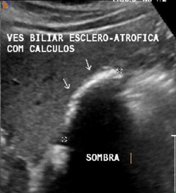
This diagnostic image is an abdominal ultrasound in the right upper quadrant demonstrating a scleroatrophic gallbladder. The primary visual finding is a shrunken, contracted gallbladder lumen that is largely replaced by echogenic material. Hyperechoic (bright) structures, indicated by white arrows, represent multiple gallstones (cholelithiasis) densely packed within the organ. A prominent posterior acoustic shadow, labeled as 'SOMBRA', is visible distal to the calculi, a hallmark sonographic sign of gallstones. The image illustrates chronic cholecystitis leading to a 'Wall-Echo-Shadow' (WES) sign or scleroatrophic appearance, where the gallbladder wall and the echo from the stones are closely apposed. This finding is of high clinical significance in hepatobiliary imaging as it confirms cholelithiasis in a chronically diseased, contracted gallbladder, which can complicate surgical interventions such as cholecystectomy.
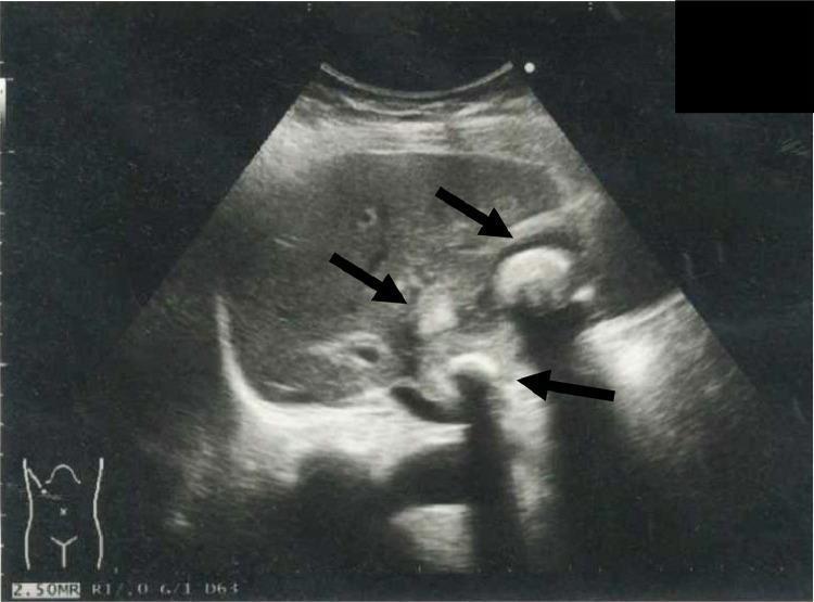
This abdominal ultrasonography image demonstrates the gallbladder in a patient with cholelithiasis. The primary findings consist of multiple hyperechoic structures (indicated by black arrows) within the gallbladder lumen, consistent with cholelithiasis (gallstones). These stones exhibit high echogenicity and are accompanied by prominent posterior acoustic shadowing, a hallmark diagnostic feature of dense calcified structures on ultrasound. The gallbladder wall appears thickened, suggesting underlying inflammation or cholecystitis. The surrounding liver parenchyma shows a relatively homogeneous echotexture, providing a clear acoustic window for the gallbladder. This diagnostic image is used to assess biliary pathology and plan clinical interventions such as percutaneous transhepatic gallbladder drainage (PTGBD). The orientation is shown via the body habitus icon in the bottom-left corner, indicating a right upper quadrant scan. This visual serves as a primary tool for medical students and clinicians to identify the classic sonographic presentation of gallstones and associated pericholecystic changes.
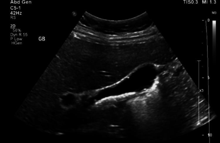
This diagnostic image is a 2D grayscale abdominal ultrasound focusing on the gallbladder (labeled 'GB'). The gallbladder is visualized as an anechoic (black) fluid-filled sac. Within the dependent portion of the gallbladder lumen, there are multiple hyperechoic (bright) mobile structures consistent with cholelithiasis (gallstones). These stones demonstrate posterior acoustic shadowing, a hallmark diagnostic feature where the sound waves are blocked by the dense stones. The gallbladder wall appears thin and regular, without obvious evidence of wall thickening, pericholecystic fluid, or other signs of acute cholecystitis. In the clinical context of situs inversus totalis, this ultrasound identifies the gallbladder positioned on the left side of the upper abdomen. This visual serves as a primary diagnostic tool for evaluating biliary colic and symptomatic cholelithiasis.
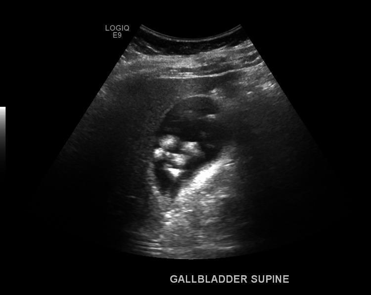
This diagnostic image is a gray-scale ultrasound of the right upper quadrant (RUQ) showing the gallbladder (GB) in a supine position. The visual focus is on the gallbladder lumen, which contains multiple distinct, hyperechoic (bright) foci. These structures exhibit classic posterior acoustic shadowing, where ultrasound waves are blocked, confirming the presence of cholelithiasis (gallstones). The gallbladder itself is non-distended. The gallbladder wall appears thin and smooth, measuring approximately 3 mm, which is within the normal physiological range. There is a notable absence of sonographic signs associated with acute cholecystitis, such as gallbladder wall thickening, pericholecystic fluid, or wall hyperemia. The adjacent hepatic parenchyma is visible but unremarkable. This image serves as a clinical example of symptomatic or asymptomatic cholelithiasis without secondary inflammatory changes of the gallbladder wall.
"gallstones" AND "cholecystectomy"
Gallbladder Stones (Cholelithiasis)
Overview
Types of Stones
| Type | Composition | Key Associations |
|---|---|---|
| Cholesterol / mixed (~80% in West) | 50-99% pure cholesterol + calcium salts, bile pigments, phospholipids | Obesity, high-calorie diet, oral contraceptives/estrogen, pregnancy, rapid weight loss, ileal disease/resection |
| Black pigment | Insoluble bilirubin polymer + calcium phosphate/bicarbonate | Chronic hemolysis (hereditary spherocytosis, sickle cell), cirrhosis |
| Brown pigment | Calcium bilirubinate/palmitate/stearate + cholesterol | Bile stasis, biliary infection, stents, parasites (Clonorchis, Ascaris) - more common in the bile ducts itself |
Pathophysiology
Clinical Presentation
- Silent/asymptomatic: majority of cases, often found incidentally on imaging.
- Biliary colic: right upper quadrant or epigastric pain (often more dull/continuous than "colicky," despite the name), may radiate to the back or right scapula, lasting minutes to hours, with nausea/vomiting; classically starts at night and resolves as the stone falls back into the gallbladder body.
- Acute cholecystitis: persistent pain with fever, and a positive Murphy's sign (RUQ tenderness worsened by inspiration during subcostal palpation).
- Courvoisier's sign: a palpable, non-tender gallbladder with jaundice suggests malignant distal bile duct obstruction rather than stones.
Complications
- Biliary colic, acute/chronic cholecystitis
- Empyema, mucocele, or perforation of the gallbladder
- Mirizzi syndrome (stone compresses/erodes into the common bile duct causing jaundice)
- Cholecystoduodenal fistula -> gallstone ileus (bowel obstruction from a large stone)
- Choledocholithiasis with obstructive jaundice, acute cholangitis, and gallstone pancreatitis
Diagnosis
- Ultrasound (USG) is first-line: shows hyperechoic stones with posterior acoustic shadowing, ± gallbladder wall thickening/pericholecystic fluid in cholecystitis.
- If jaundice or deranged liver enzymes (ALP) are present, MRCP is used to exclude choledocholithiasis.
- Tokyo Guidelines 2018 criteria combine local signs (Murphy's sign, RUQ tenderness/mass), systemic signs (fever, raised CRP/WBC), and imaging findings for a definite diagnosis of acute cholecystitis.
Management
- Asymptomatic stones: generally no intervention needed. Prophylactic cholecystectomy is reserved for specific high-risk situations: stones >3 cm, coexisting choledocholithiasis, chronic hemolytic disease, gallbladder polyps >1 cm, porcelain gallbladder, high-risk ethnic/geographic groups, transplant or bariatric surgery patients.
- Symptomatic gallstones: cholecystectomy (usually laparoscopic) is the definitive treatment.
- Acute cholecystitis initial medical management: NPO + IV fluids, analgesia, broad-spectrum antibiotics covering Gram-negative aerobes (e.g., cefazolin, cefuroxime, ciprofloxacin), followed by interval or early cholecystectomy once inflammation settles.