
Teratoma of Testis - gross, microscopic features 2. Seminoma***** - gross and microscopic features; Labelled diagram; Classical Seminoma - morphology, modes of spread 3. Classify Testicular Tumors* 4. Teratoma - define, classify, Extragonadal sites of appearance 5. Testicular Germ Cell Tumors - ennumerate, describe any one answer in detail covering imp points as welll in exaam point of view 2mbbs pathology refer harsh moham add diagrams as well cover required points for muhs exam Add histo pathology diagram and gross as well
Reading File
Now let me search the medical library for all the relevant topics in parallel.
Finding Sources
Finding Sources
Finding Sources
Finding Sources
Finding Sources
Finding Sources
I found the key sources. Let me now read the Robbins Pathology block on testicular germ cell tumors and the Campbell Walsh block with histology images in parallel.
Reading File
Reading File
Excellent - I have very rich content from Robbins. The first read_file already returned extensive material. Let me now fetch the key histology images and read more of the Robbins content on seminoma and teratoma specifically.
Reading File
Loading Image
Now I have the pathogenesis diagram. The detailed Robbins content from the first large read was truncated. Let me retrieve the seminoma and teratoma specific morphology sections, and also fetch the gross/micro images from Campbell Walsh.
Reading File
Loading Image
I have excellent content now. Let me fetch the key images from Robbins - the classification flowchart and the seminoma/teratoma gross/micro images.
Loading Image
Reading File
Now let me fetch the teratoma histology image and also get the seminoma gross/micro images.
Loading Image
Reading File
Excellent! Now let me fetch the seminoma gross image and the microscopic image, and also read the teratoma section.
Loading Image
Reading File
Now let me get the seminoma histology image and the teratoma section with its content.
Reading File
Now let me fetch the seminoma histology image and get the teratoma section.
Loading Image
Reading File
I now have all the key content. Let me fetch the teratoma gross image and then compile the comprehensive answer.
Loading Image
I now have all the images and text I need. Let me compile the comprehensive, exam-oriented answer.
Testicular Tumors - Complete MUHS Exam Notes (2nd MBBS Pathology)
Source: Robbins, Cotran & Kumar - Pathologic Basis of Disease (2022 edition)
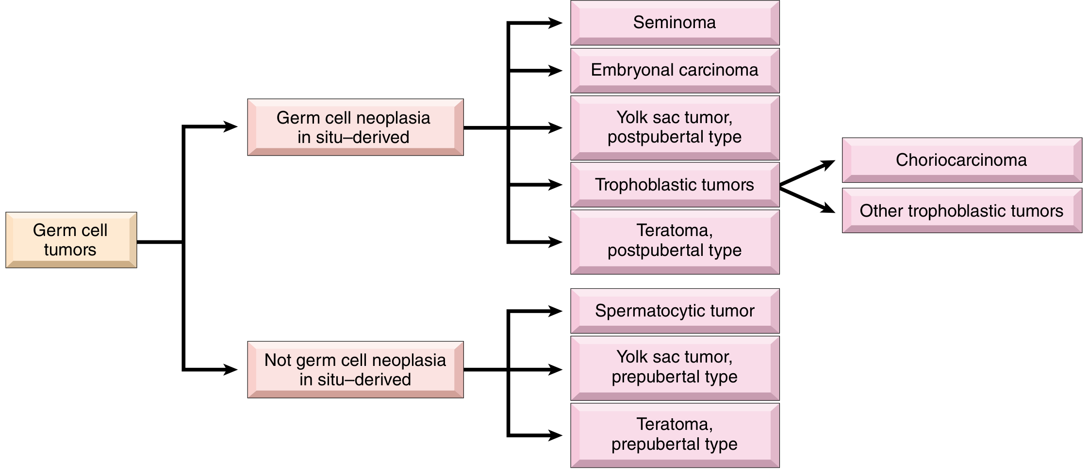
3. CLASSIFY TESTICULAR TUMORS ⭐
WHO 2022 Classification of Testicular Tumors
A. Germ Cell Tumors (GCTs) - 95% of all testicular tumors
I. Derived from Germ Cell Neoplasia In Situ (GCNIS)
Pure (single histologic type):
- Seminoma
Nonseminomatous Germ Cell Tumors (NSGCTs):
- Embryonal carcinoma
- Yolk sac tumor (postpubertal type)
- Choriocarcinoma
- Teratoma (postpubertal type)
- Teratoma with somatic-type malignancy
Mixed GCT - >1 histologic type (30-40% of GCTs)
II. NOT derived from GCNIS
- Spermatocytic tumor
- Teratoma (prepubertal type)
- Yolk sac tumor (prepubertal type)
- Mixed teratoma and yolk sac tumor (prepubertal type)
B. Sex Cord-Stromal Tumors
- Leydig cell tumor
- Sertoli cell tumor
- Granulosa cell tumor (adult and juvenile types)
- Mixed and unclassified sex cord-stromal tumors
Classification Flowchart (Robbins Fig. 21.19):
5. TESTICULAR GERM CELL TUMORS - DETAILED (EXAM ANSWER)
Enumerate
| Tumor | Peak Age | Key Marker |
|---|---|---|
| Seminoma | 4th decade (30-40 yrs) | KIT, OCT3/4, podoplanin |
| Embryonal carcinoma | 20-30 yrs | OCT3/4, SALL4, CK+ |
| Yolk sac tumor | Infants (<3 yrs) | AFP, glypican-3 |
| Choriocarcinoma | 20-30 yrs | hCG (very high) |
| Teratoma | Any age | - |
| Mixed GCT | - | Variable |
| Spermatocytic tumor | >65 yrs | KIT, SALL4 |
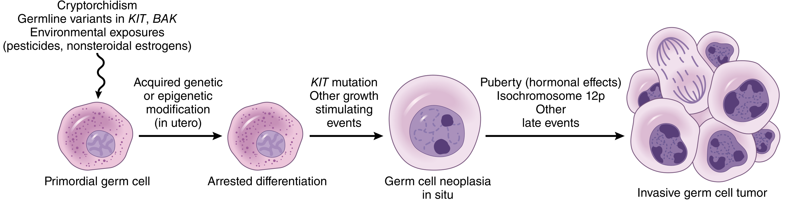
Pathogenesis of GCTs
Pathogenesis diagram (Robbins Fig. 21.18):
Steps:
- Primordial germ cell undergoes acquired genetic/epigenetic modification in utero (due to cryptorchidism, KIT/BAK germline variants, pesticides, nonsteroidal estrogens)
- This leads to arrested differentiation
- KIT mutation + other growth stimulating events → Germ Cell Neoplasia In Situ (GCNIS)
- At puberty: isochromosome 12p + hormonal effects → Invasive GCT
Risk Factors:
- Cryptorchidism (10% of GCTs)
- Klinefelter syndrome (50x risk for mediastinal GCTs)
- Family history (8-10x risk in brothers)
- Testicular dysgenesis syndrome
Key Genetic Event: Isochromosome 12p [i(12p)] - present in virtually ALL GCNIS-derived GCTs - a key diagnostic marker
2. SEMINOMA ⭐⭐⭐⭐⭐
Introduction
- Most common GCT (~50% of all GCTs)
- Peak age: 4th decade (30-40 years)
- Identical tumor in ovary = Dysgerminoma; in CNS = Germinoma
- Most radiosensitive of all GCTs
- Best prognosis among GCTs
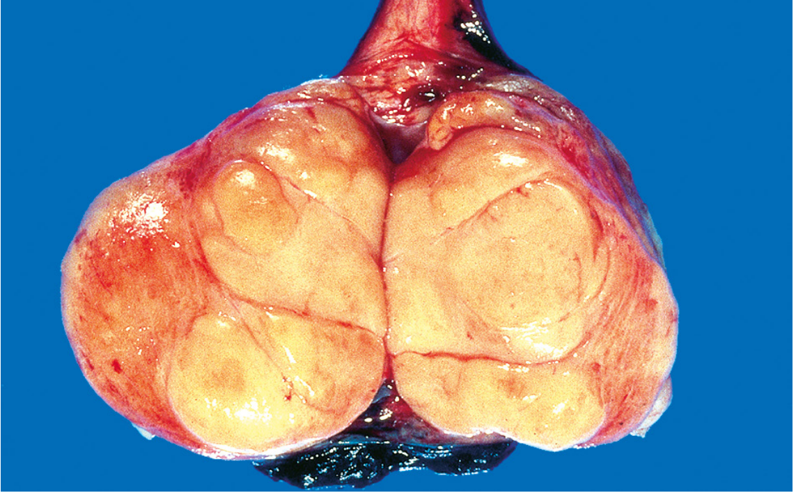
GROSS FEATURES
Gross specimen (Robbins Fig. 21.20):
| Feature | Description |
|---|---|
| Size | Bulky - up to 10x normal testis |
| Cut surface | Homogeneous, gray-white, lobulated |
| Consistency | Firm, fleshy |
| Hemorrhage/necrosis | Absent (characteristic!) |
| Tunica albuginea | Usually intact; occasionally invaded |
| Extension | May extend to epididymis, spermatic cord, scrotal sac |
Exam tip: The absence of hemorrhage and necrosis on cut surface is a classic distinguishing feature of seminoma compared to embryonal carcinoma.
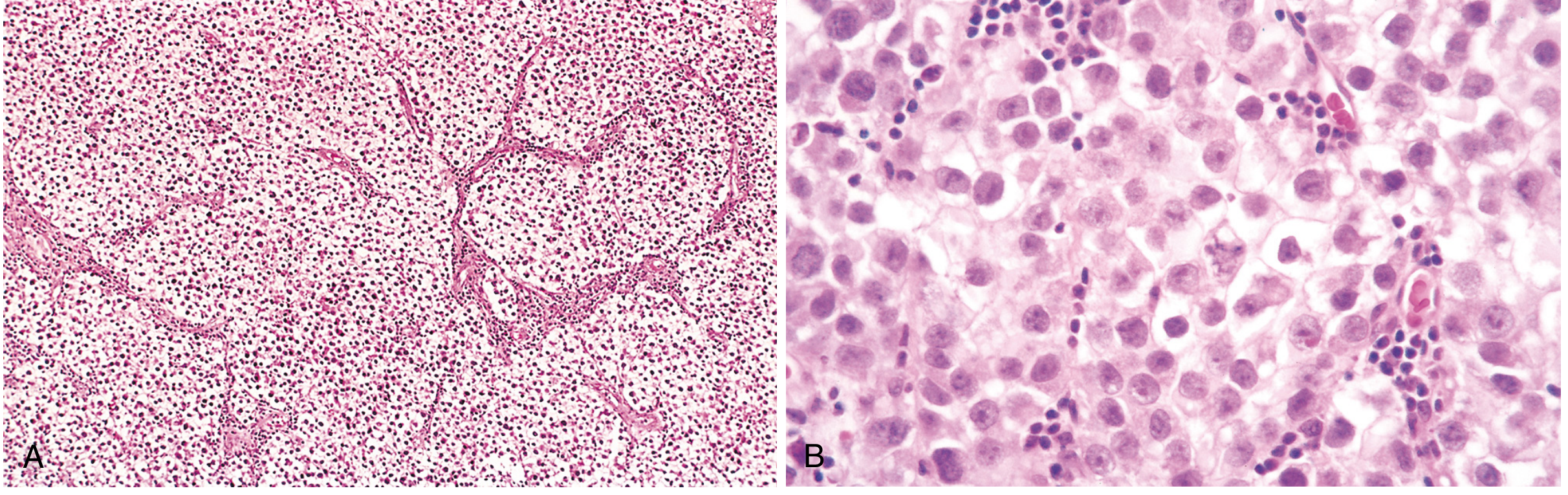
MICROSCOPIC FEATURES (Classical Seminoma Morphology)
Histology (Robbins Fig. 21.21 A & B):
Low power (A):
- Sheets of uniform cells divided into poorly demarcated lobules
- Lobules separated by delicate fibrous septa
- Septa contain lymphocytic infiltrate (T-lymphocytes)
- Ill-defined granulomas may be present (host response)
High power (B) - Classic Seminoma Cell:
- Round to polyhedral cells
- Distinct cell membrane
- Clear/watery cytoplasm (contains glycogen)
- Large, central nucleus with one or two prominent nucleoli
- Cells uniform in size (no significant pleomorphism)
Immunohistochemistry:
- Positive: KIT (CD117), OCT3/4, podoplanin (D2-40)
- Negative: cytokeratin (helps distinguish from embryonal carcinoma)
- ~15% have syncytiotrophoblasts → elevated serum hCG (but not as high as choriocarcinoma)
LABELLED DIAGRAM OF SEMINOMA (Schematic)
SEMINOMA - MICROSCOPIC (Schematic Diagram)
┌─────────────────────────────────────────┐
│ FIBROUS SEPTUM with LYMPHOCYTES │
│ │ │ │
│ ╔════╧═════════╧════╗ │
│ ║ LOBULE ║ │
│ ║ ┌──────────────┐ ║ │
│ ║ │ ○ ○ ○ ○ ○ │ ║ ← Uniform │
│ ║ │ ○ ○ ○ ○ ○ │ ║ tumor cells │
│ ║ │ ○ ○ ○ ○ ○ │ ║ │
│ ║ └──────────────┘ ║ │
│ ╚════════════════════╝ │
│ │
│ SINGLE CELL DETAIL: │
│ ┌────────────────────┐ │
│ │ Round cell │ │
│ │ ┌──────────┐ │ │
│ │ │ Nucleus │◄─── Large, central │
│ │ │ ●●Nucleol│ nucleus │
│ │ └──────────┘ │ │
│ │ Clear cytoplasm │ │
│ │ (glycogen) │ │
│ │ Distinct membrane │ │
│ └────────────────────┘ │
└─────────────────────────────────────────┘
MODES OF SPREAD OF SEMINOMA
1. Lymphatic spread (PRIMARY route)
- First: Retroperitoneal para-aortic lymph nodes
- Then: Mediastinal lymph nodes
- Then: Supraclavicular lymph nodes (especially left-sided - Virchow's node)
- Note: Seminomas typically spread via lymphatics FIRST
2. Hematogenous spread (LATER)
- Occurs later in course compared to NSGCTs
- Sites: Lungs (most common), liver, brain, bone
Exam Mnemonics:
- "Seminoma = Slow, Lymphatics first, Later hematogenous"
- Lymph node drainage of testis: Para-aortic (NOT inguinal - testis drains to para-aortic because of its embryological origin from the abdomen)
1 & 4. TERATOMA - DEFINITION, CLASSIFICATION, GROSS, MICROSCOPIC, EXTRAGONADAL SITES
Definition
A teratoma is a germ cell tumor containing cellular or organoid components derived from more than one germ layer (ectoderm, mesoderm, endoderm), resembling normal derivatives of these layers in varying stages of differentiation.
Classification of Teratoma
A. Based on Age (most important):
| Feature | Prepubertal Type | Postpubertal Type |
|---|---|---|
| Age | Infants, children | Adults |
| GCNIS association | NO | YES |
| Isochromosome 12p | Absent | Present |
| Behavior | Benign | Malignant |
| Differentiation | Well-organized, mature elements | Disorganized mix |
B. Based on Differentiation (Classical classification):
- Mature teratoma - all elements well-differentiated (adult tissue types)
- Immature teratoma - contains embryonic/fetal elements (especially immature neural tissue)
- Teratoma with malignant transformation - somatic malignancy arising (squamous cell carcinoma, adenocarcinoma, sarcoma) - chemoresistant
C. Ovarian teratoma types (for reference):
- Mature cystic teratoma (Dermoid cyst) - most common benign ovarian tumor
- Immature teratoma (malignant)
- Monodermal teratoma (Struma ovarii, Carcinoid)
GROSS FEATURES of Testicular Teratoma
Gross specimen (Robbins Fig. 21.25):
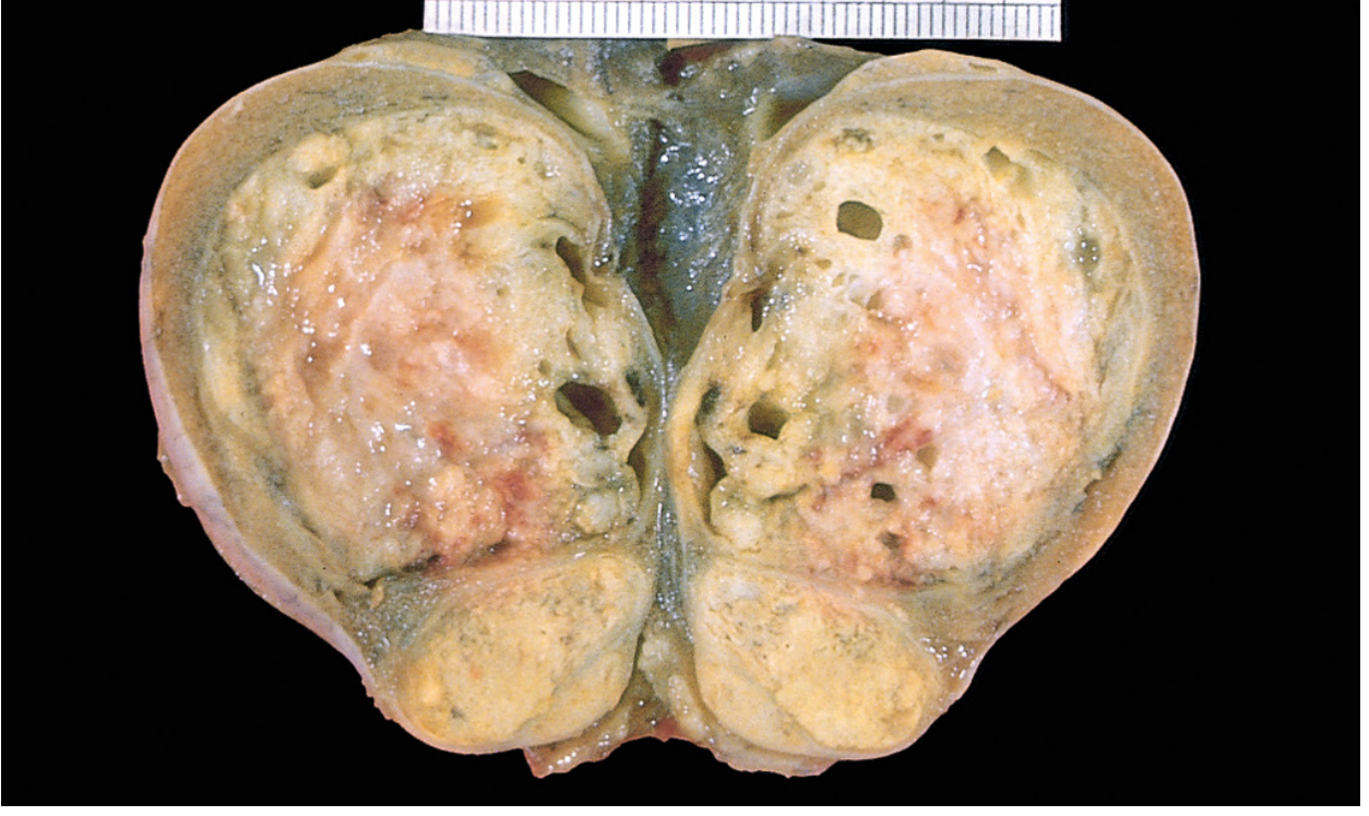
| Feature | Description |
|---|---|
| Size | 5-10 cm in adults |
| Cut surface | Variegated/heterogeneous appearance |
| Areas | Solid AND cystic areas |
| Consistency | Mixed - firm (cartilage), soft, cystic |
| Special | May contain cartilage, hair, teeth (dermoid cysts in prepubertal type) |
Contrast with seminoma: teratoma has a heterogeneous cut surface whereas seminoma is homogeneous
MICROSCOPIC FEATURES of Teratoma
Histology (Robbins Fig. 21.26):
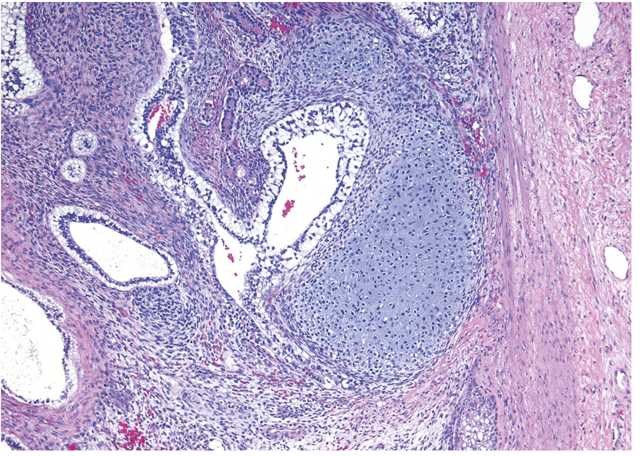
(Section showing disorganized glands, islands of cartilage, smooth muscle bundles, and immature stroma - the hallmark of postpubertal testicular teratoma)
Microscopic features:
- Collections of differentiated cells or organoid structures from multiple germ layers:
- Ectoderm: Neural tissue, squamous epithelium, skin adnexae (hair follicles, sebaceous glands)
- Mesoderm: Muscle bundles (smooth/skeletal), cartilage islands, connective tissue
- Endoderm: Bronchial epithelium, intestinal wall, thyroid-like structures, glandular elements
- Elements embedded in fibrous or myxoid stroma
- Disorganized arrangement (unlike normal tissue)
- May be mature or immature
Prepubertal type: More organized, closely resembles normal tissue; special type = dermoid cyst (hair, teeth, skin)
EXTRAGONADAL SITES OF TERATOMA
Teratomas arise from primordial germ cells that migrate during embryonic development. Sites of extragonadal occurrence follow the midline migration path of primordial germ cells:
| Site | Notes |
|---|---|
| Sacrococcygeal region | Most common extragonadal site; most common in infants/neonates |
| Anterior mediastinum | Second most common; associated with Klinefelter syndrome |
| Retroperitoneum | |
| Pineal gland (CNS) | Called germinoma in CNS |
| Neck/oral cavity | Rare |
| Stomach | Very rare |
Exam tip: Sacrococcygeal teratoma is the most common solid tumor of the newborn. Klinefelter syndrome has 50x increased risk for mediastinal GCTs specifically.
SERUM TUMOR MARKERS (Very Important for Exams)
| Marker | Tumor | Notes |
|---|---|---|
| AFP (Alpha-fetoprotein) | Yolk sac tumor | Elevated in yolk sac tumor; NEVER elevated in pure seminoma |
| hCG | Choriocarcinoma (very high); ~15% seminomas (mild elevation via syncytiotrophoblasts) | |
| LDH | All GCTs | Correlates with tumor bulk/burden |
| PLAP (Placental alkaline phosphatase) | Seminoma |
Key point: Pure seminoma does NOT elevate AFP. If AFP is elevated in a "seminoma," it contains non-seminomatous elements (mixed GCT).
MODES OF SPREAD - GENERAL (ALL GCTs)
| Route | Seminoma | NSGCTs |
|---|---|---|
| Lymphatic | First, predominant | Present |
| Hematogenous | Late | Early, predominant |
| First LN | Retroperitoneal para-aortic nodes | Same |
| Distant sites | Lungs, liver, brain, bone | Same, but earlier |
Note on testicular lymphatic drainage: The testis drains to para-aortic (lumbar) lymph nodes (NOT inguinal nodes), because the testis descended from the abdomen embryologically. Inguinal nodes only involved if scrotal skin or tunica is breached.
KEY CONCEPTS SUMMARY (Quick Revision)
| Feature | Seminoma | Teratoma | Embryonal Ca. | Choriocarcinoma |
|---|---|---|---|---|
| % of pure GCTs | 50% | 2-3% (pure) | Common | Rare pure |
| Age | 30-40 yrs | Any age | 20-30 yrs | 20-30 yrs |
| Gross | Homogeneous, gray-white, no hemorrhage | Variegated, cystic + solid | Hemorrhage/necrosis | Small, hemorrhagic |
| Lymphocytes | Yes (in septa) | No | No | No |
| AFP | Normal | Normal | May elevate | Normal |
| hCG | Normal (15% mild ↑) | Normal | Variable | Very high |
| Radiosensitivity | Very sensitive | Resistant | Moderate | Resistant |
| Prognosis | Best | Variable | Worse | Worst (pure) |
Sources: Robbins, Cotran & Kumar - Pathologic Basis of Disease (2022), Table 21.6 and Chapters on Male Genital Pathology; Campbell-Walsh Urology. All histology images from Robbins.
This is a shared conversation. Sign in to Orris to start your own chat.