
Explain circle of Willis
Reading File
Now let me search the medical library for Circle of Willis content, in parallel with a web search for images.
Finding Sources
Searching the Web
circle of Willis anatomy diagram labeled
Good results. Now let me read the most detailed textbook sections in parallel.
Reading File
Reading File
Excellent content found. Let me now fetch the key diagrams and read more of the neuroanatomy text in parallel.
Loading Image
Loading Image
Reading File
I now have excellent content and images. Let me read the continuation of the neuroanatomy text to get the territorial supply information.
Reading File
I have comprehensive information now. Let me compile the full answer.
Circle of Willis
The circle of Willis (also called the circulus arteriosus cerebri or cerebral arterial circle) is an anastomotic ring of arteries located at the base of the brain, in the subarachnoid space above the sella turcica. It connects the two major arterial systems supplying the brain - the internal carotid system anteriorly and the vertebrobasilar system posteriorly - and is named after English physician Thomas Willis who described it in 1664.
Diagrams
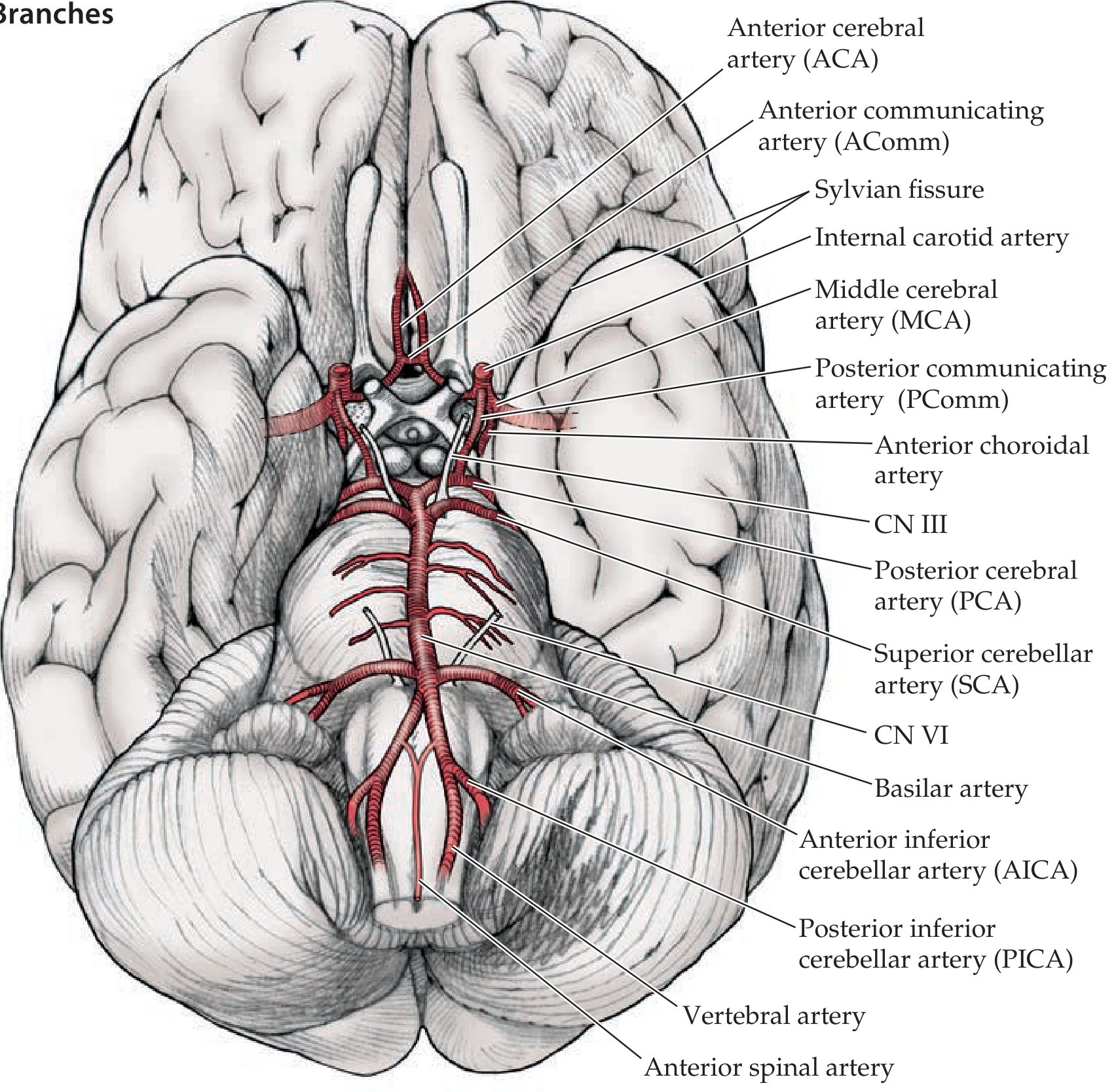
Here is a labeled inferior view of the brain showing the circle of Willis and all its main branches:
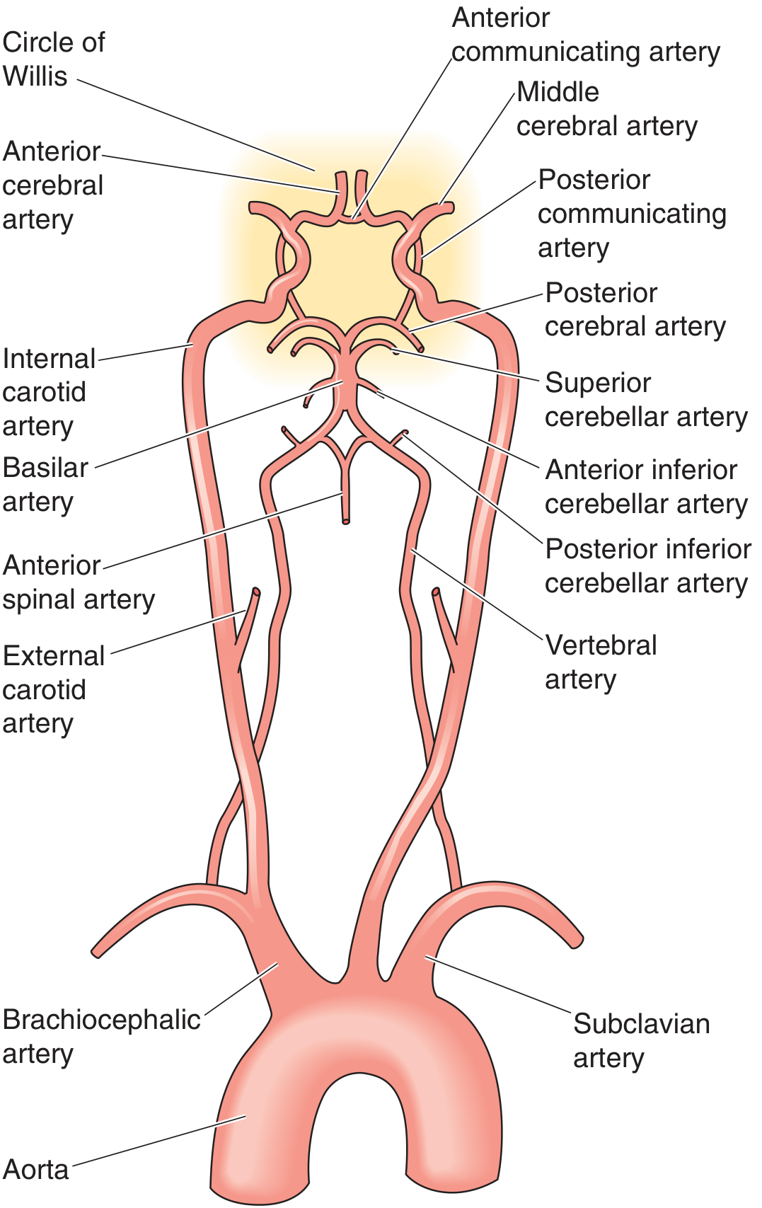
And a schematic diagram showing the full arterial supply from the aorta up to the circle of Willis:
Arterial Inflow: The Two Source Systems
Anterior circulation - supplied by the bilateral internal carotid arteries (ICAs). The common carotid arteries arise from the aorta (or the brachiocephalic artery on the right), bifurcate in the neck into the external and internal carotid arteries, and the ICAs pass through the carotid canal, then the cavernous sinus (carotid siphon/S-shaped bend), and then pierce the dura to enter the subarachnoid space as the supraclinoid segment.
Posterior circulation - supplied by the bilateral vertebral arteries, which arise from the subclavian arteries, ascend through the foramina transversaria of the cervical vertebrae, enter the foramen magnum, and join at the ponto-medullary junction to form the single basilar artery. The basilar artery runs along the ventral surface of the pons before bifurcating at its apex into the two posterior cerebral arteries (PCAs).
Components of the Circle
The circle itself is formed by seven arterial segments connecting these two systems:
| Vessel | Number | Origin | Role |
|---|---|---|---|
| Anterior cerebral artery (ACA) | Paired | Terminal branch of ICA | Anterior part of circle |
| Anterior communicating artery (AComm) | Single | Connects left and right ACA | Anterior bridge |
| Internal carotid artery (supraclinoid) | Paired | Continuation of ICA | Lateral entry points |
| Posterior communicating artery (PComm) | Paired | ICA to PCA junction | Links anterior + posterior |
| Posterior cerebral artery (PCA) | Paired | Apex of basilar artery | Posterior part of circle |
The AComm bridges the two ACAs anteriorly. The PComms connect each ICA to the ipsilateral PCA posteriorly, completing the ring on each side.
Branches Arising from the Circle
The main branches of the supraclinoid ICA are remembered by the mnemonic OPAAM:
- O - Ophthalmic artery (retinal blood supply)
- P - Posterior communicating artery (PComm)
- A - Anterior choroidal artery (supplies choroid plexus, internal capsule, hippocampus)
- A - Anterior cerebral artery (ACA)
- M - Middle cerebral artery (MCA)
The three main cerebral arteries that distribute blood to the hemispheres are:
1. Anterior Cerebral Artery (ACA)
- Passes forward and into the interhemispheric fissure, sweeping over the corpus callosum
- Major branches: pericallosal and callosomarginal arteries
- Territory: medial surface of the frontal and anterior parietal lobes, including the medial sensorimotor cortex (leg/foot area)
- Deep branches: supply the anterior limb of the internal capsule and head of caudate nucleus
2. Middle Cerebral Artery (MCA)
- Turns laterally into the Sylvian (lateral) fissure, usually bifurcating into superior and inferior divisions
- Territory: most of the lateral surface of the cerebral hemisphere - frontal, parietal, and temporal lobes
- Includes primary motor/sensory cortex for the face and arm, Broca's and Wernicke's speech areas
- Deep branches (lenticulostriate arteries): supply the putamen, caudate, and posterior limb of the internal capsule
- This is the most commonly occluded artery in stroke
3. Posterior Cerebral Artery (PCA)
- Arises from basilar apex; loops around the midbrain
- Territory: occipital lobe (primary visual cortex), medial temporal lobe (hippocampus), thalamus
- Deep branches supply the thalamus and posterior internal capsule
Function: Collateral Protection
The primary function of the circle of Willis is collateral blood flow. If one of the feeder vessels becomes stenotic or occluded (e.g., ICA occlusion), blood can be redistributed from the contralateral or posterior circulation to maintain perfusion of the affected territory. This is a vital protective mechanism against ischemic stroke.
However, a critical caveat: a complete, full-caliber circle of Willis is present in only approximately 34% of individuals (Neuroanatomy through Clinical Cases, 3rd ed.). Anatomical variants are extremely common and include hypoplastic or absent PComms, fetal-type PCA (where the PCA arises directly from the ICA rather than the basilar artery), and absent AComm. These variants reduce the effectiveness of collateral compensation.
Clinical Relevance
| Condition | Connection to Circle of Willis |
|---|---|
| Berry (saccular) aneurysm | Most commonly form at arterial junctions within the circle - especially the AComm (most common site), PComm origin, and MCA bifurcation. Rupture causes subarachnoid hemorrhage (SAH) - "worst headache of life." |
| Ischemic stroke | Occlusion of MCA, ACA, or PCA causes predictable deficits based on vascular territory. |
| CN III palsy | The PComm aneurysm compresses the adjacent oculomotor nerve (CN III), causing a painful, "down and out" pupil-involving third nerve palsy. |
| Watershed infarcts | Occur at the border zones between ACA/MCA and MCA/PCA territories during hypoperfusion. |
| Carotid stenosis | If severe, the AComm and PComm may provide collateral supply; their adequacy determines stroke risk. |
Vertebrobasilar System Branches (Posterior Circulation)
Beyond the PCAs, the basilar artery gives off several branches:
- Superior cerebellar artery (SCA) - just before bifurcation; cerebellar supply
- Anterior inferior cerebellar artery (AICA) - from mid-basilar; supply to pons and cerebellum
- Posterior inferior cerebellar artery (PICA) - from the vertebral arteries; supply to lateral medulla and inferior cerebellum (occlusion = Wallenberg/lateral medullary syndrome)
Sources: Neuroanatomy through Clinical Cases, 3rd Edition; Medical Physiology (Boron & Boulpaep); Goldman-Cecil Medicine.
This is a shared conversation. Sign in to Orris to start your own chat.