
spindle cell carcinoma in detail for 20 marks with diagram and flow charts from Scott Brown, Cummings, stell and marans, Zakir Hussain, dhingra, hazarika and related articles with recent advances according to rguhs university
Reading File
Finding Sources
Searching PubMed
"spindle cell carcinoma" AND "head and neck"
Searching the Web
spindle cell carcinoma larynx ENT RGUHS 20 marks recent advances sarcomatoid carcinoma
Finding Sources
Reading Article
PMID: 41453574
Reading Web Page
https://www.frontiersin.org/journals/oncology/articles/10.33…
Finding Sources
Finding Sources
Reading File
Reading File
Loading Image
Finding Sources
Finding Sources
Loading Image
Reading File
Reading File
SPINDLE CELL CARCINOMA (SARCOMATOID CARCINOMA)
A Comprehensive 20-Mark Answer for RGUHS Examination
INTRODUCTION & DEFINITION
Spindle Cell Carcinoma (SpCC) is a biphasic malignant neoplasm arising from the surface epithelium of the upper aerodigestive tract, composed of two distinct components:
- A squamous cell carcinoma element (in situ or invasive)
- A malignant spindle cell (sarcomatoid/mesenchymal-appearing) component that predominates
Synonyms: Sarcomatoid carcinoma | Carcinosarcoma | Pseudosarcoma | Lane tumor | Pleomorphic carcinoma
It constitutes 2-3% of all laryngeal cancers and 4.2% of laryngeal squamous cell carcinomas. (Cummings Otolaryngology, p. 2005)
HISTORICAL BACKGROUND / NOMENCLATURE EVOLUTION
┌──────────────────────────────────────────────────────────────────┐
│           EVOLUTION OF NOMENCLATURE                              │
│                                                                  │
│  Lane (1957)        → "Pseudosarcoma" (benign-seeming)          │
│  Goethals (1963)    → "Carcinosarcoma" (dual malignant elements)│
│  Hyams (1971)       → "Spindle cell carcinoma"                  │
│  WHO 2005/2017      → "Spindle Cell Carcinoma / Sarcomatoid     │
│                        Carcinoma" — monoclonal origin confirmed  │
└──────────────────────────────────────────────────────────────────┘
Three competing theories of histogenesis:
THEORY 1: MONOCLONAL / EPITHELIAL-MESENCHYMAL TRANSITION (EMT)
─────────────────────────────────────────────────────────────────
Surface Epithelium (Squamous)
        │
        ▼ (Oncogenic mutation - TP53, EGFR, PIK3CA)
   SCC in situ / Invasive SCC
        │
        ▼ (EMT — loss of E-cadherin, gain of vimentin)
   Spindle Cell (Sarcomatoid) Component
        │
        └──► BIPHASIC TUMOR ← [ACCEPTED BY WHO 2017]

THEORY 2: COLLISION TUMOR
─────────────────────────
   Independent SCC + Independent Sarcoma meeting in same site
   (Now DISCREDITED — monoclonal origin proven by IHC & molecular studies)

THEORY 3: DUAL DIFFERENTIATION FROM STEM CELL
──────────────────────────────────────────────
   Uncommitted pluripotent stem cell → dual epithelial + mesenchymal lineage
(Scott-Brown's ORL H&N Surgery, p. 480; Frontiers in Oncology 2025)
EPIDEMIOLOGY
| Parameter | Detail |
|---|---|
| Incidence | 2-3% of all laryngeal malignancies |
| Age | Middle-aged to elderly (peak: 6th-7th decade) |
| Sex | Male >> Female (M:F ratio ~6:1) |
| Site | Larynx (most common) > Oral cavity > Oropharynx > Hypopharynx |
| Laryngeal subsite | Glottis > Supraglottis > Subglottis |
(Gonçalves et al., Systematic Review, J Stomatol Oral Maxillofac Surg 2026, PMID 41453574 — 2,250 cases across 39 studies)
ETIOPATHOGENESIS & RISK FACTORS
┌─────────────────────────────────────────────────────────────────────┐
│                    RISK FACTORS FOR SpCC                            │
├──────────────────────┬──────────────────────────────────────────────┤
│  PRIMARY             │  - Tobacco (smoking / chewing) — STRONGEST   │
│                      │  - Alcohol abuse                             │
├──────────────────────┼──────────────────────────────────────────────┤
│  SECONDARY           │  - Previous RADIATION THERAPY (important)    │
│                      │    [SpCC can arise post-RT, though RT is not │
│                      │     a major etiologic factor per se]         │
├──────────────────────┼──────────────────────────────────────────────┤
│  OTHERS              │  - HPV (in oropharyngeal cases)              │
│                      │  - Chronic laryngeal trauma / inflammation   │
└──────────────────────┴──────────────────────────────────────────────┘
(Cummings Otolaryngology, p. 2005)
Molecular Alterations (Recent Advances - 2026):
- TP53 mutations (most common)
- EGFR overexpression
- PIK3CA mutations
- ALK rearrangements
- STAT3 activation (via SUMOylation/SENP3 pathway)
- Loss of E-cadherin → epithelial-mesenchymal transition (EMT)
- HIF-1α translational activation (via YB-1) promoting sarcomatoid transformation
(Gonçalves et al. 2026 systematic review; Frontiers in Oncology 2025)
PATHOLOGY
A. Macroscopic / Gross Features
┌────────────────────────────────────────────────────────────┐
│              GROSS APPEARANCE OF SpCC                      │
│                                                            │
│  ┌─────────────────┐    ┌──────────────────────────────┐   │
│  │   POLYPOID /    │    │  BROAD-BASED /               │   │
│  │   PEDUNCULATED  │    │  SESSILE (less common)       │   │
│  │   (MOST COMMON) │    └──────────────────────────────┘   │
│  └────────┬────────┘                                       │
│           │                                                │
│           ▼ Surface: ULCERATED                             │
│     May AUTO-AMPUTATE and be expectorated by patient       │
│                                                            │
│  Size: 0.5 – 8 cm (average ~2 cm in glottic tumors)       │
│  Color: Gray-white to tan, with areas of necrosis         │
└────────────────────────────────────────────────────────────┘
(Scott-Brown's ORL H&N Surgery, p. 480)
B. Microscopic / Histopathological Features
The image below is from Scott-Brown's Otorhinolaryngology (Figure 26.11) - showing the biphasic histology:
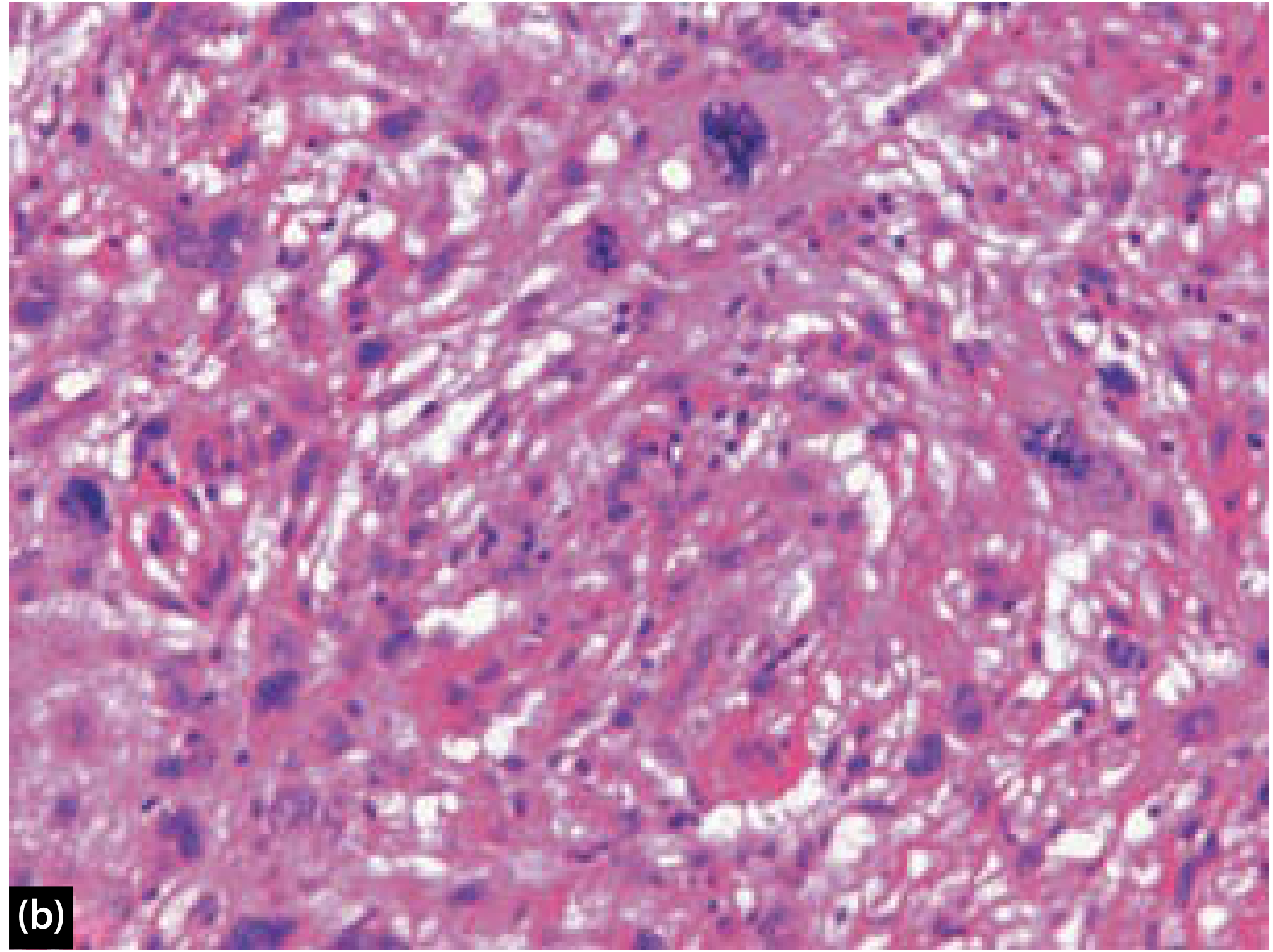
Figure 26.11 from Scott-Brown's: Cross-section of epiglottic resection specimen. This is ulcerated and polypoidal, also infiltrating through epiglottic cartilage. Representative field beneath the tumour surface exhibiting highly pleomorphic spindle and bizarre giant cells with abnormal mitoses, reminiscent of undifferentiated pleomorphic sarcoma (H&E stain, high magnification).
HISTOPATHOLOGICAL COMPONENTS OF SpCC
─────────────────────────────────────────────────────────────────────
COMPONENT 1: SQUAMOUS ELEMENT
  - Surface squamous epithelium with CIS or invasive SCC
  - May be subtle or absent if ulceration is prominent
  - Identification CRITICAL for correct diagnosis
  - Often found at the base of the pedicle

COMPONENT 2: SPINDLE CELL ELEMENT (PREDOMINANT)
  - Fascicular / storiform / myxoid patterns
  - Atypical, pleomorphic spindle cells
  - Hyperchromatic nuclei, prominent nucleoli
  - Bizarre giant cells with abnormal mitoses
  - May show heterologous sarcomatous differentiation:
    ► Chondrosarcoma
    ► Rhabdomyosarcoma
    ► Osteosarcoma
C. Immunohistochemistry (IHC) - CRITICAL FOR DIAGNOSIS
┌─────────────────────────────────────────────────────────────────────┐
│                  IHC PROFILE OF SpCC                                │
├────────────────────────┬──────────────────┬─────────────────────────┤
│  MARKER                │  EXPRESSION      │  SIGNIFICANCE           │
├────────────────────────┼──────────────────┼─────────────────────────┤
│  Pan-CK (AE1/AE3)      │  + (focal)       │  Confirms epithelial     │
│  CAM5.2                │  + (focal)       │  origin of spindle cells │
│  p40 / p63             │  + (key marker)  │  Squamous differentiation│
│  CK5/6                 │  + (variable)    │  Squamous origin         │
│  Vimentin              │  + (strong)      │  Mesenchymal phenotype   │
│  Smooth muscle actin   │  + (variable)    │  Myofibroblastic change  │
│  S-100 / Desmin        │  Focal positive  │  Heterologous elements   │
│  CD34                  │  Negative        │  Excludes solitary        │
│                        │                  │  fibrous tumor           │
│  Ki-67                 │  HIGH (>40%)     │  High proliferative index│
└────────────────────────┴──────────────────┴─────────────────────────┘

KEY: Spindle cells are often NEGATIVE for epithelial markers on
     superficial biopsy — hence a significant minority paradoxically
     co-express aberrant positivity for mesenchymal markers.
(Scott-Brown's, p. 480; Cummings, p. 2005; NYU Pathology Case Report 2021)
Note: p40 positivity is the single most helpful marker for confirming SpCC diagnosis and separating it from true sarcoma. (PMC 11314860)
CLINICAL FEATURES
Sites of Occurrence (in order of frequency):
  LARYNX (most common - 50-60% of H&N SpCC)
      ↓
  Glottis > Supraglottis > Subglottis
      ↓
  ORAL CAVITY (tongue, floor of mouth, lip)
      ↓
  OROPHARYNX (tonsil, base of tongue)
      ↓
  HYPOPHARYNX / SINONASAL / TRACHEA (rare)
Symptoms by Site:
| Site | Presenting Symptoms |
|---|---|
| Glottis | Hoarseness (EARLIEST), dysphonia, voice change |
| Supraglottis | Dysphagia, odynophagia, muffled voice, neck mass |
| Oral cavity | Non-healing ulcer, pain, dysphagia |
| Larynx (advanced) | Dyspnea, stridor, weight loss, hemoptysis |
(Cummings Otolaryngology; PMC 3539458 - case: 67-yr male with hoarseness, dysphagia, odynophagia, 20-lb weight loss)
DIAGNOSIS
Diagnostic Flowchart:
                    SUSPECTED SpCC
                          │
           ┌──────────────┼──────────────┐
           ▼              ▼              ▼
     CLINICAL          IMAGING        ENDOSCOPY
     HISTORY           (CT/MRI)       (Laryngoscopy)
           │              │              │
     Elderly male   Polypoid mass   Polypoid/pedunculated
     Smoker/alcohol  Larynx/OC      exophytic lesion,
     ± Prior RT      No calcification  ulcerated surface
           │              │              │
           └──────────────┴──────────────┘
                          │
                          ▼
                      BIOPSY
                  (Deep / Multiple)
                          │
                  ┌───────┴───────┐
                  ▼               ▼
           H&E STAINING      IHC PANEL
          Biphasic tumor?    p40, Pan-CK,
          Spindle cells?     Vimentin, Ki-67
                  │               │
                  └───────┬───────┘
                          ▼
                   SpCC CONFIRMED
                          │
                 ┌────────┴────────┐
                 ▼                 ▼
          STAGING (TNM)     MULTIDISCIPLINARY
          CT Chest/PET       TEAM REVIEW
          (exclude mets)
Differential Diagnosis:
┌─────────────────────────────────────────────────────────────────────┐
│              DIFFERENTIAL DIAGNOSIS OF SpCC                         │
├────────────────────┬────────────────────────────────────────────────┤
│  DIAGNOSIS         │  DISTINGUISHING FEATURE                        │
├────────────────────┼────────────────────────────────────────────────┤
│  Fibrosarcoma      │  CD34+, negative for CK/p40                    │
│  Malignant fibrous │  Negative CK; no surface SCC component         │
│  histiocytoma (MFH)│                                                │
│  Nodular fasciitis │  Benign; low mitoses; no atypia; self-limiting │
│  Synovial sarcoma  │  TLE1+, SYT-SSX fusion on FISH                │
│  Leiomyosarcoma    │  Desmin+, SMA+, CK negative                   │
│  Rhabdomyosarcoma  │  Desmin+, MyoD1+, myogenin+                   │
│  Carcinosarcoma    │  True dual malignancy (historical term,        │
│  (true)            │  now included under SpCC umbrella)             │
└────────────────────┴────────────────────────────────────────────────┘
(Cummings, p. 2005)
IMAGING
| Modality | Findings |
|---|---|
| CT scan | Polypoid soft tissue mass; extension to paraglottic space, cartilage invasion |
| MRI | Better soft tissue delineation; T1 intermediate, T2 high signal |
| PET-CT | Used for staging; detect occult nodal/distant metastases |
| Laryngoscopy | Gold standard for visualization + biopsy |
| Chest X-ray / CT chest | Rule out distant metastases |
STAGING (TNM - AJCC 8th Edition)
Standard T/N/M staging applies - same as conventional SCC for larynx:
- T1: Tumor limited to one subsite, normal cord mobility
- T2: Tumor invades >1 subsite or adjacent region
- T3: Cord fixation, or invasion of postcricoid / pre-epiglottic space
- T4a/b: Moderately/very advanced local disease
TREATMENT
Treatment Flowchart:
                    DIAGNOSIS OF SpCC CONFIRMED
                               │
           ┌───────────────────┼───────────────────┐
           ▼                   ▼                   ▼
      EARLY STAGE         INTERMEDIATE          ADVANCED
      (T1-T2, N0)          STAGE (T3)           (T4/N+)
           │                   │                   │
           ▼                   ▼                   ▼
   Transoral laser        Partial              Total
   microsurgery (TLM)     laryngectomy +       laryngectomy +
   OR endoscopic          Adjuvant RT          Neck dissection +
   excision               (IMRT preferred)     Adjuvant CRT
           │                   │                   │
           └───────────────────┴───────────────────┘
                               │
                   IMPORTANT PRINCIPLES:
                 ┌─────────────────────────┐
                 │ Surgery is PRIMARY Rx    │
                 │ RT alone is INEFFECTIVE  │
                 │ Adjuvant RT does NOT     │
                 │ improve DSS significantly│
                 │ (unlike conventional SCC)│
                 └─────────────────────────┘
Key Treatment Principles:
- Surgery is the recommended primary treatment modality - RT alone is NOT effective for SpCC (Cummings, p. 2005)
- Extent of surgery (partial vs. total laryngectomy, neck dissection) is similar to conventional SCC at same site and stage
- Adjuvant RT has not been demonstrated to improve disease-specific survival (DSS) (Cummings)
- IMRT (intensity-modulated radiation therapy) preferred when adjuvant RT is given
- CO₂ laser resection (TLM) effective for early-stage glottic lesions (Frontiers Oncology 2025)
- Neck dissection: For T3/T4 or clinically node-positive cases
- Chemotherapy: Role as adjuvant/concurrent in advanced unresectable cases
Points Specific to SpCC vs. Conventional SCC:
| Feature | SpCC | Conventional SCC |
|---|---|---|
| Response to RT alone | Poor | Moderate |
| Primary treatment | Surgery | Surgery or CRT |
| Adjuvant RT benefit | Not proven | Proven |
| Surgical approach | Same as SCC at same stage | - |
(Cummings Otolaryngology, p. 2005)
PROGNOSIS
┌────────────────────────────────────────────────────────────────────┐
│                    PROGNOSTIC FACTORS IN SpCC                      │
│                                                                    │
│  FAVORABLE:                                                        │
│  ✓ Glottic location (T1 polypoid tumors)                           │
│  ✓ Lower T-stage                                                   │
│  ✓ No prior irradiation                                            │
│  ✓ Polypoid growth pattern                                         │
│  ✓ Reduced cytokeratin expression in spindle cells                 │
│  ✓ Early detection + complete surgical excision                    │
│                                                                    │
│  UNFAVORABLE:                                                      │
│  ✗ Oral cavity location (WORST prognosis - per 2026 SR)           │
│  ✗ Advanced T-stage (T3/T4)                                        │
│  ✗ Previous radiation therapy                                      │
│  ✗ Regional lymph node metastasis (~25% incidence)                 │
│  ✗ Distant metastasis (5-15% - lung most common)                   │
│  ✗ High cytokeratin/IHC positivity in spindle cells               │
└────────────────────────────────────────────────────────────────────┘

5-YEAR SURVIVAL RATES:
  • Overall: 63% - 94% (Cummings Otolaryngology)
  • Favorable (T1 glottic polypoid): 65 - 95%
  • Advanced/Oral cavity: Poor (<50%)
Regional metastases: ~25% cases
Distant metastases: 5-15% (lung, bone, liver)
(Cummings, p. 2005; PMC 11314860)
RECENT ADVANCES (2022-2026) - RGUHS EXAM FOCUS
1. WHO 2022 Classification Update
- SpCC is firmly classified under sarcomatoid carcinomas in the WHO 2022 Classification of Head and Neck Tumors
- Confirmed as monoclonal neoplasm with EMT as primary mechanism
- Collision tumor concept has been abandoned
2. Molecular Targets & Biomarkers (Frontiers Oncology 2025; Colizza et al. 2022)
MOLECULAR LANDSCAPE OF SpCC (RECENT ADVANCES)
─────────────────────────────────────────────────────────────────────
• TP53 mutation → Loss of tumor suppressor activity → SpCC genesis
• EGFR amplification → Potential target for cetuximab therapy
• PIK3CA mutation → PI3K/AKT/mTOR pathway activation
• ALK rearrangement → Potential for ALK inhibitor (crizotinib) therapy
• STAT3 activation (via SUMOylation/SENP3) → JAK-STAT pathway target
• HIF-1α / YB-1 axis → Promotes sarcomatoid transformation
• Loss of E-cadherin + gain of N-cadherin → EMT confirmed
3. Epithelial-Mesenchymal Transition (EMT) as Central Mechanism
- EMT is now the universally accepted mechanism for spindle cell transformation
- Key molecular event: downregulation of E-cadherin, upregulation of vimentin
- This explains why spindle cells lose epithelial markers on IHC
4. Improved Immunohistochemistry Panel
- p40 (ΔNp63) is now the preferred marker (more sensitive and specific than p63) for confirming squamous differentiation in spindle cells
- Combined panel: p40 + Pan-CK + Vimentin + Ki-67 is the current recommended approach
5. Transoral Robotic Surgery (TORS) and TLM
- TORS and CO₂ transoral laser microsurgery (TLM) now offer minimally invasive surgical options for early-stage SpCC
- Comparable oncological outcomes with better functional preservation
6. Targeted Therapy and Immunotherapy
- Anti-PD-1/PD-L1 checkpoint inhibitors (pembrolizumab, nivolumab) being evaluated - rationale: SpCC shows high PD-L1 expression in some series
- EGFR-targeted therapy (cetuximab) being explored as adjunct in unresectable disease
- No randomized controlled trial data available yet for SpCC specifically
7. Systematic Review 2026 (Gonçalves et al., PMID 41453574)
- Largest pooled analysis: 2,250 cases from 39 studies
- Confirmed larynx is most common site; oral cavity has worst prognosis
- Surgery remains primary treatment
- Local recurrences, regional and distant metastases are frequent despite treatment
- Genetic alterations in TP53, EGFR, ALK, PIK3CA are key molecular drivers
KEY TEACHING POINTS / EXAM SUMMARY
┌────────────────────────────────────────────────────────────────────┐
│         SPINDLE CELL CARCINOMA - HIGH-YIELD EXAM POINTS            │
├────────────────────────────────────────────────────────────────────┤
│ Definition  │ Biphasic tumor: SCC + spindle cell (sarcomatoid)     │
│             │ component                                            │
├────────────────────────────────────────────────────────────────────┤
│ Origin      │ Squamous epithelium undergone EMT                    │
├────────────────────────────────────────────────────────────────────┤
│ Most common │ Larynx (glottis) - polypoid/pedunculated lesion      │
│ site        │                                                       │
├────────────────────────────────────────────────────────────────────┤
│ IHC         │ p40+, Pan-CK+ (focal), Vimentin+ (strong),          │
│             │ Ki-67 high                                           │
├────────────────────────────────────────────────────────────────────┤
│ Treatment   │ Surgery (primary); RT alone ineffective              │
│             │ Adjuvant RT does NOT improve DSS                     │
├────────────────────────────────────────────────────────────────────┤
│ Prognosis   │ 5-year survival: 63-94%                              │
│             │ Best: T1 glottic polypoid; Worst: oral cavity        │
├────────────────────────────────────────────────────────────────────┤
│ Key DD      │ True sarcomas (fibrosarcoma, MFH, RMS)               │
│             │ Distinguished by IHC (CK/p40 positive in SpCC)      │
└────────────────────────────────────────────────────────────────────┘
SOURCES & REFERENCES
- Cummings Otolaryngology Head and Neck Surgery (9780323612173) - Ch. 105, p. 2005: Spindle Cell Carcinoma section
- Scott-Brown's Otorhinolaryngology Head & Neck Surgery (9781138094642) - Ch. 26, p. 480-481: HNSCC Variants - Spindle cell carcinoma; Figure 26.11
- Dhingra/Hazarika - Laryngeal malignancy variants; SCC classification (covered under laryngeal carcinoma variants chapter)
- Stell & Maran's - Laryngeal SCC variants; surgical management principles
- Gonçalves MWA et al. Clinicopathological analysis and survival outcomes of head and neck spindle cell carcinoma: A systematic review. J Stomatol Oral Maxillofac Surg. 2026. PMID: 41453574
- Colizza A et al. Carcinosarcomas of the larynx: systematic review. Eur Arch Otorhinolaryngol. 2022. PMID: 34351467
- Laryngeal sarcomatoid carcinoma: molecular targets for therapeutic opportunities. Frontiers in Oncology 2025
- NYU Pathology: ENT Pathology: Spindle Cell Carcinoma of the Larynx - Case Report
Exam Tip for RGUHS 20-Mark Questions: Structure your answer as: Definition → Synonyms → Histogenesis → Epidemiology → Pathology (macro + micro + IHC) → Clinical features → Diagnosis (flowchart) → Differential diagnosis → Staging → Treatment (flowchart) → Prognosis → Recent advances. Always mention that surgery is primary treatment and RT alone is ineffective - this is a high-yield distinguishing fact examiners expect.
This is a shared conversation. Sign in to Orris to start your own chat.